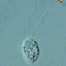
Description of Cryothecomonas
(
Inglês
)
fornecido por BioPedia
Circumscription: Biflagellated heterotrophic flagellate moving by swimming or gliding, with one anteriorly directed and one posteriorly directed flagellum, body surface coated in delicate theca except at site of emergence of flagella and in area of food ingestion. Theca not visible by light microscopy. Feeding by ingestion by ventral face of body often involving pseudopodia. Ultrastructural identity: Mitochondria with tubular cristate, two flagella inserting as inclined basal bodies interconnected by a striated band, giving rise to several microtubular arrays, one of which links basal bodies to nucleus, transition zone with constriction, cell with dictyosomes and osmiophilic bodies. Cell surface with thin organic coating. During mitosis, the nuclear envelope disintegrates, spindle microtubules arise from basal bodies. Synapomorphy: Not specified-this is a tubulocristate flagellate with body enclosed by delicate mucoid theca and a large ventral groove, but neither feature is unique to this taxon. Composition: One genus, several species.
Description of Cryothecomonas armigera
(
Inglês
)
fornecido por BioPedia
Cells egg-shaped and dorso-ventrally flattened (12-32 x 7-23 x about 5 microns). The cytostome is located at the end of the cell at the bottom of a lateral groove. The theca is multilayered, with an electron-dense inner layer and an outer most layer in which the material is arranged to form regularly spaced ridges (not visible by light microscopy). Extrusomes are present throughout the cell.
Description of Cryothecomonas inermis
(
Inglês
)
fornecido por BioPedia
Cells egg-shaped and slightly flattened (10-15 x 7-10 microns). The cytostome is located at the end of the cell at the bottom of a lateral groove. The theca is multilayered (not visible by light microscopy), with an electron-dense inner layer and an outer most layer in which the material is arranged to form regularly spaced ridges. Without extrusomes.
Description of Cryothecomonas scybalophora
(
Inglês
)
fornecido por BioPedia
Cells variable in shape (9-14 x 4.5-9 microns). The theca consists of a single electron-dense layer which externally supports regularly spaced protuberances (not visible by light microscopy). Cells surrounded by debris outside the theca. A few large extrusomes are present at the anterior end of the cell. Complex pseudopodia emanate from the cytostome.
Description of Cryothecomonas vesiculata
(
Inglês
)
fornecido por BioPedia
Cells are elongate (9-14 x 4.5-9 microns). The theca consists of two distinct layers (not visible by light microscopy). Muciferous bodies are regularly packed (hexagonal close packing) underneath the entire cell surface. Lysosome-like bodies very conspicuous.