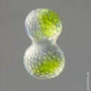
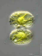
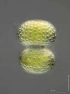
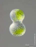
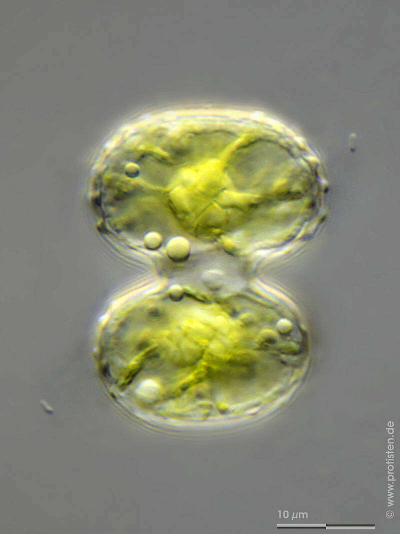
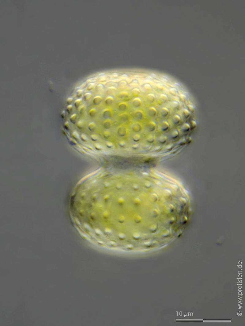
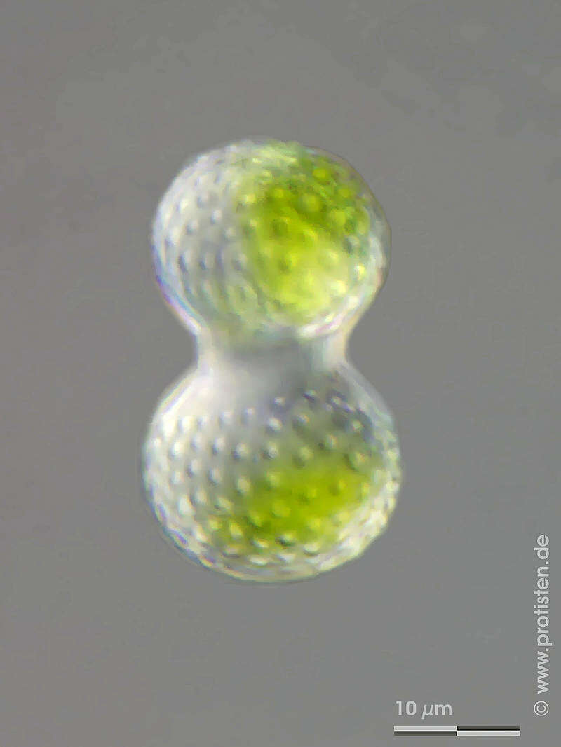
Sampling date 06/2013. Scale bars indicate 10 µm.Three images.First:Synoptic representation of the cell surface (frontal view).Second:Optical cross-section showing nucleus, chloroplasts, assimilate globules and pyrenoids.Third:Synoptic representation of the cell surface (lateral view).Please click on < or > on the image edges or on the dots at the bottom edge of the images to browse through the slides!Place name: Wetland Mittermoos near Fieberbrunn (Tyrol, Austria)Latitude: 47.47998695 Longitude: 12.5240922Microscope Zeiss Universal, camera Canon EOS 600D. DOF images.© Wolfgang Bettighofer,images under Creative Commons License V 3.0 (CC BY-NC-SA).For permission to use of (high resolution) images please contact
postmaster@protisten.de.For further information about the image, please click here:
Link to protisten.de page