-
-
-
-
-
-
-
-
-
Mahide, Castille and Leon, Spain
-
Mellanes, Castilla y Len, Espaa
-
Los Cotos, Madrid, Spain
-

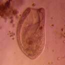
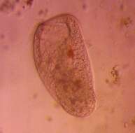
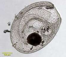
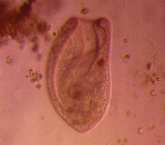
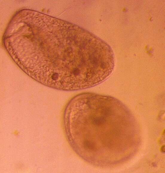
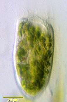
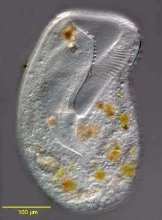
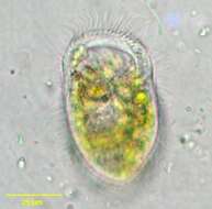
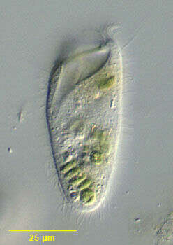
Paracondylostoma setigerum, a colpodid ciliate originally recovered from an alpine pond in central Austria and described by Foissner and Kreutz (Foissner, W., Kreutz, M.: Systematic position and phylogentic relationship of the genus Bursaridium, Paracondylostoma, Thylakidium, Bryometopus, and Bursaria (Ciliphora: Colpoda). Acta Protozoologica 37, 227 - 240 (1998). Overall shape is fusiform with truncate anterior. Oral aperture is anterior, funnel-shaped and extends about 1/3 the length of the cell. A line of membranelles extends completely along the margin of the aperture on the left. Somatic ciliature is uniform in longitudinal kineties. Prominent long bristle-like cilia radiate from the anterior end. The cells occupy a mucus sheath which is transparent and very difficult to see unless bacteria and debris adhere. Cells often flee the sheath when placed on the slide. This individual contains large numbers of zoochlorellae. Centrally located macronucleus is spherical. From freshwater pond near Boise, Idaho. Brightfield illumination.
-
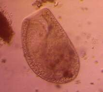
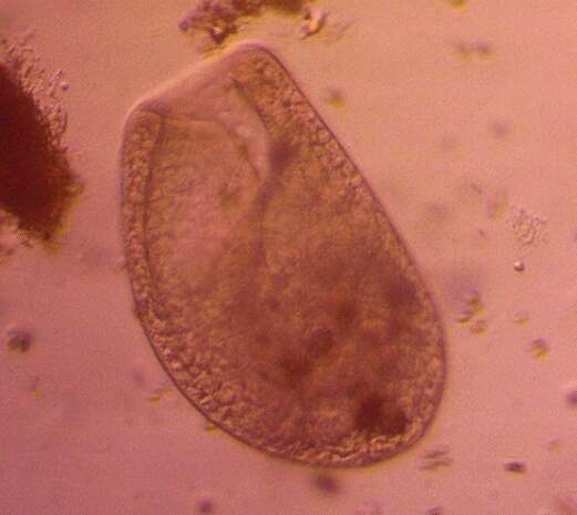
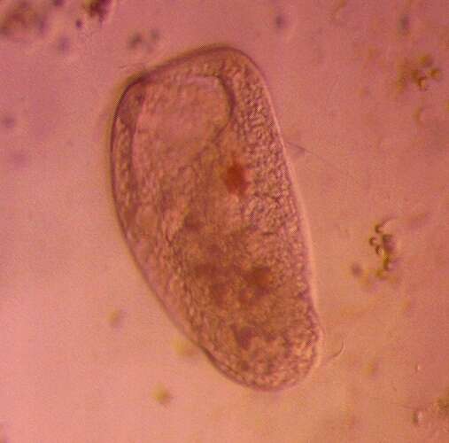
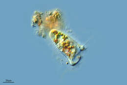
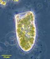
Paracondylostoma setigerum. This image demonstrates the membranelles around the funnel-shaped oral aperture and the long anterior bristle cilia (indistinct here but seen on the organism's right). This individual contains many zoochlorellae. From freshwater pond near Boise, Idaho. Oblique illumination.
-
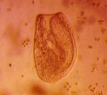
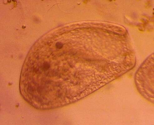
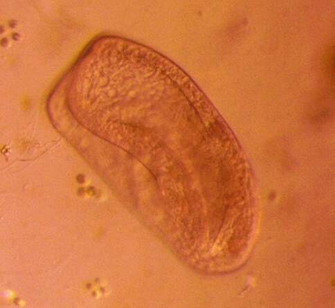
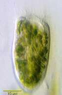
Paracondylostoma setigerum. This image demonstrates the membranelles around the funnel-shaped oral aperture and the long anterior bristle cilia (indistinct here but seen on the organism's right). The delicate, transparent mucus sheath is apparent only due adherent debris. This individual contains many zoochlorellae. From freshwater pond near Boise, Idaho. Phase contrast illumination.
-
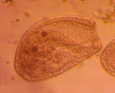
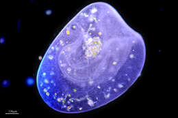
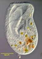
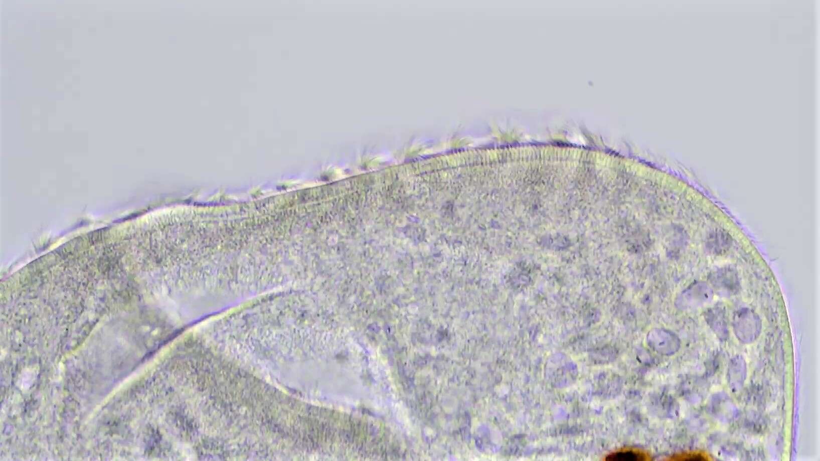
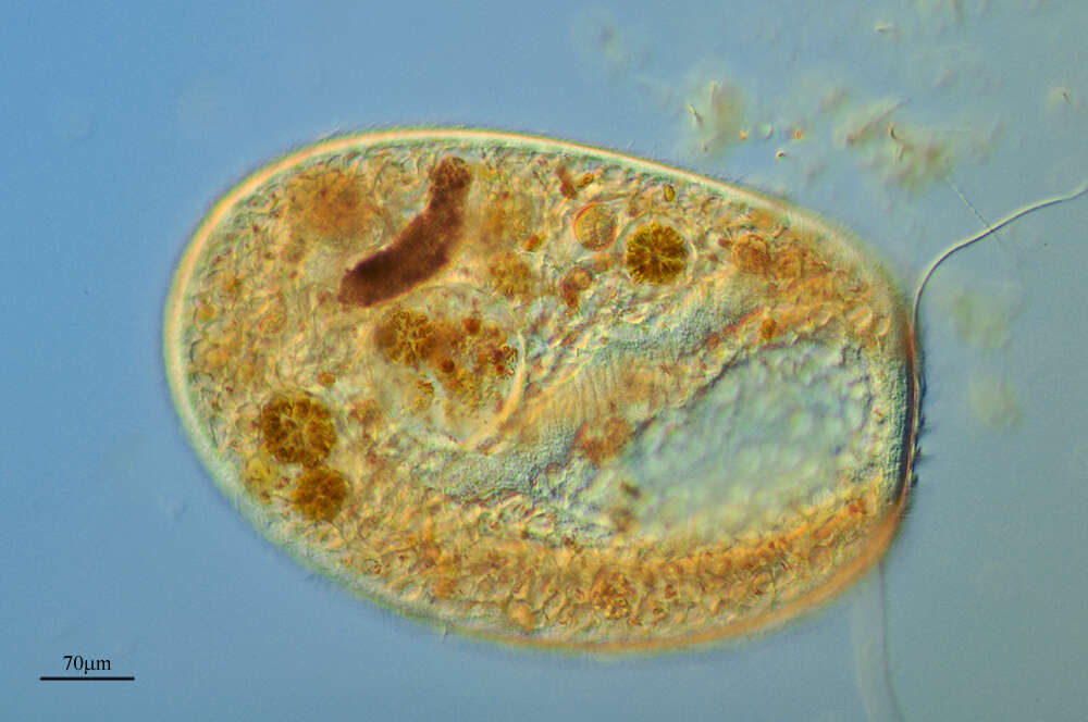
Portrait of slightly contracted Paracondylostoma setigerum (Foissner, 1980), a colpodid ciliate originally recovered from an alpine pond in central Austria and described by Foissner and Kreutz (Foissner, W., Kreutz, M.: Systematic position and phylogentic relationship of the genus Bursaridium, Paracondylostoma, Thylakidium, Bryometopus, and Bursaria (Ciliophora: Colpoda). Acta Protozoologica 37, 227 - 240 (1998). P. Setigerum is the type species. Overall shape is fusiform with truncate anterior. Contracts quickly closing oral aperture when disturbed. Oral aperture is anterior, funnel-shaped and extends about 1/3 the length of the cell. A line of membranelles extends completely along the margin of the aperture on the left. Somatic ciliature is uniform in longitudinal kineties. Prominent long bristle-like cilia radiate from the anterior end. The cells occupy a mucus sheath which is transparent and very difficult to see unless bacteria and debris adhere. Cells often flee the sheath when placed on the slide.Centrally located macronucleus is spherical. The contractile vacuole is located in the posterior half.From freshwater pond near Boise, Idaho. DIC optics.
-
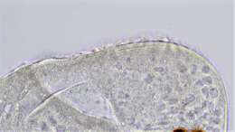
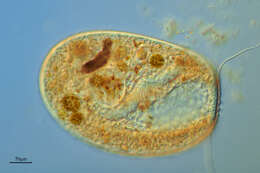
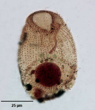
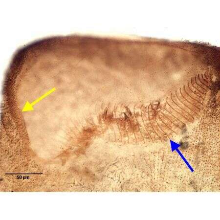
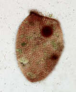
Ventral infraciliature of the colpodid ciliate, Paracondylostoma setigerum chlorelligerum(Foissner&Kreutz,1998). There are approximately 50 longitudinal somatic kineties. The anterior most kinetids of these are very closely spaced. Long stiff bristle-like cilia originate from these closely spaced kineties. The anterior vestibular opening is bordered by a narrow paraoral membrane except for an interruption adjacent to the sigmoid adoral zone of membranelles. The densely stained macronucleus and 3 adjacent micronuclei are visible in this image. Multiple zoochlorellae are visible in the cytoplasm. P. setigerum v. chlorelligerum is identical to P. setigerum except that it contains many zoochlorellae. Foissner feels this characteristic warrants separation as a subspecies. Stained by the silver carbonate technic (see Foissner, W.Europ. J. Protistol.27, 313-330;1991). Brightfield.
-
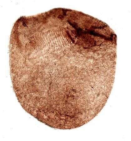
Dorsal infraciliature of the colpodid ciliate, Paracondylostoma setigerum chlorelligerum(Foissner&Kreutz,1998). There are approximately 50 longitudinal somatic kineties. The anterior most kinetids of these are very closely spaced. Long stiff bristle-like cilia originate from these closely spaced kineties. The anterior vestibular opening is bordered by a narrow paraoral membrane except for an interruption adjacent to the sigmoid adoral zone of membranelles. The densely stained macronucleus and 3 adjacent micronuclei are visible in this image. Multiple zoochlorellae are visible in the cytoplasm. Stained by the silver carbonate technic (see Foissner, W.Europ. J. Protistol.27, 313-330;1991). Brightfield.
-
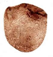
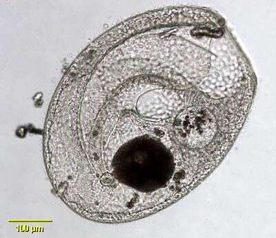
Bursaria truncatella (Müller,1773), a large ciliate classified within the Colpodea. Broadly rounded posteriorly and truncate anteriorly with oral cavity opening apically. The posterior end of the funnel-shaped cytostome curves to left. Cirromembranelles wind in clock-wise fashion around the opening of the oral cavity and descend along the left side of the cytostome. Many small peripheral contractile vacuoles. The somatic ciliation is uniform. The macronucleus is long and band-like. There are many micronuclei. Bursaria is predatory, feeding on other protists and rotifers. Dark food vacuoles are seen in this image. From temporary rainwater pool in grassy field near Boise, Idaho. Brightfield.
-
-
-
-
-
Stained by the methyl green pyronin-Y technique (see Foissner, W.Europ. J. Protistol.27:313-330;1991).Brightfield.
-
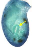
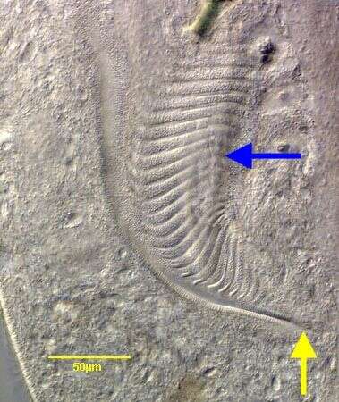
The yellow arrow indicates the closely spaced dikinetids of the "paraoral rows". The blue arrow indicates the adoral membranelles, each composed of three rows of kinetids.Only the anterior two rows bear cilia. Stained by the silver carbonate technique (see Foissner, W.Europ. J. Protistol.27:313-330;1991).Brightfield.