-
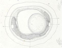
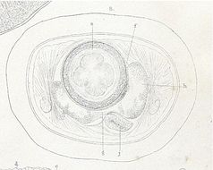
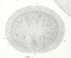
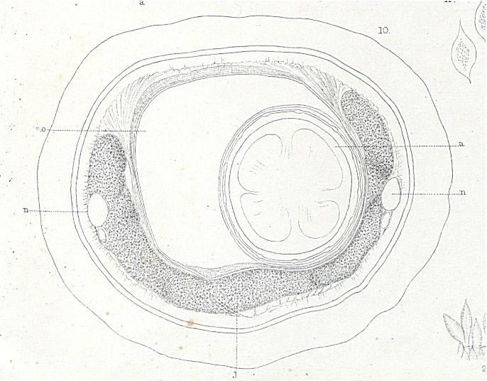
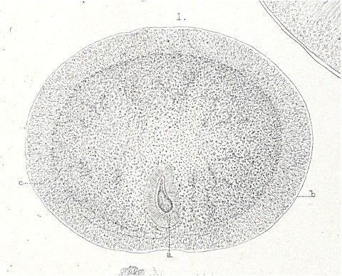
Plate10.10 Transverse section of the body of a Amphiporus lactifloreus, in which no reproductive elements are visible.
-
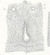
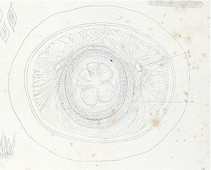
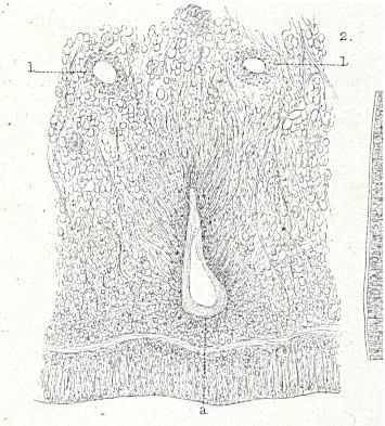
Amphiporus lactifloreus. The invagination of the proboscis and the changes in the region surrounding it are well shown.
-
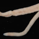
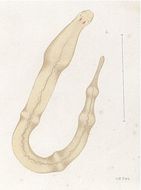
Plate1.1 Amphiporus lactifloreus, Johnston pinkish variety.
-
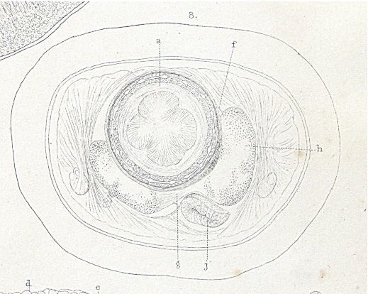
Plate10.8 Amphiporus lactifloreus Transverse section of the anterior part of the cephalic ganglia.
-
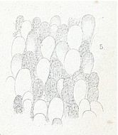
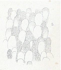
Plate10.5 Amphiporus lactifloreus View of a portion of skin snipped from living specimen, under moderate pressure.
-
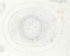
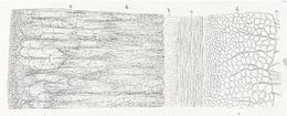
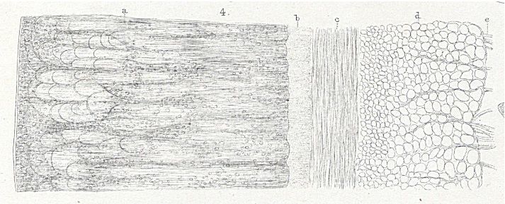
Plate10.4 Transverse section of the body wall of an Amphiporus lactifloreus.
-
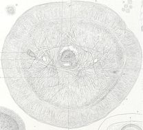
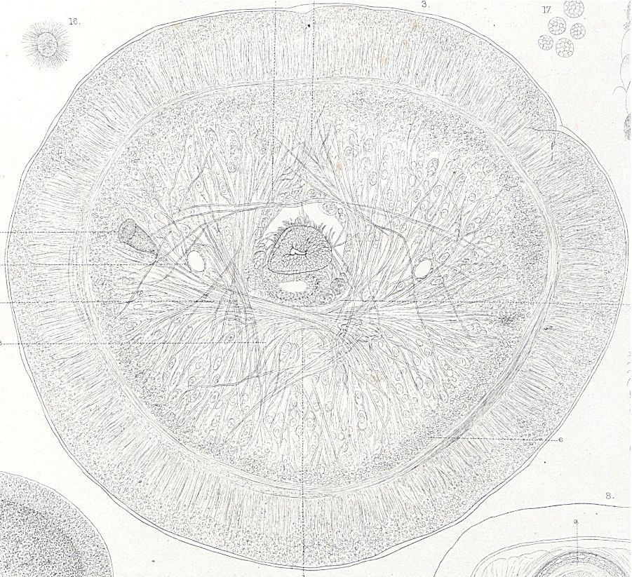
Plate10.3 Transverse section of the snout of Amphiporus lactifloreus in front of the ganglia, somewhat flattened from pressure.
-
Plate10.2 Amphiporus lactifloreus. Section of the snout.
-
Plate10.1 First transverse section of the snout of Amphiporus lactifloreus.
-
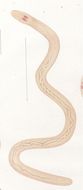
Plate1.2 Amphiporus lactifloreus whitish variety.
-
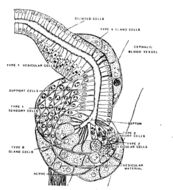
Diagram of the cerebral organ of Amphiporus lactifloreus in frontal section; the top of the diagram is anteriorFrom: Fine structure of the cerebral organs in hoplonemerteans (Nemertini), with a discussion of their function Helen M. Amerongen Zoomorphology (1987) 10: 145-159
-
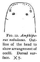
Outline of the head to show the arrangement of ocelli. Dorsal surface.Coe, W. R. (1901). The Nemerteans of the Expedition. Proceedings of the Washington Academy of Sciences, Vol. 3, 1-110.
-
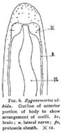
Outline of anterior portion of body to show the arrangement of ocelli. Coe, W. R. (1901). The Nemerteans of the Expedition. Proceedings of the Washington Academy of Sciences, Vol. 3, 1-110.
-
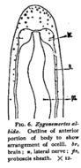
Zygonemertes albida: Outline of anterior portion of body to show arrangement of ocelli.
-
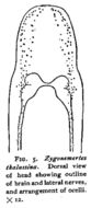
Dorsal view of anterior portion of body showing outline of brain and lateral nerves, and arrangement of ocelli.Coe, W. R. (1901). The Nemerteans of the Expedition. Proceedings of the Washington Academy of Sciences, Vol. 3, 1-110.
-
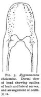
Zygonemertes thalassina: Dorsal view of head showing outline of brain and lateral nerves, and arrangement of ocelli x12
-
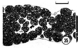
Photomicrograph of a longitudinal section through the posterior end of a female that was apparently depositing cleaving... Photomicrograph of a longitudinal section through the posterior end of a female that was apparently depositing cleaving embryos into an egg string. The single arrowhead marks the caudal end of the epidermis. Scale bar, 100 um.
-
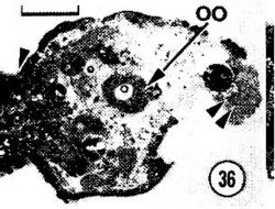
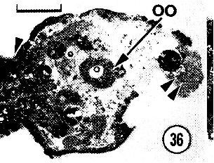
Photomicrograph of a longitudinal section through the posterior end of a female that was fixed while laying an egg string...Photomicrograph of a longitudinal section through the posterior end of a female that was fixed while laying an egg string. The single arrowhead marks a subterminal constriction, and the double arrowheads point to a mass of spermatozoa. Cellular debris occurring toward the end of the worm probably represents artifactual damage incurred when the worm was removed from its egg string. Ruthenium red/sodium cacodylate primary fixative. 00, oocyte. Scale bar, 100 um.
-
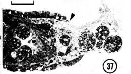
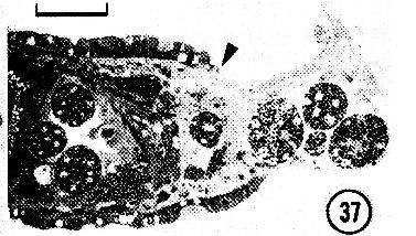
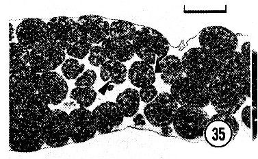
A section of an egg string containing blastulae (arrowhead). Note that extraembryonic cells are lacking within the egg string...A section of an egg string containing blastulae (arrowhead). Note that extraembryonic cells are lacking within the egg string. Ruthenium red - sodium cacodylate primary fixative. Scale bar, 100 um.
-
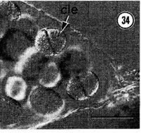
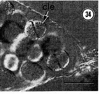
The distal end of an egg string with zygotes undergoing first cleavage (cle). Scale bar, 100 um.
-
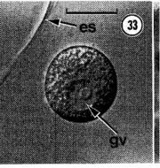
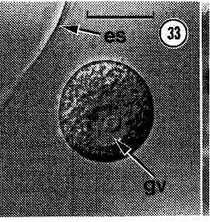
The peripheral region of a newly deposited egg string (es) showing an oocyte with an intact germinal vesicle (gv)...the peripheral region of a newly deposited egg string (es) showing an oocyte with an intact germinal vesicle (gv). Scale bar, 50 u.m.
-
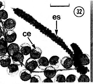
An egg string (es) containing numerous developing embryos. ce, crab egg. Scale bar, 0.5 mm.
-
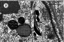
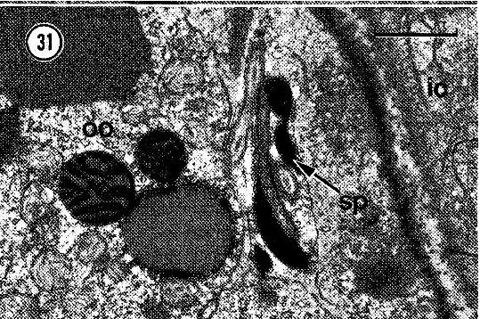
Spermatozoa (sp) tightly appressed to an intraovarian oocyte (00). ic, intestinal cell. Scale bar, l um.
-
Spermatozoa (sp) tightly appressed to an intraovarian oocyte (00). ic, intestinal cell. Scale bar, l um.