-
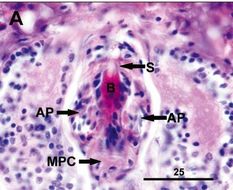
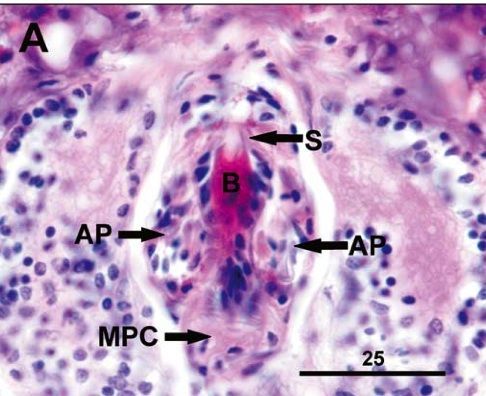
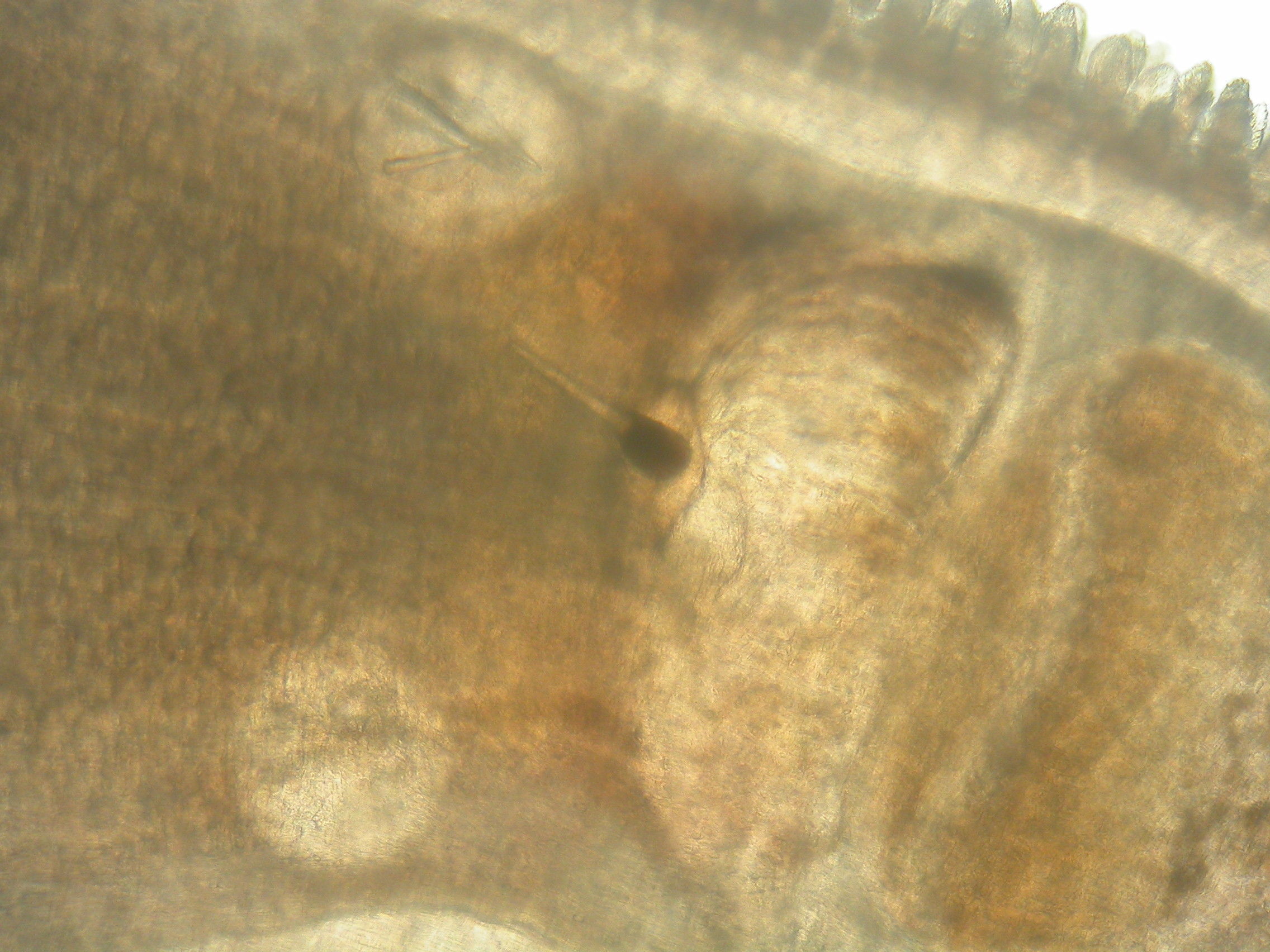
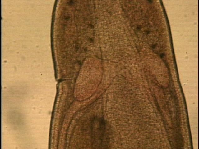
Holotype (Carcino B.l. (1), slide 3, worm 1). Stylet bulb region of worm showing pyriform stylet (S) on the basis (B) with two..Holotype (Carcino B.l. (1), slide 3, worm 1). Stylet bulb region of worm showing pyriform stylet (S) on the basis (B) with two accessory stylet pouches (AP) anterior to the middle proboscis chamber (MPC).
-
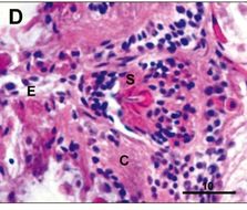
Portion of the stylet in the anterior proboscis chamber showing the stylet hub, a portion of the esophagus.
-
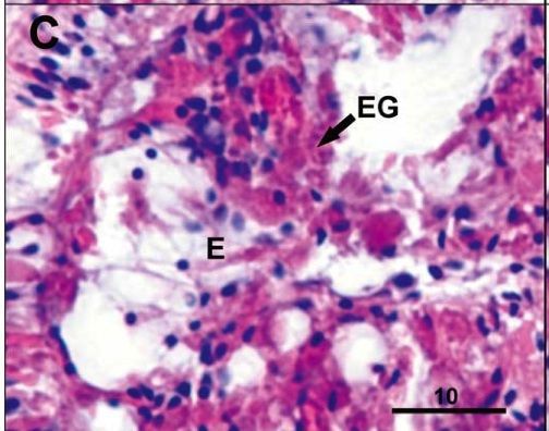
Eosinophilic glands (EG) lining the anterior esophagus.
-
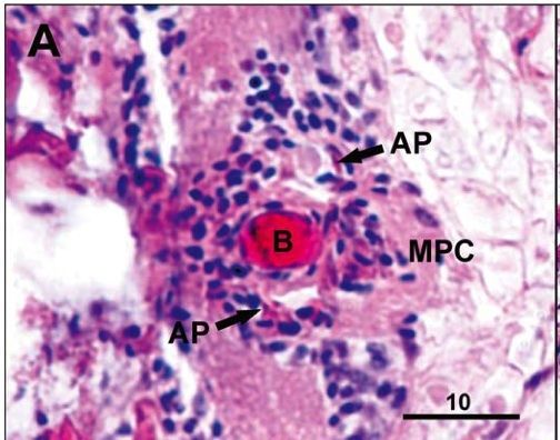
Stylet bulb region of worm showing two accessory stylet pouches (AP) adjacent to the basis.
-
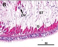
Medial section of body with single row of submuscular glands (G) and a presumptive ovum undergoing resorption (Ov).
-
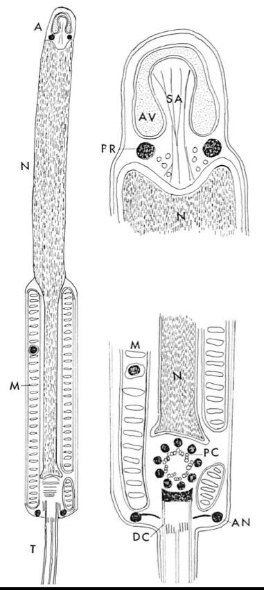
Schematic diagram of the Malacabdella grossa spermatozoon and its acrosomal region and neck region.From: The spermatozoon of the nemertine Malacobdella grossa Bjorn Afzelius J. Submicro. Cytol., 3, 181-192, 1971 (A) Acrosome (AN) Annulus (AV) Acrosomal vesicle (DC) Distal cenriole (M) Mitochondrial sheath (N) Nucleus (PC) Proximal centriole (PR) Post-acrosomal ring (SA) Subacrosomal material (T) TailWith these considerations as a background a fine structural investigation was started using the sperm of the parasitic nemertine Malacobdella grossa (O. F. Müller). The sperm of this species can be characterized as advanced as evidenced by the studies of Retzius (1904) and Franzén (1956). The mode of fertilization is incompletely known. (A) Acrosome (AN) Annulus (AV) Acrosomal vesicle (DC) Distal cenriole (M) Mitochondrial sheath (N) Nucleus (PC) Proximal centriole (PR) Post-acrosomal ring (SA) Subacrosomal material (T) TailWith these considerations as a background a fine structural investigation was started using the sperm of the parasitic nemertine Malacobdella grossa (O. F. Müller). The sperm of this species can be characterized as advanced as evidenced by the studies of Retzius (1904) and Franzén (1956). The mode of fertilization is incompletely known.From: The spermatozoon of the nemertine Malacobdella grossa Bjorn Afzelius J. Submicro. Cytol., 3, 181-192, 1971(A) Acrosome (AN) Annulus (AV) Acrosomal vesicle (DC) Distal cenriole (M) Mitochondrial sheath (N) Nucleus (PC) Proximal centriole (PR) Post-acrosomal ring (SA) Subacrosomal material (T) TailWith these considerations as a background a fine structural investigation was started using the sperm of the parasitic nemertine Malacobdella grossa (O. F. Müller). The sperm of this species can be characterized as advanced as evidenced by the studies of Retzius (1904) and Franzén (1956). The mode of fertilization is incompletely known.
-
-
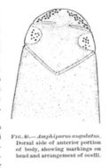
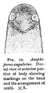
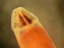
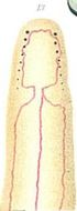
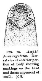
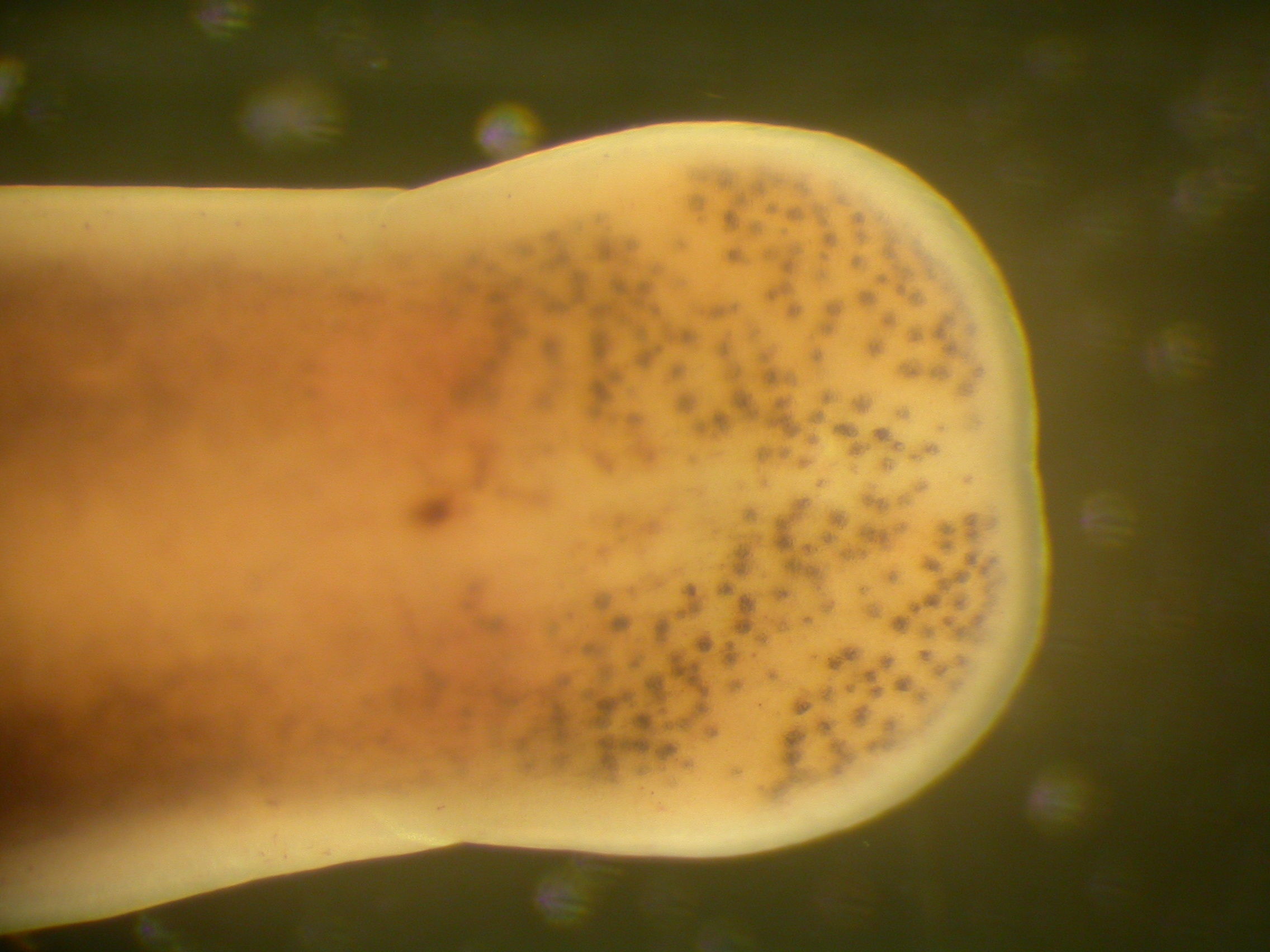
Dorsal view of anterior portion of body showing markings on the head and the arrangement of ocelli.Coe, W. R. (1901). The Nemerteans of the Expedition. Proceedings of the Washington Academy of Sciences, Vol. 3, 1-110.
-
-
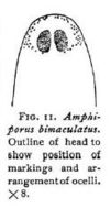
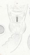
Outline of head to show position of markings and arrangement of ocelli.Coe, W. R. (1901). The Nemerteans of the Expedition. Proceedings of the Washington Academy of Sciences, Vol. 3, 1-110.
-
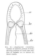
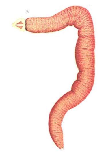
Amphiporus bimaculatus: head
-
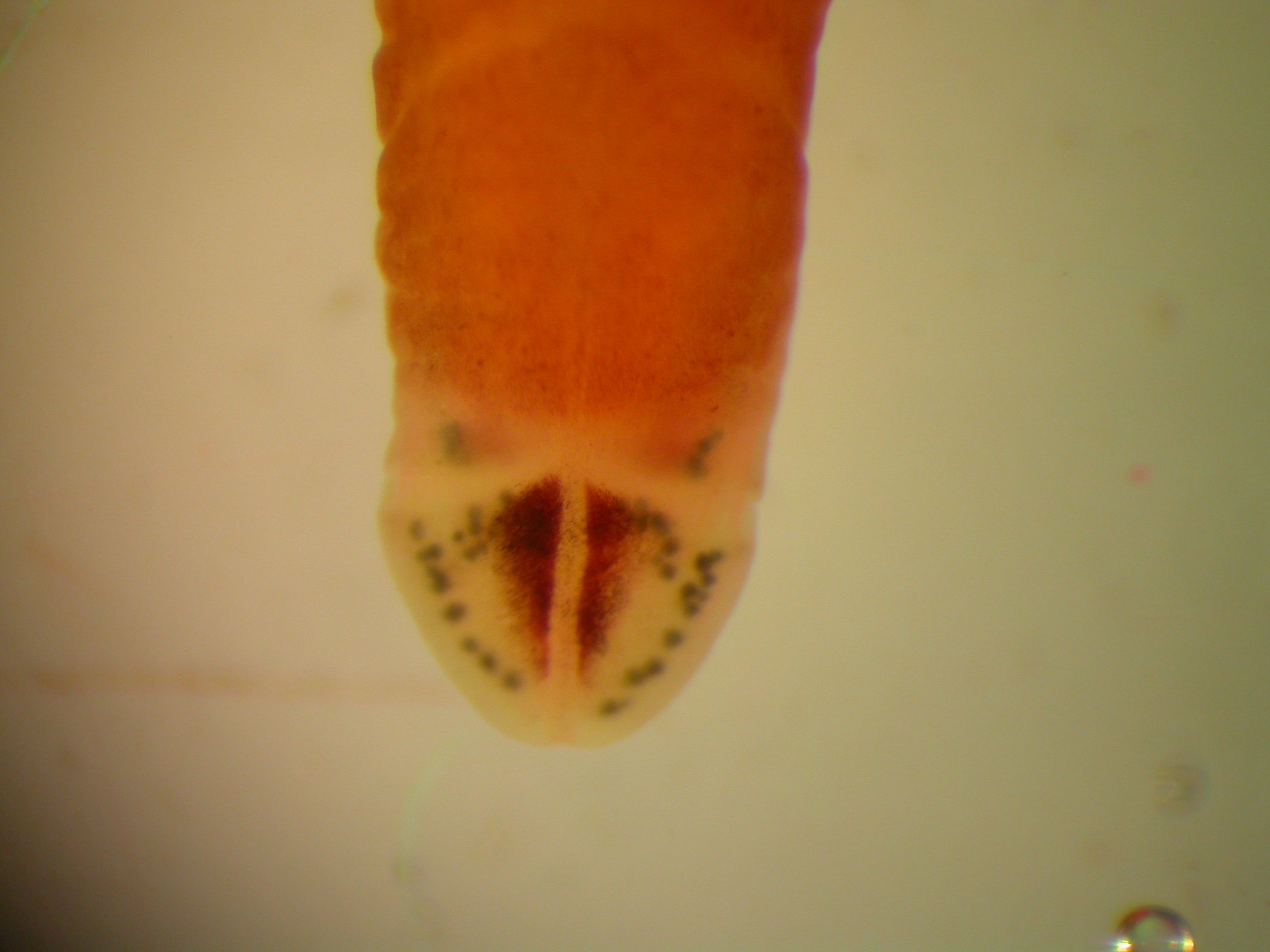
Amphiporus bimaculatus: head
-
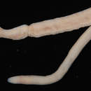
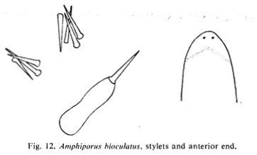
Stylets and anterior endBrunberg, L. On the Nemertean Fauna of Danish Waters. Ophelia, 1(1), 77-111.
-
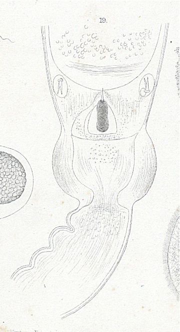
Plate13.19 Stylet-region of Amphiporus bioculatus
-
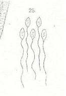
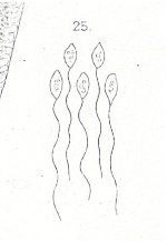
Plate7.25 Spermatozoa of Amphiporus bioculatus
-
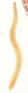
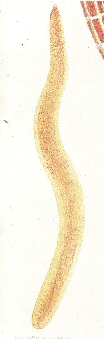
Plate8.3 Amphiporus bioculatus n. s.
-
-
-
-
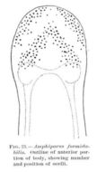
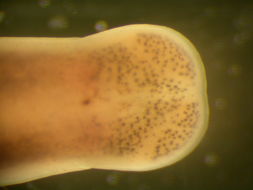
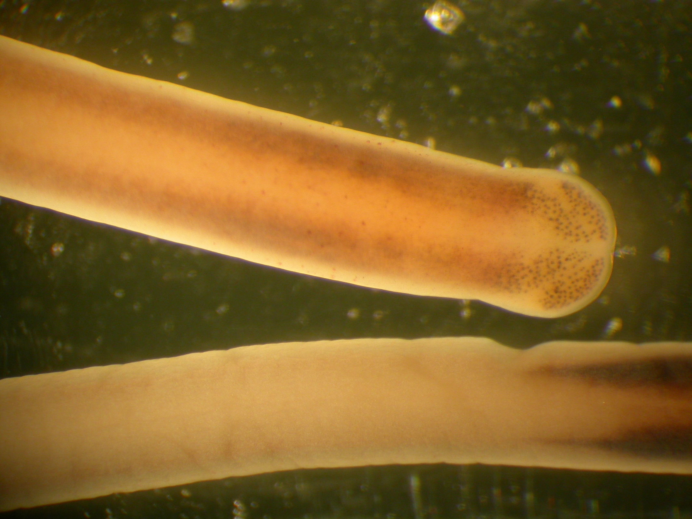
Amphiporus formidabilis: Close-up of head
-
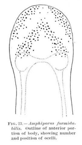
Amphiporus formidabilis: Head showing eye pattern
-
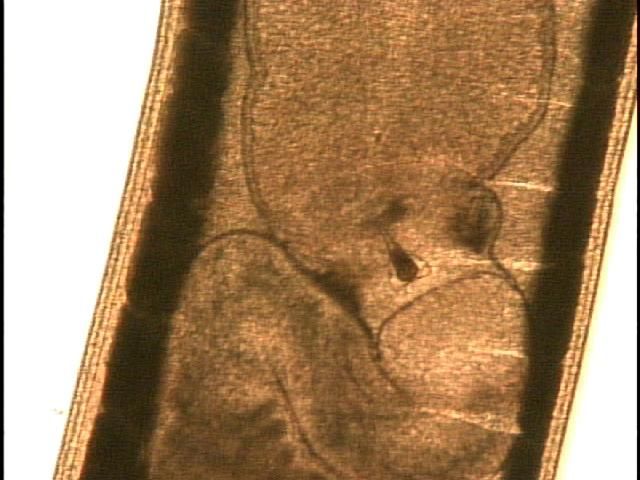
Amphiporus formidabilis: stylet
-
Amphiporus formidabilis: Stylet (squash prep)
-
Amphiporus formidabilis: Brain region (squash prep)