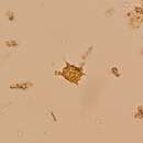
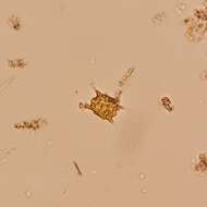
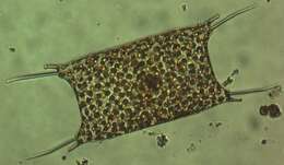
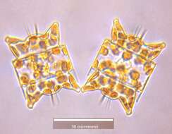
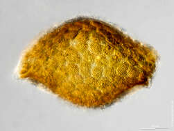
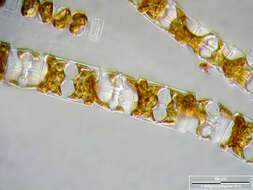
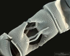
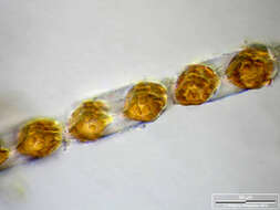
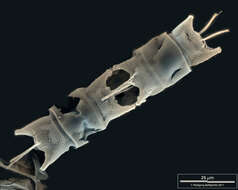
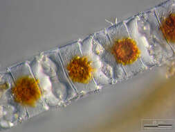
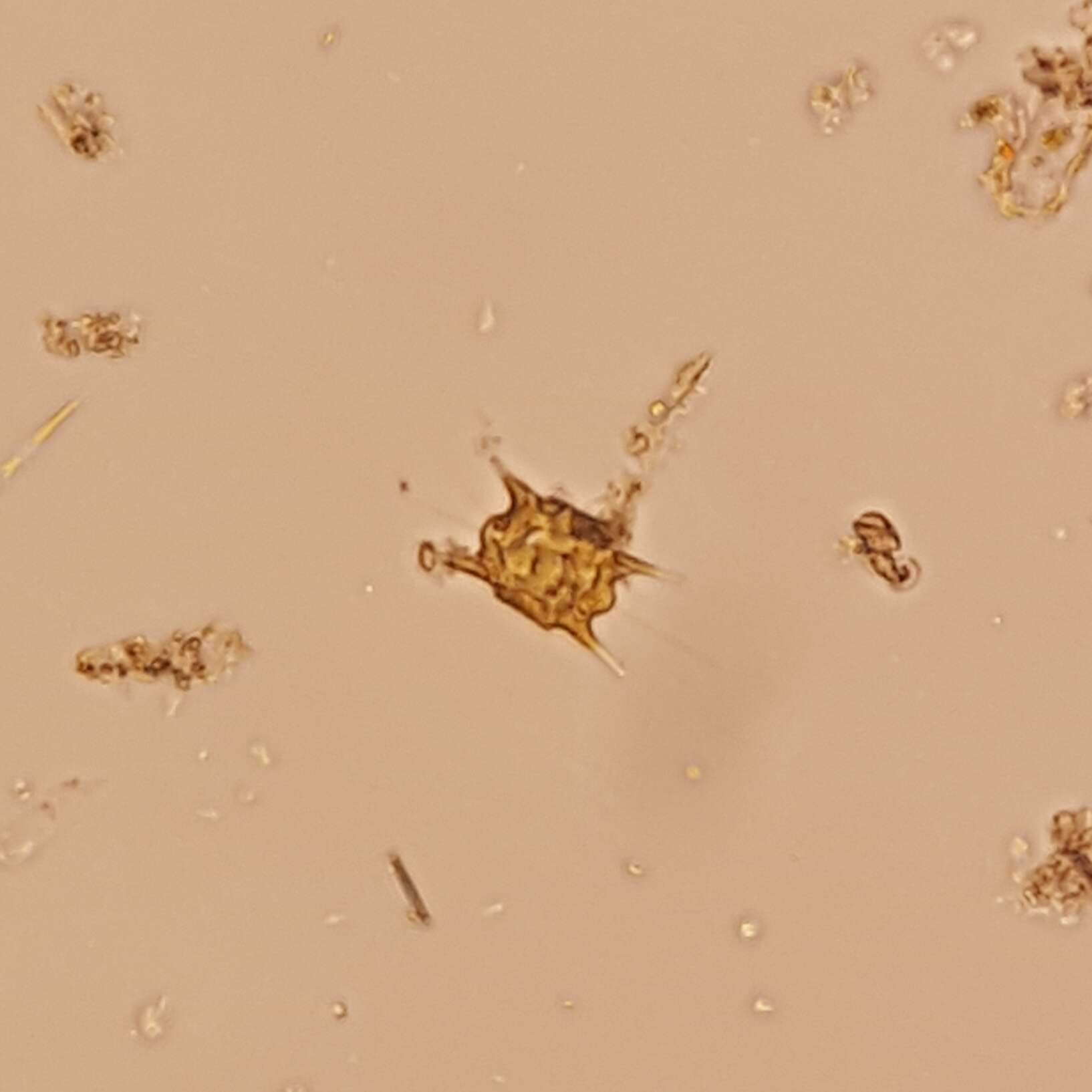
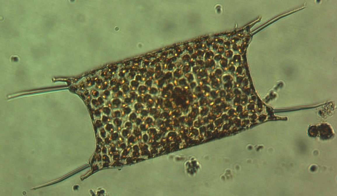
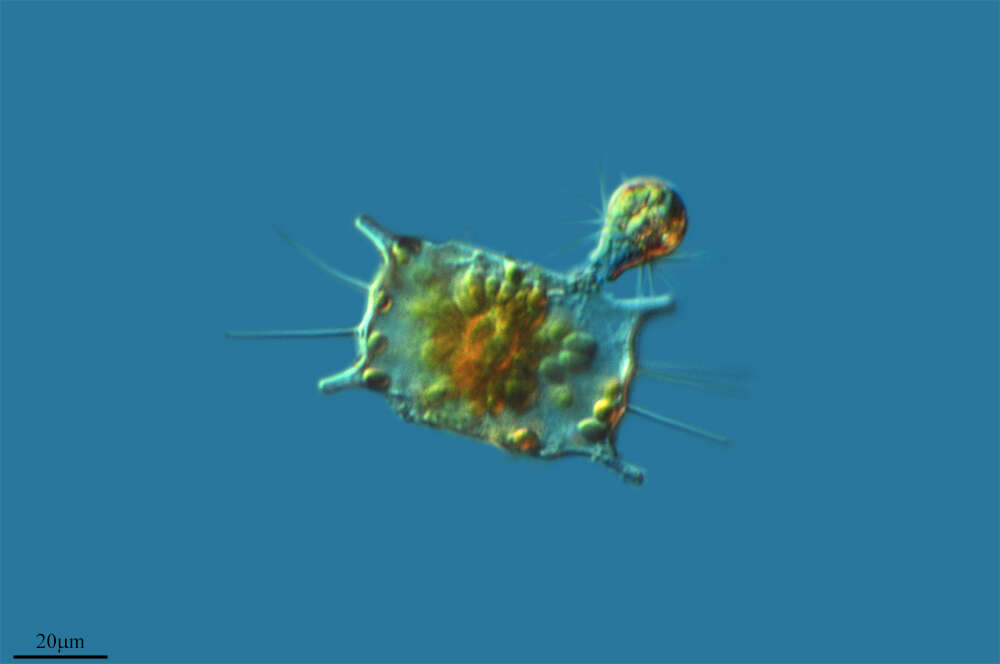
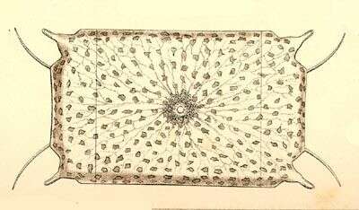
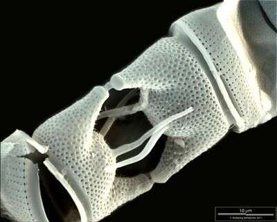
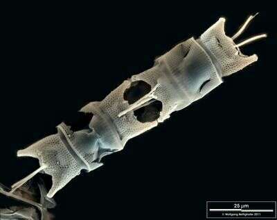
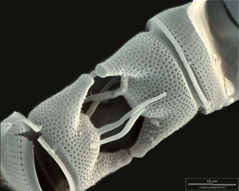
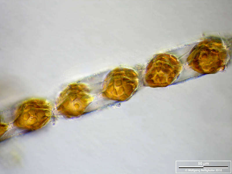
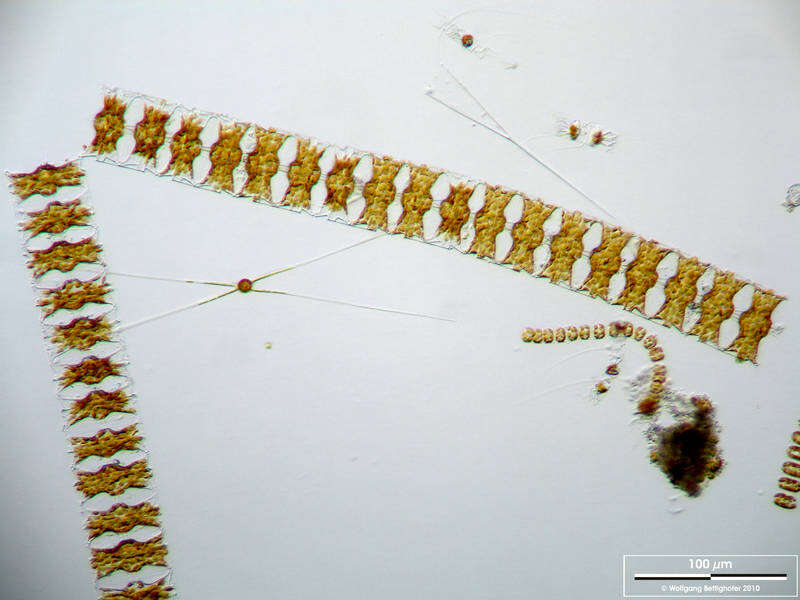
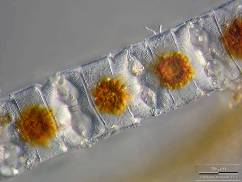
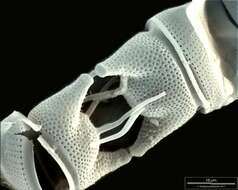
Sampling date 04/2011. Scale bars indicate 25 µm (1), 50 µm (2, 3), 100 µm (4).Four images.First and second:Depth-of-focus image exhibiting structure of the frustules. In the first image, chloroplasts are concentrated in cell centers due to long lasting exposition with the microscope’s illumination. Third:Depth-of-focus image exhibiting structure of the frustules (apical view). Brightfield, oblique light.Fourth:Long chains of
Odontella aurita accompanied by
Chaetoceros danicus and
Thalassiosira nordenskjoeldii.Please click on < or > on the image edges or on the dots at the bottom edge of the images to browse through the slides!Place name: North Sea around HeligolandLatitude: 54.186311 Longitude: 7.895034Microscope Zeiss Universal, camera Olympus C7070WZ. DOF images.© Wolfgang Bettighofer,images under Creative Commons License V 3.0 (CC BY-NC-SA).For permission to use of (high resolution) images please contact
postmaster@protisten.de.For further information about the image, please click here:
Link to protisten.de page