-
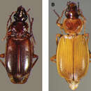
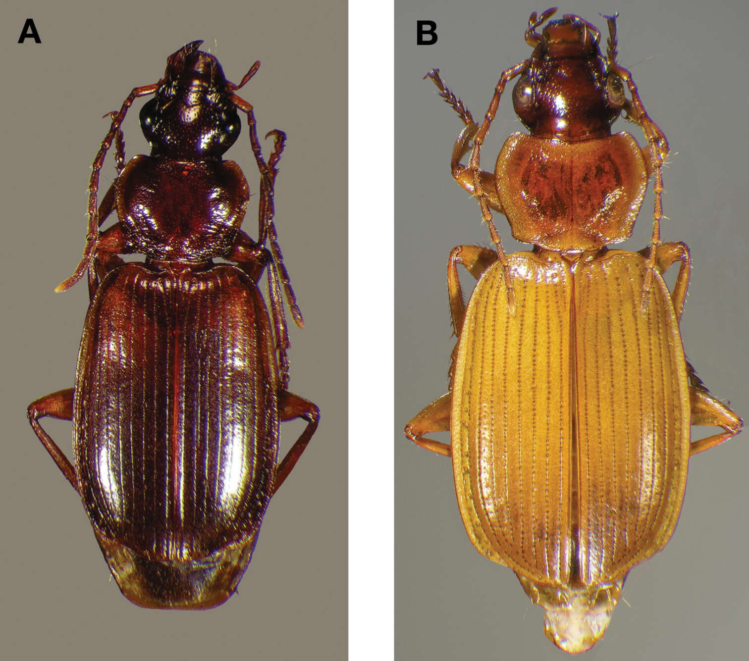
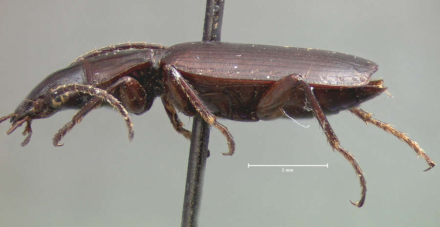
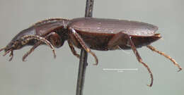
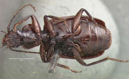
Figure 22. Dorsal habitus and color pattern of Cymindis punctigera punctigera LeConte: A, showing typical dorsal coloration (OBL 10.17 mm); B, showing coloration of some specimens from the Big Bend area of southwestern Texas, U.S.A. (OBL 11.83 mm).
-
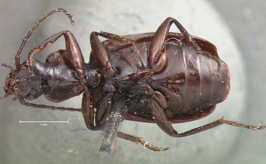
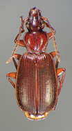
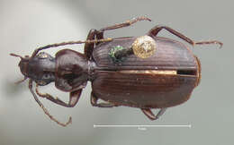
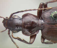
Figure 23. Dorsal habitus and color pattern of Cymindis punctigera sulcipennis (Horn) (OBL 10.83 mm).
-
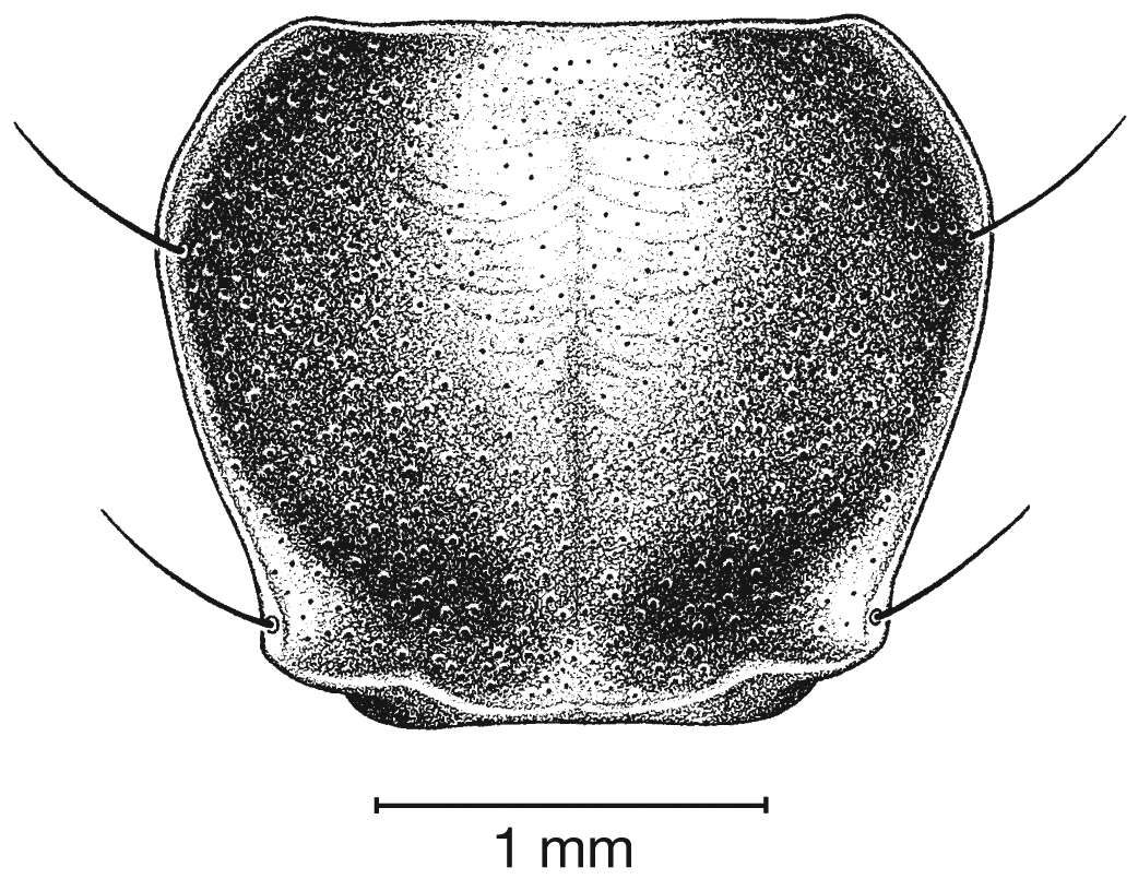
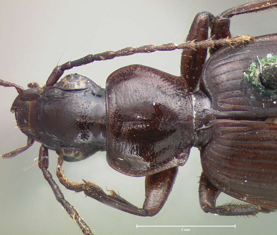
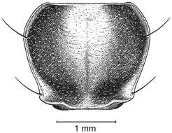
Figure 24. Pronotum, dorsal aspect, of Cymindis punctigera sulcipennis (Horn).
-
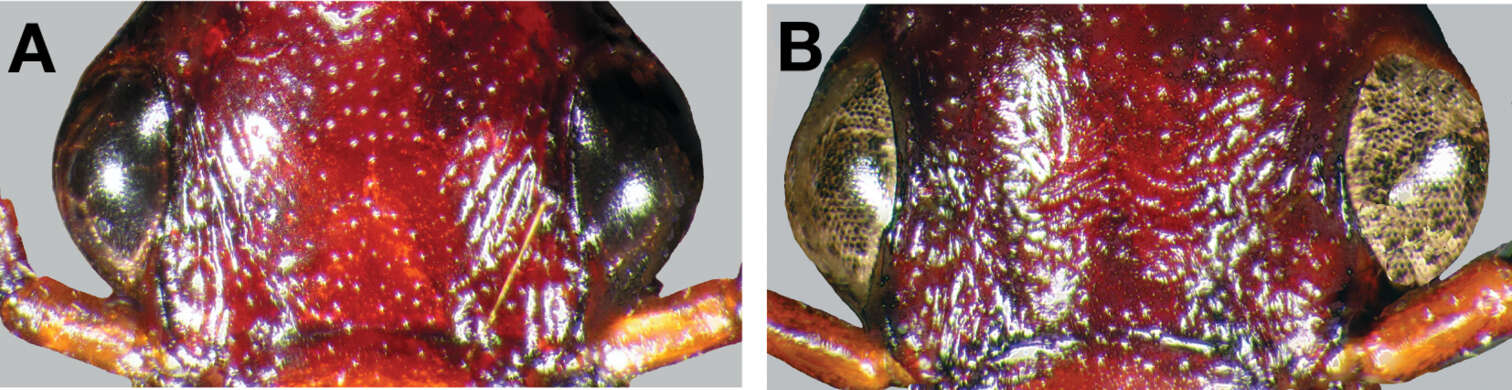
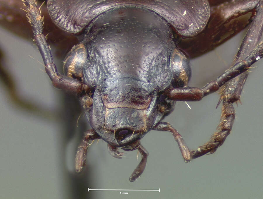
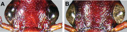
Figure 25. Head capsule, dorsal aspect, showing frontal macrosculpture: A, Cymindis punctigera punctigera LeConte; B, Cymindis punctigera sulcipennis (Horn), note transverse lines between eyes.
-
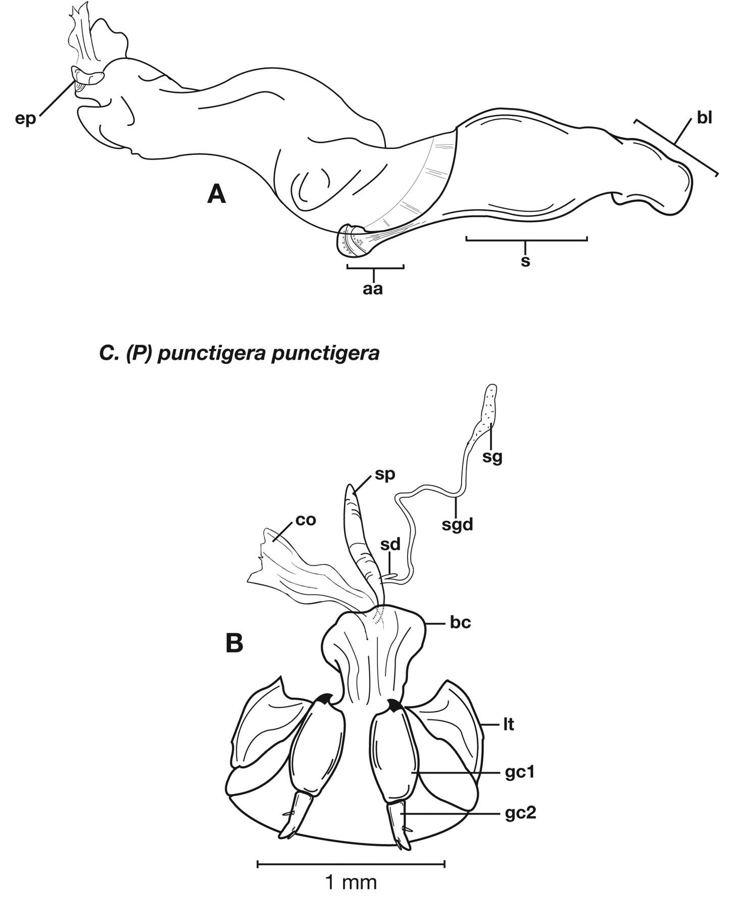
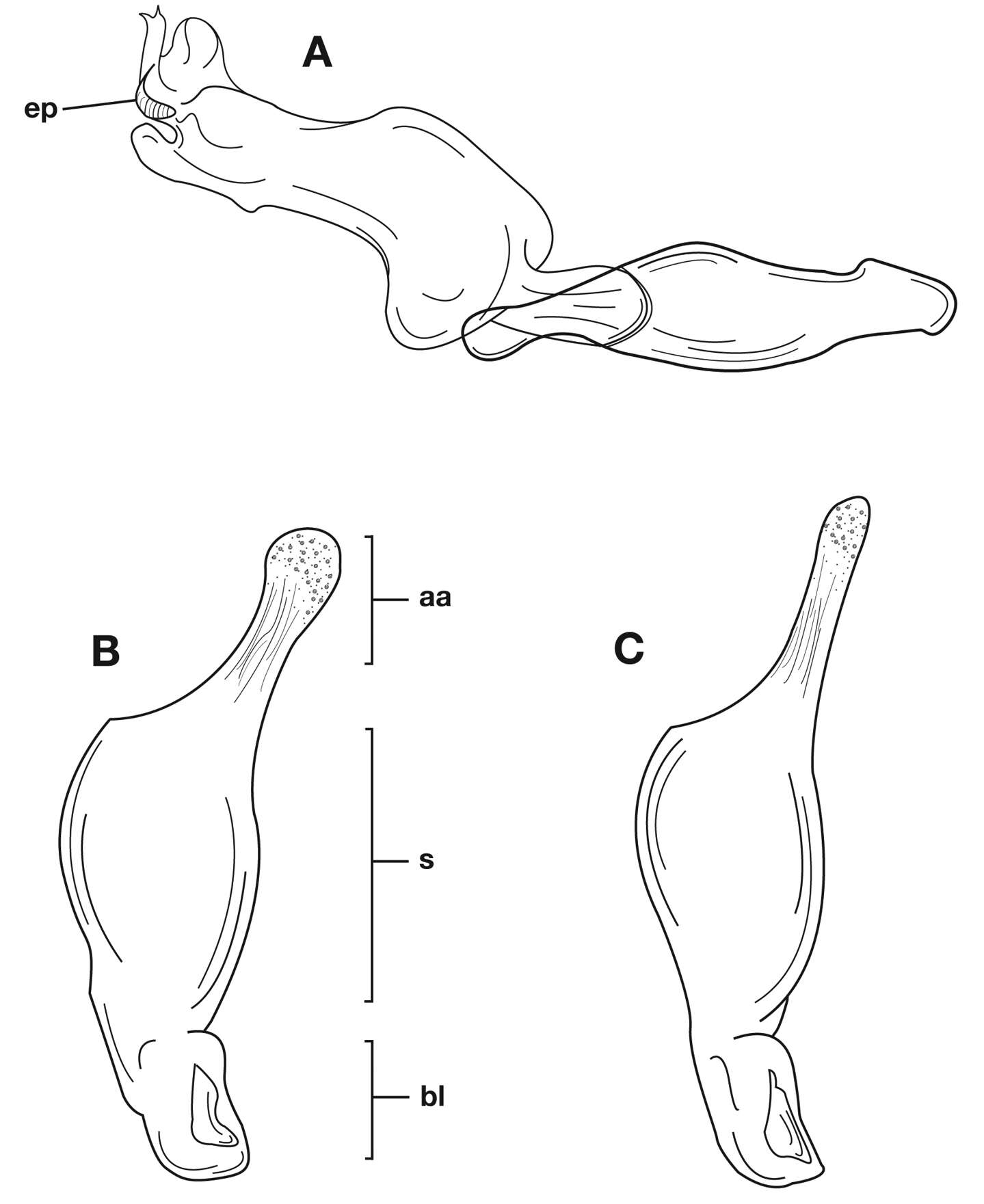
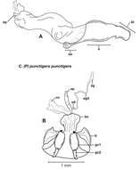
Figure 26. Structural features of Cymindis punctigera punctigera LeConte: A, phallus and everted endophallus, right lateral aspect; B, female reproductive tract and ovipositor, ventral aspect. Legend: aa, apical area; bc, bursa copulatrix; bl, basal lobe; co, common oviduct; ep, endophallic plate; gc1, gonocoxite 1; gc2, gonocoxite 2; lt, lateral tergite; s, shaft; sd, spermathecal diverticulum; sg, spermathecal gland; sgd, spermathecal gland duct; sp, spermatheca.
-
Figure 27. Male genitalia of Cymindis punctigera punctigera LeConte: A, phallus and everted endophallus, dorsal aspect, showing right-hand placement of endophallus when everted; B-C, left lateral aspect of phallus, endophallus inverted, showing intrapopulation variation (Jeff Davis Co., Texas, U.S.A.) in form of phallic apex; texture and distinctive striations extended from mid-apical area toward shaft. Legend: aa, apical area; bl, basal lobe; ep, endophallic plate; s, shaft.
-
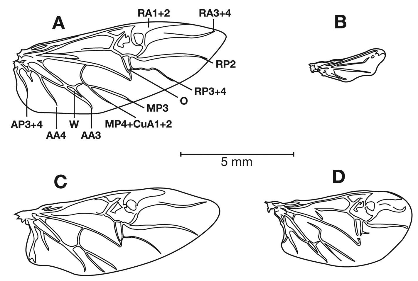
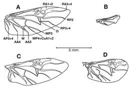
Figure 28. Right hindwings of Cymindis punctigera punctigera LeConte, dorsal aspect: A-B, extremes of intraspecific variation from functional (A) to markedly atrophied (B); C-D, showing intrapopulation variation (Cloudcroft, New Mexico, U.S.A.) from functional (C) to somewhat atrophied (D). Legend: (from Kukalova-Peck and Lawrence 2004) AA3, anterior anal 3; AA4, anterior anal 4; AP3+4, posterior anal 3+4; MP3, posterior median 3; MP4+CuA1+2, posterior median 4 + anterior cubitus 1+2; O, Oblongum cell; RA1+2, anterior radius 1+2; RA3+4, anterior radius 3+4; RP2, posterior radius 2; RP3+4, posterior radius 3+4; W, wedge cell.
-
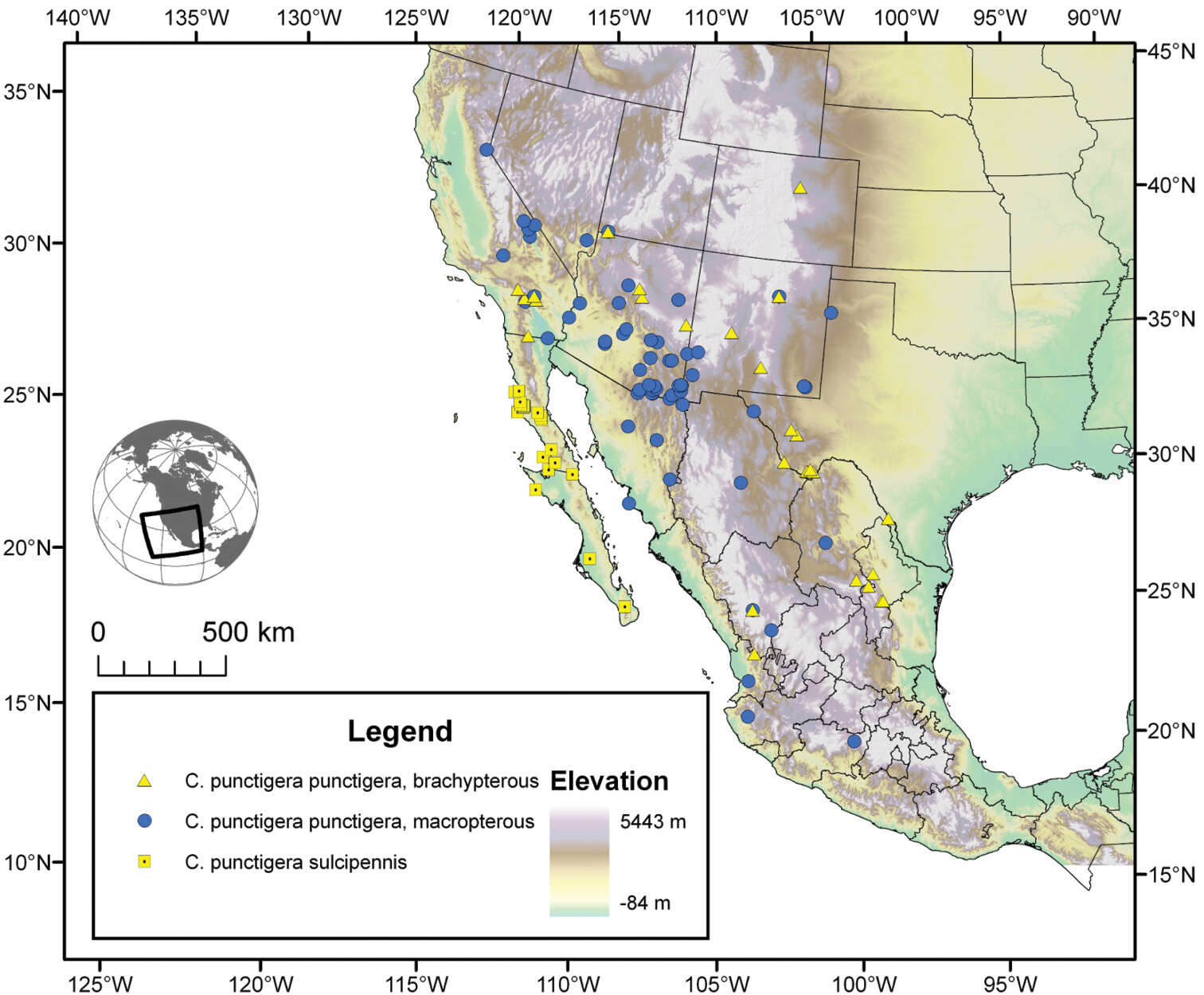
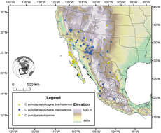
Figure 29. Map of southwestern U.S.A. and northern Mexico, showing position of localities of the subspecies of Cymindis punctigera LeConte, and distribution of macropterous and brachypterous individuals of Cymindis punctigera punctigera.
-
Figure 22. Dorsal habitus and color pattern of Cymindis punctigera punctigera LeConte: A, showing typical dorsal coloration (OBL 10.17 mm); B, showing coloration of some specimens from the Big Bend area of southwestern Texas, U.S.A. (OBL 11.83 mm).
-
Figure 25. Head capsule, dorsal aspect, showing frontal macrosculpture: A, Cymindis punctigera punctigera LeConte; B, Cymindis punctigera sulcipennis (Horn), note transverse lines between eyes.
-
Figure 26. Structural features of Cymindis punctigera punctigera LeConte: A, phallus and everted endophallus, right lateral aspect; B, female reproductive tract and ovipositor, ventral aspect. Legend: aa, apical area; bc, bursa copulatrix; bl, basal lobe; co, common oviduct; ep, endophallic plate; gc1, gonocoxite 1; gc2, gonocoxite 2; lt, lateral tergite; s, shaft; sd, spermathecal diverticulum; sg, spermathecal gland; sgd, spermathecal gland duct; sp, spermatheca.
-
Figure 27. Male genitalia of Cymindis punctigera punctigera LeConte: A, phallus and everted endophallus, dorsal aspect, showing right-hand placement of endophallus when everted; B-C, left lateral aspect of phallus, endophallus inverted, showing intrapopulation variation (Jeff Davis Co., Texas, U.S.A.) in form of phallic apex; texture and distinctive striations extended from mid-apical area toward shaft. Legend: aa, apical area; bl, basal lobe; ep, endophallic plate; s, shaft.
-
Figure 28. Right hindwings of Cymindis punctigera punctigera LeConte, dorsal aspect: A-B, extremes of intraspecific variation from functional (A) to markedly atrophied (B); C-D, showing intrapopulation variation (Cloudcroft, New Mexico, U.S.A.) from functional (C) to somewhat atrophied (D). Legend: (from Kukalova-Peck and Lawrence 2004) AA3, anterior anal 3; AA4, anterior anal 4; AP3+4, posterior anal 3+4; MP3, posterior median 3; MP4+CuA1+2, posterior median 4 + anterior cubitus 1+2; O, Oblongum cell; RA1+2, anterior radius 1+2; RA3+4, anterior radius 3+4; RP2, posterior radius 2; RP3+4, posterior radius 3+4; W, wedge cell.
-
Figure 29. Map of southwestern U.S.A. and northern Mexico, showing position of localities of the subspecies of Cymindis punctigera LeConte, and distribution of macropterous and brachypterous individuals of Cymindis punctigera punctigera.
-
Figure 23. Dorsal habitus and color pattern of Cymindis punctigera sulcipennis (Horn) (OBL 10.83 mm).
-
Figure 24. Pronotum, dorsal aspect, of Cymindis punctigera sulcipennis (Horn).
-
Figure 25. Head capsule, dorsal aspect, showing frontal macrosculpture: A, Cymindis punctigera punctigera LeConte; B, Cymindis punctigera sulcipennis (Horn), note transverse lines between eyes.
-
Figure 29. Map of southwestern U.S.A. and northern Mexico, showing position of localities of the subspecies of Cymindis punctigera LeConte, and distribution of macropterous and brachypterous individuals of Cymindis punctigera punctigera.
-
-
-
-
-
-