-
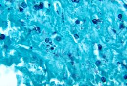
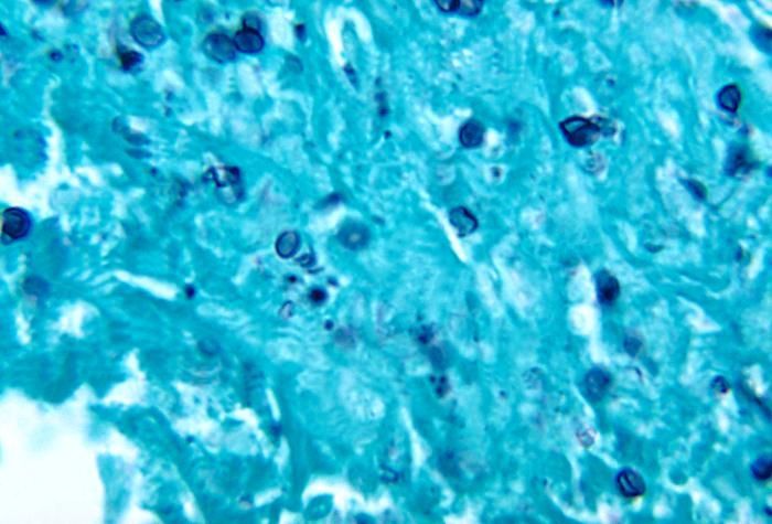
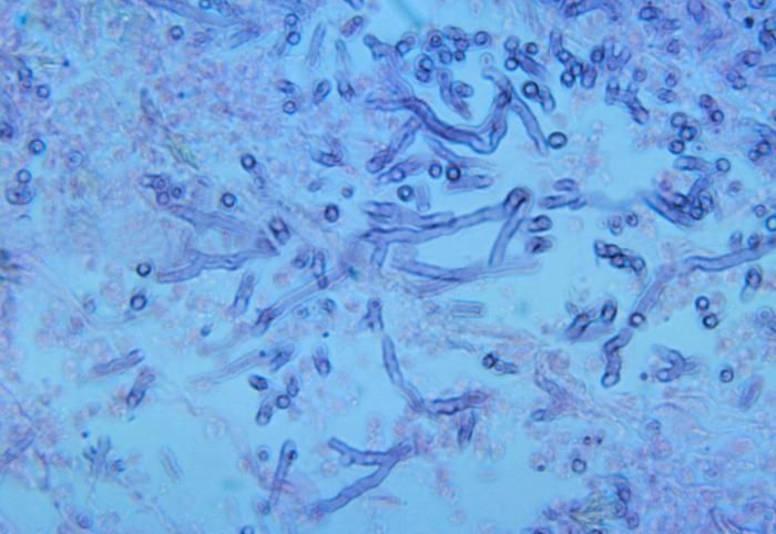
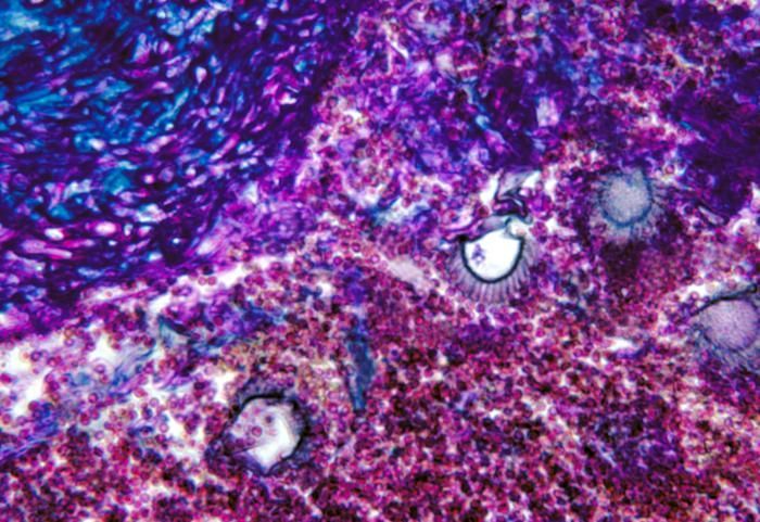
Note the histopathologic changes seen in histoplasmosis due to Histoplasma capsulatum using methenamine silver stain.Created: 1972
-
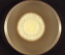
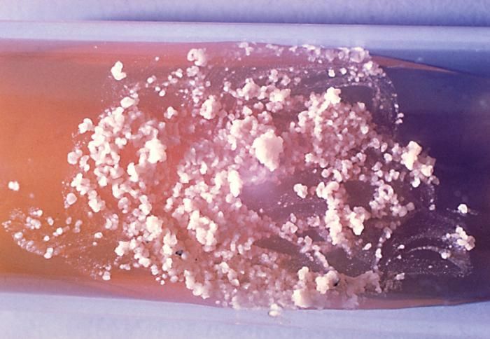
These two slant cultures grew Histoplasma capsulatum colonies: (Lt tube) Sabourauds agar; (Rt tube) Sabhi agar.Created: 1979
-
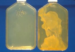
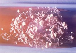
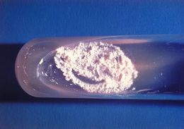
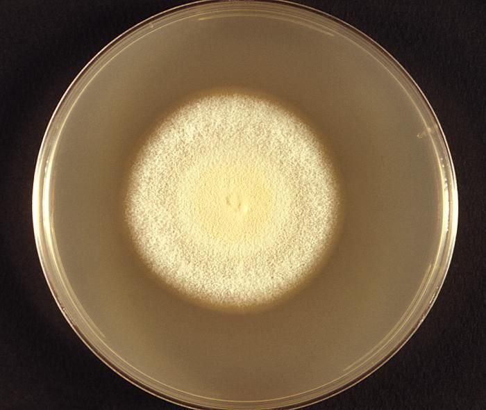
These were two differently concentrated sputum specimen cultures of Histoplasma capsulatum.Created: 1979
-
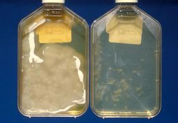
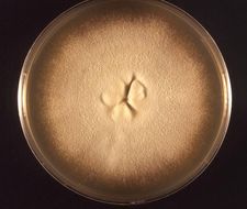
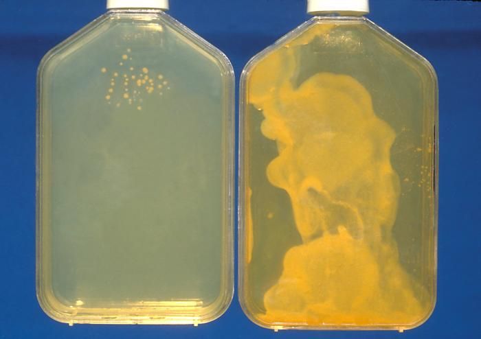
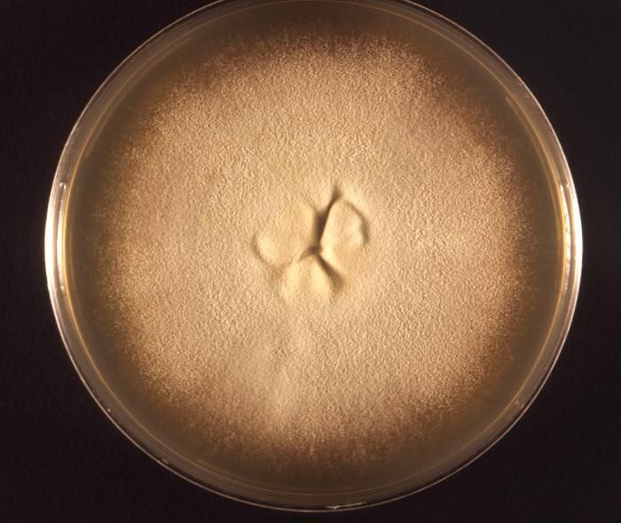
The left bottle Histoplasma capsulatum culture growth is shown at 8wks and the right bottle is shown at 3wks.Created: 1979
-
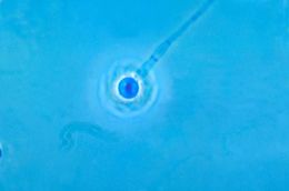
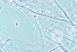
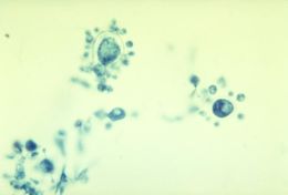
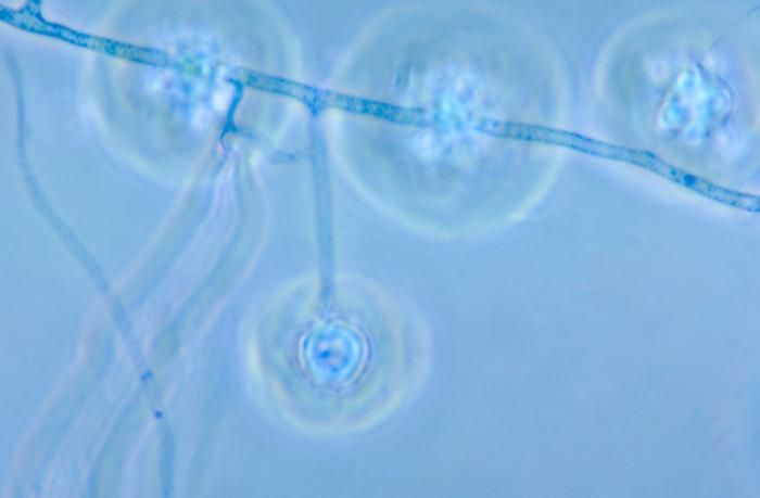
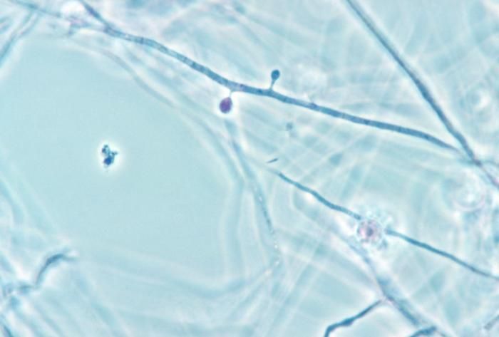
This photomicrograph reveals a conidiophore of the fungus Histoplasma capsulatum.Created: 1963
-
This photomicrograph reveals a conidiophore of the fungus Histoplasma capsulatum.Created: 1963
-
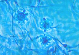
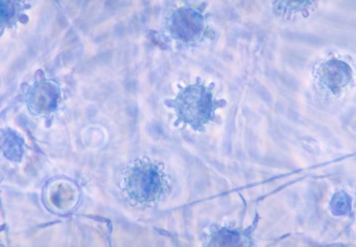
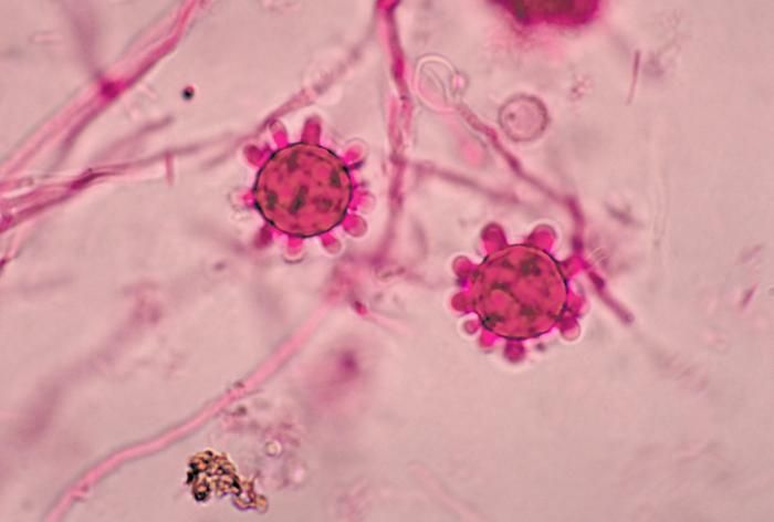
This photomicrograph reveals a number of macroconidia of the fungus Histoplasma capsulatum.Created: 1963
-
This photomicrograph reveals a number of macroconidia of the fungus Histoplasma capsulatum.Created: 1963
-
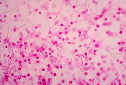
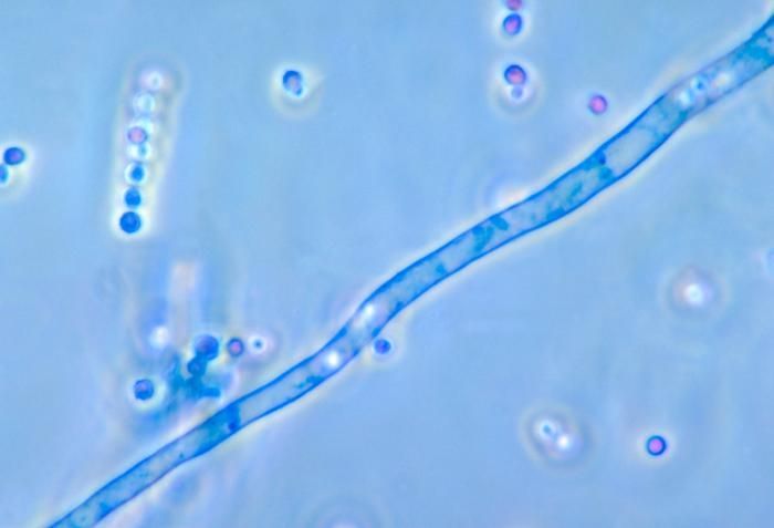
This photomicrograph shows the microconidia of the fungal species Histoplasma capsulatum.Created: 1961
-
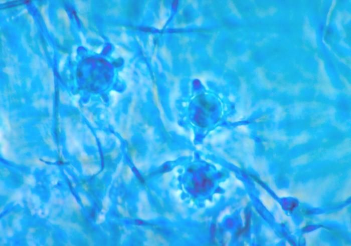
This photomicrograph shows two tuberculate macroconida of the Jamaican isolate of Histoplasma capsulatum.Created: 1968
-
This photomicrograph shows the smooth macroconidia of the Jamaican isolate of Histoplasma capsulatum.Created: 1968
-
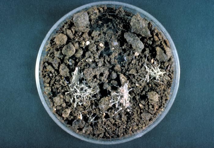
This is a photograph of a hair plate culture of the fungus Histoplasma capsulatum.Created: 1963
-
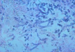
This photomicrograph depicts the appearance of a conidiophore of the fungus Aspergillus fumigatus.Created: 1963
-
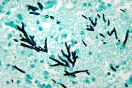
Note the aspergillosis associated histopathologic changes in this turkey poult brain tissue due to Aspergillus fumigatus.Created: 1972
-
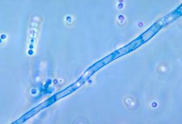
This photomicrograph reveals a conidiophore filament of the fungus Aspergillus fumigatus.Created: 1963
-
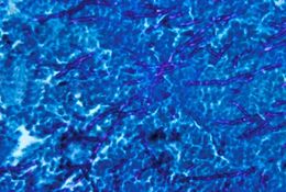
This image depicts histopathologic changes indicating aspergillosis of the lung of a caged parrot caused by A. fumigatus.Created: 1963
-
This image depicts histopathologic changes indicating aspergillosis of the lung of a caged parrot caused by A. fumigatus.Created: 1963
-
Histopathology of aspergillosis of the lung of a caged sulfur-crested cockatoo caused by Aspergillus fumigatus.Created: 1963
-
This photomicrograph revealed some of the morphology exhibited by a Trichophyton tonsurans fungal colony. Note the glaborous, or smooth velvety appearance of this colony, and early changes at its center, which in time will lead to its characteristically raised appearance, as well as its yellowish-beige colorationT. tonsurans and T. rubrum are two common dermatophytes. These two species are usually transmitted from person to person. Another common dermatophyte is Microsporum canis, which is transmitted from animals, including cats and dogs, to people. Dermatophytes like to live on moist areas of the skin, such as places where there are skin folds. They can also contaminate items in the environment, such as clothing, towels and bedding.Created: 1974
-
This photomicrograph revealed some of the morphology exhibited by a Trichophyton tonsurans fungal colony. Note the glaborous, or smooth velvety appearance of this colony, and its characteristically raised center, and yellowish-beige colorationT. tonsurans and T. rubrum are two common dermatophytes. These two species are usually transmitted from person to person. Another common dermatophyte is Microsporum canis, which is transmitted from animals such as cats and dogs to people. Dermatophytes like to live on moist areas of the skin, such as places where there are skin folds. They can also contaminate items in the environment, such as clothing, towels and bedding.Created: 1974
-
This photomicrograph depicts budding cells of the fungus Paracoccidioides brasiliensis during its yeast phase.Created: 1963
-
This photomicrograph depicts budding cells of the fungus Paracoccidioides brasiliensis during its yeast phase.Created: 1963
-
This is a slant culture growing the fungus Paracoccidioides brasiliensis during its yeast phase.Created: 1963
-
This is a slant culture growing the fungus Paracoccidioides brasiliensis during its yeast phase.Created: 1963