-
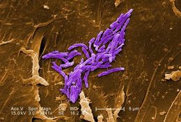
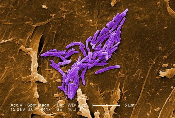
Under a magnification of 3841X, this scanning electron micrograph SEM) revealed some of the ultrastructural morphologic details exhibited by a number of Gram-positive bacilli, or rod-shaped, Mycobacterium fortuitum bacteria. See PHIL 11032 for a black and white version of this image.M. fortuitum is classified as a rapidly-growing Mycobacterium, due to the fact that it can be grown on laboratory culture medium in less than 7 days. As a human pathogen, this organism has been determined to be the cause of skin infections, including furunculosis, i.e., boils, on the legs of people receiving pedicures in nail salons.Created: 2009
-
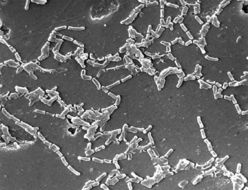
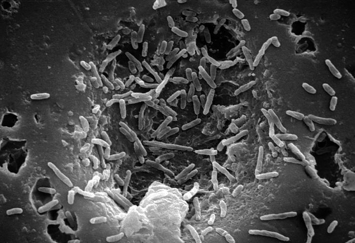
Scanning Electron Micrograph of Mycobacterium chelonaeCreated:
-
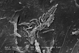
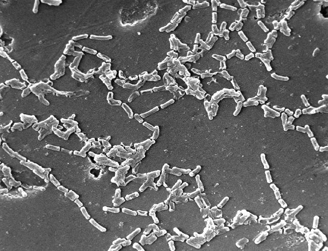
Under a magnification of 3841X, this scanning electron micrograph SEM) revealed some of the ultrastructural morphologic details exhibited by a number of Gram-positive bacilli, or rod-shaped, Mycobacterium fortuitum bacteria. See PHIL 11033 for a colorized version of this image.M. fortuitum is classified as a rapidly-growing Mycobacterium, due to the fact that it can be grown on laboratory culture medium in less than 7 days. As a human pathogen, this organism has been determined to be the cause of skin infections, including furunculosis, i.e., boils, on the legs of people receiving pedicures in nail salons.Created: 2009
-
Scanning Electron Micrograph of Mycobacterium chelonaeCreated:
-
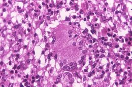
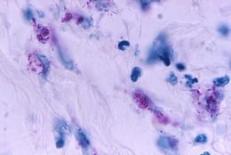
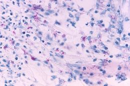
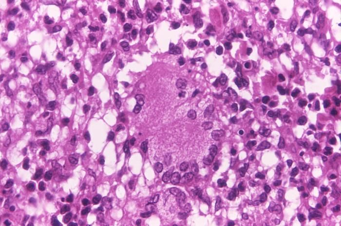
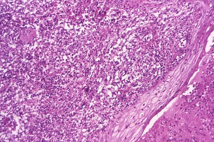
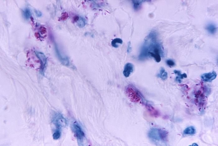
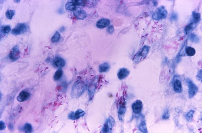
This light photomicrograph revealed some of the histopathologic cytoarchitectural characteristics seen in a mycobacterial skin infection.Created: 1972
-
This light photomicrograph revealed some of the histopathologic cytoarchitectural characteristics seen in a mycobacterial skin infection.Created: 1972
-
This light photomicrograph revealed some of the histopathologic cytoarchitectural characteristics seen in a mycobacterial skin infection.Created: 1972
-
This light photomicrograph revealed some of the histopathologic cytoarchitectural characteristics seen in a mycobacterial skin infection.Created: 1972
-
This light photomicrograph revealed some of the histopathologic cytoarchitectural characteristics seen in a mycobacterial skin infection.Created: 1972
-
This 1971 photomicrograph of a sputum smear, which had been stained using the Morse method of fluorescent acid-fast staining technique, revealed the presence of two Mycobacterium tuberculosis, rod-shaped bacteria, otherwise known as bacilli.Created: 1971
-
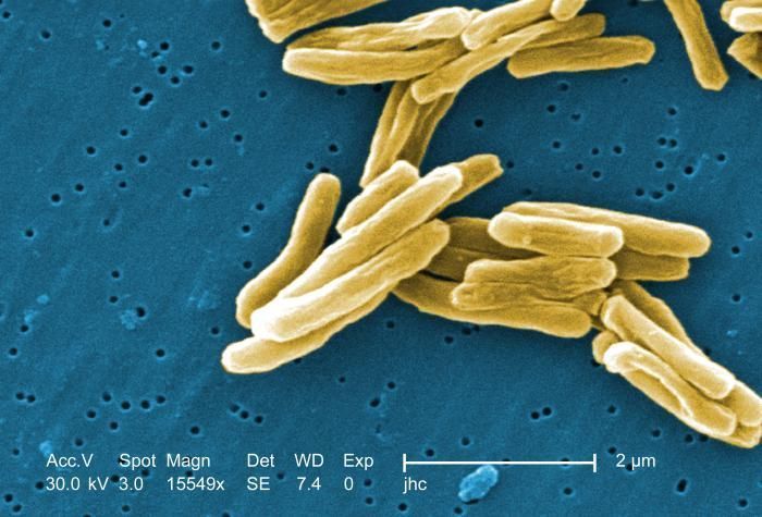
Under a high magnification of 15549x, this colorized scanning electron micrograph (SEM) depicted some of the ultrastructural details seen in the cell wall configuration of a number of Gram-positive Mycobacterium tuberculosis bacteria. As an obligate aerobic organism M. tuberculosis can only survive in an environment containing oxygen. This bacterium ranges in length between 2 - 4 microns, and a width between 0.2 - 0.5 microns. See PHIL 8438 for a black and white version of this image.Created: 2006
-
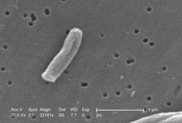
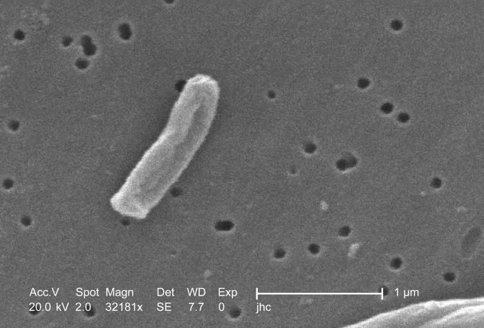
Under a very high magnification of 32181x, this scanning electron micrograph (SEM) depicted some of the ultrastructural details seen in the cell wall configuration of a Gram-positive Mycobacterium tuberculosis bacterium. As an obligate aerobic organism M. tuberculosis can only survive in an environment containing oxygen. This bacterium ranges in length between 2 - 4µm, and a width between 0.2 - 0.5µm.Created: 2006
-
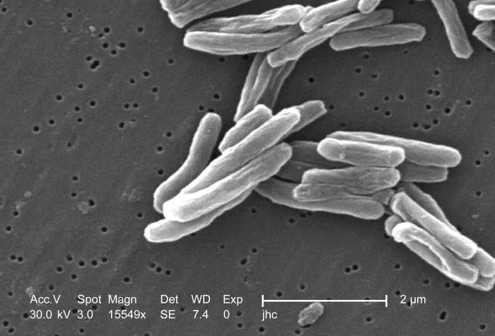
Under a high magnification of 15549x, this scanning electron micrograph (SEM) depicted some of the ultrastructural details seen in the cell wall configuration of a number of Gram-positive Mycobacterium tuberculosis bacteria. As an obligate aerobic organism M. tuberculosis can only survive in an environment containing oxygen. This bacterium ranges in length between 2 - 4 microns, and a width between 0.2 - 0.5 microns. See PHIL 9997 for a colorized version of this image.Created: 2006
-
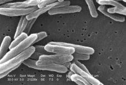
Under a high magnification of 21228x, this scanning electron micrograph (SEM) depicted some of the ultrastructural details seen in the cell wall configuration of a number of Gram-positive Mycobacterium tuberculosis bacteria. As an obligate aerobic organism M. tuberculosis can only survive in an environment containing oxygen. This bacterium ranges in length between 2 - 4µm, and a width between 0.2 - 0.5µm.Created: 2006
-
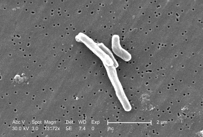
At a magnification of 13172x, this scanning electron micrograph (SEM) depicted a number of Gram-positive Mycobacterium tuberculosis bacteria. As an obligate aerobic organism M. tuberculosis can only survive in an environment containing oxygen. This bacterium ranges in length between 2 - 4µm, and a width between 0.2 - 0.5µm.Created: 2006
-
At a magnification of 13172x, this scanning electron micrograph (SEM) depicted a single Gram-positive Mycobacterium tuberculosis bacterium. As an obligate aerobic organism M. tuberculosis can only survive in an environment containing oxygen. This bacterium ranges in length between 2 - 4µm, and a width between 0.2 - 0.5µm.Created: 2006
-
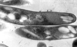
This thin section transmission electron micrograph (TEM) depicted the ultrastructural details displayed by a number of Gram-positive Mycobacterium tuberculosis bacilli, the causative agent for tuberculosis.Created: