-
Figures 41–48.Ventral view of male genital capsule.
-
Figures 49–56.Lateral view of male genital capsule.
-
Figures 1–12.1–8 Front view of male head. 9 Lateral view of male head 10–12 Ventral view of female head. Abbreviations: LID = least interocular distance, OcC = occipital carina; OOL = ocellocular distance, THC = transverse hypostomal carina, UID = upper interocular distance.
-
Figures 33–36.Distribution maps of Colocistis species.
-
Figures 41–48.Ventral view of male genital capsule.
-
Figures 49–56.Lateral view of male genital capsule.
-
Figures 1–12.1–8 Front view of male head. 9 Lateral view of male head 10–12 Ventral view of female head. Abbreviations: LID = least interocular distance, OcC = occipital carina; OOL = ocellocular distance, THC = transverse hypostomal carina, UID = upper interocular distance.
-
Figures 13–24.13, 14 Male forewing. 15 Ventral view of male hindcoxa 16 Dorsal view of male propodeum 17 dorsal view of male mandible 18 lateral view of mandible 19 Ventral view of male metasomal sterna I-II 20, 22 Lateral view of male metasomal segments I and II 21, 23, 24 Ventral view of female metasomal sternum I. Abbreviations: SM-1, 2, 3 = submarginal cells 1–3.
-
Figures 37–40.Distribution maps Colocistis species.
-
Figures 1–12.1–8 Front view of male head. 9 Lateral view of male head 10–12 Ventral view of female head. Abbreviations: LID = least interocular distance, OcC = occipital carina; OOL = ocellocular distance, THC = transverse hypostomal carina, UID = upper interocular distance.
-
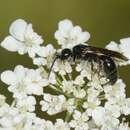
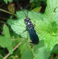
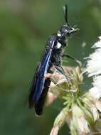
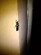
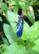
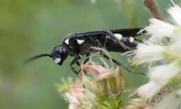
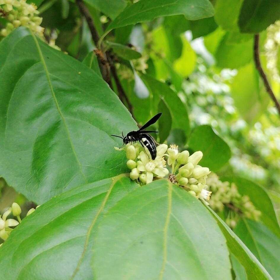
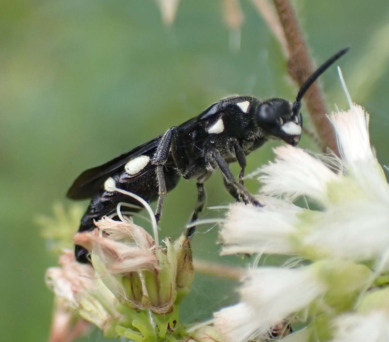
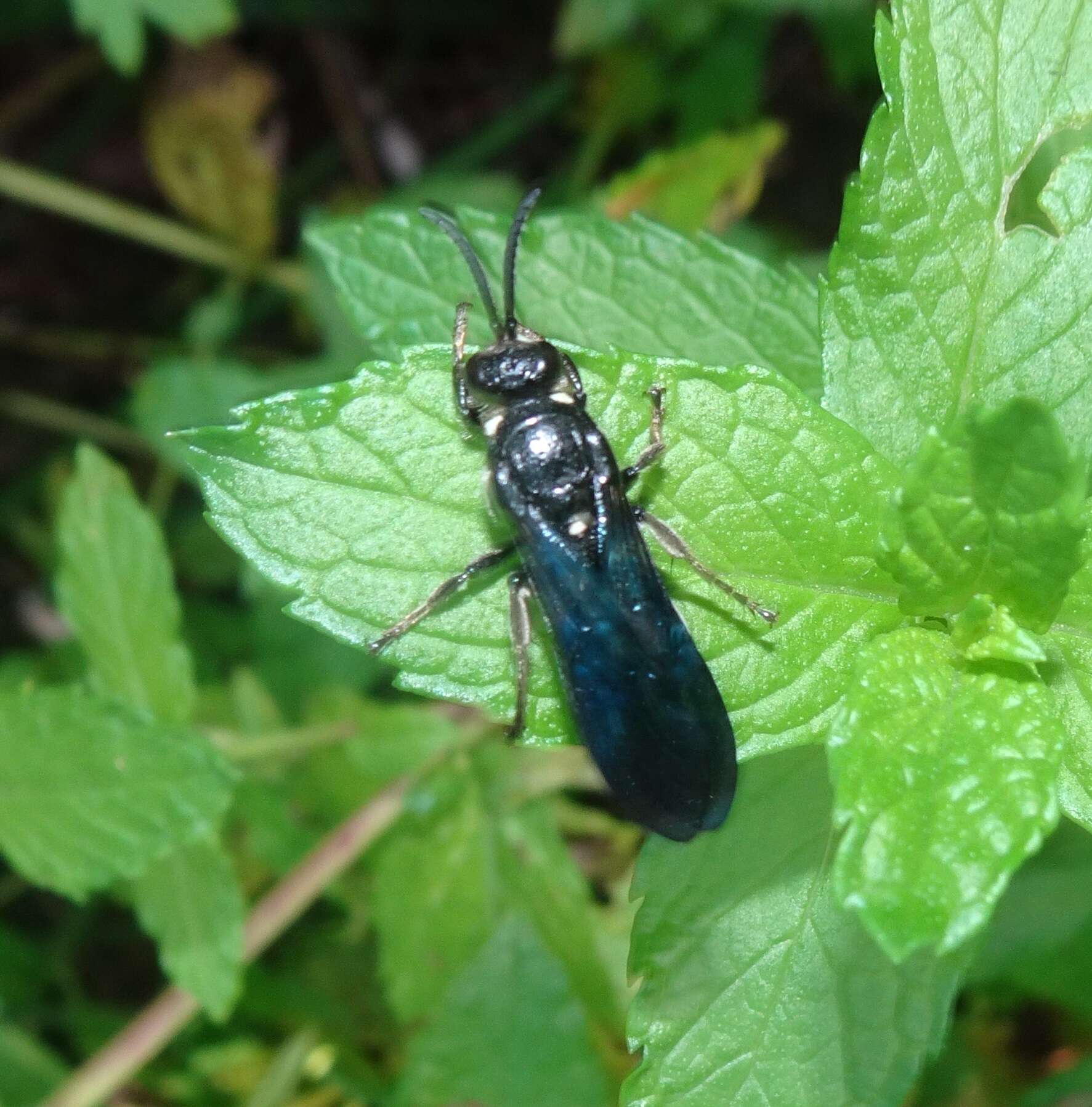
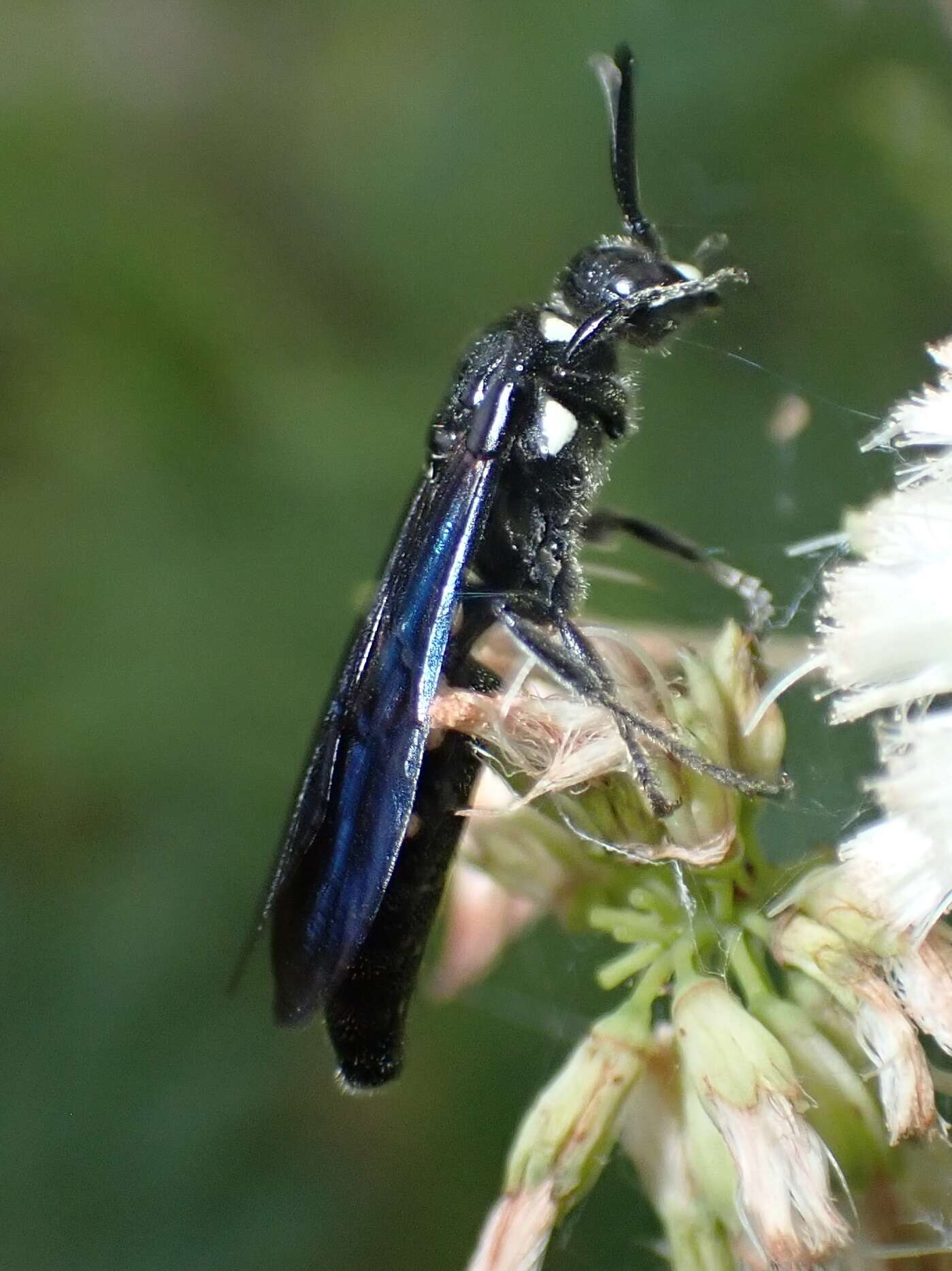
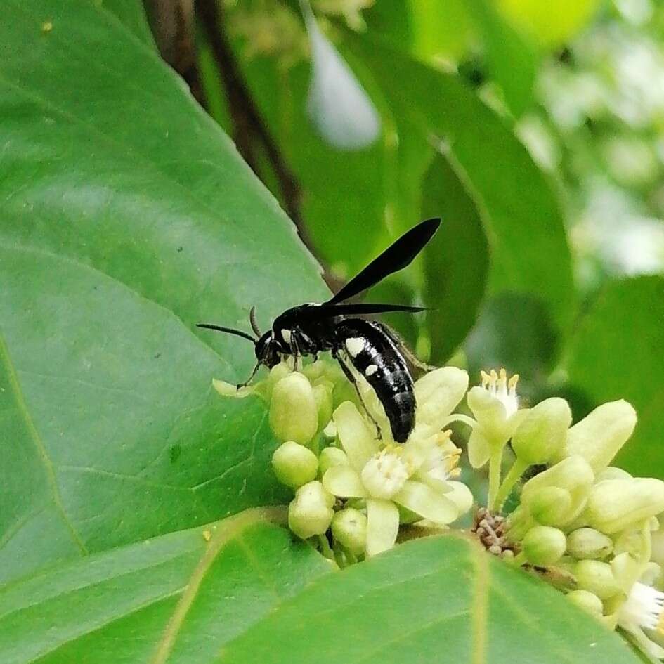
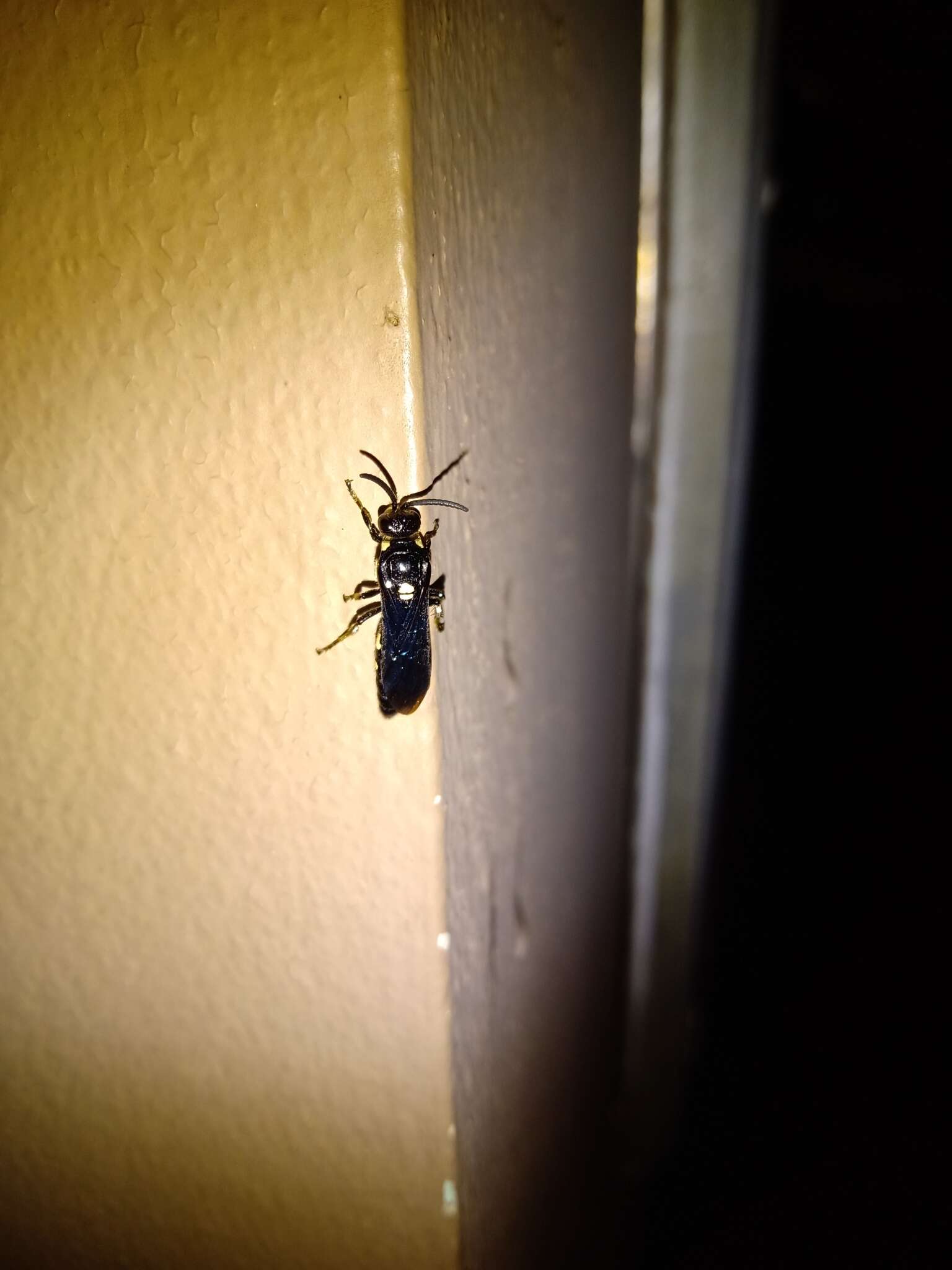
Figures 25–32.25–28. Males. 29–32 Females. 25, 27, 29, 31 Lateral view 26, 28, 30, 32 Dorsal view 25–30 are images of the holotypes.
-
Figures 37–40.Distribution maps Colocistis species.
-
Figures 41–48.Ventral view of male genital capsule.
-
Figures 49–56.Lateral view of male genital capsule.
-
Figures 13–24.13, 14 Male forewing. 15 Ventral view of male hindcoxa 16 Dorsal view of male propodeum 17 dorsal view of male mandible 18 lateral view of mandible 19 Ventral view of male metasomal sterna I-II 20, 22 Lateral view of male metasomal segments I and II 21, 23, 24 Ventral view of female metasomal sternum I. Abbreviations: SM-1, 2, 3 = submarginal cells 1–3.
-
Figures 25–32.25–28. Males. 29–32 Females. 25, 27, 29, 31 Lateral view 26, 28, 30, 32 Dorsal view 25–30 are images of the holotypes.
-
-
-
-
-
-
-
-