-
Galende, Castile and Len, Spain
-
Sobrado, Galicia, Espaa
-
Ribadelago, Castille and Leon, Spain
-
Sobrado, Galicia, Espaa
-
Ribadelago, Castilla y Len, Espaa
-
Ribadelago, Castille and Leon, Spain
-
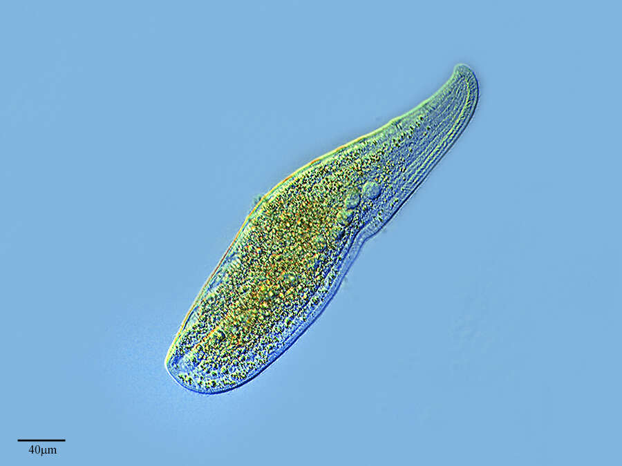
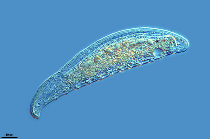
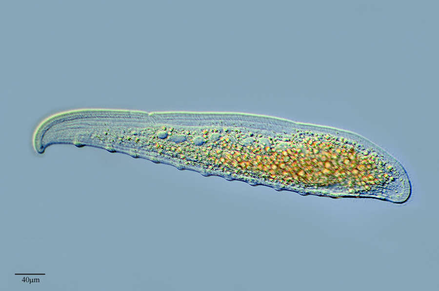
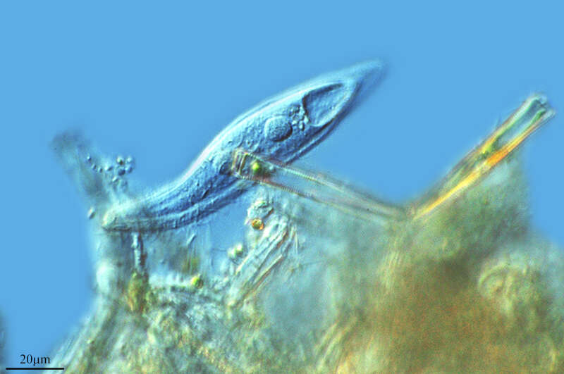
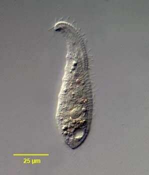
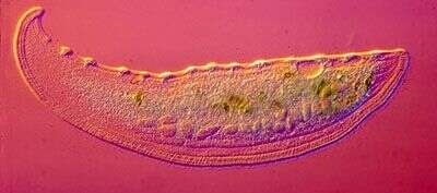
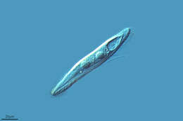
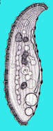
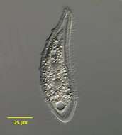
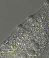
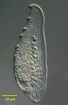
Loxophyllum (locks-o-file-um) is one of several genera of flat gliding predatory ciliates. It glides along the substrate, exploring detritus with the anterior convex margin - which is where the mouth is located. There are rod-shaped extrusomes lying just under the cell membrane and in some species in prominences along the margins of the cell - not well developed in this cell. The extrusomes can be discharged to kill potential prey - usually other ciliates. Surface view of slightly squashed cell shows the surface folds where the rows of cilia (kineties) are located. Differential interference contrast.
-
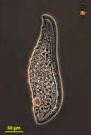
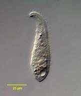
Loxophyllum (locks-o-file-um) is one of several genera of flat gliding predatory ciliates. It glides along the substrate, exploring detritus with the anterior convex margin - which is where the mouth is located. There are rod-shaped extrusomes lying just under the cell membrane and in some species in prominences along the margins of the cell - not well developed in this cell. The extrusomes can be discharged to kill potential prey - usually other ciliates. Phase contrast.
-
Loxophyllum (locks-o-file-um) a predatory ciliate. It is flattened, and glides along the substrate, exploring detritus with the anterior convex margin - which is where the mouth is located. There are extrusomes just internal to the margin of the cell, and also in some species prominences along the margins of the cell - not well developed in this cell. The extrusomes can be discharged to kill potential prey - usually other ciliates. Common. Differential interference contrast.
-
-
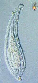
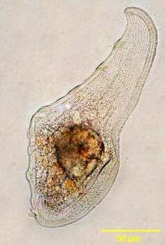
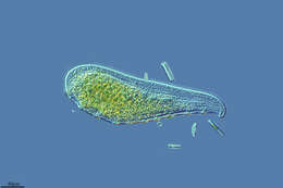
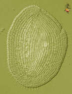
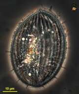
Loxophyllum, a leaf-like predatory ciliate. The mouth lies along the convex side of the body. The convex side has a number of warts and each wart contains many extrusomes. Feeds on detritus and other protists. Phase contrast micrograph.
-
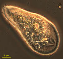
Loxophyllum. Cell observed in sandy and muddy marine sediments in the vicinity of Broome, Western Australia in September 2003. This image was taken using phase contrast optics. Â Â This work was supported by the Australian Biological Resources Study.
-
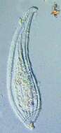
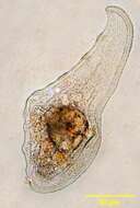
A somewhat contracted cell, extrusomes are evident as thin rods underlying the surface of the cell. Phase contrast microscopy.
-
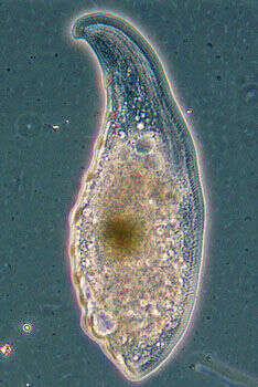
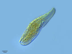
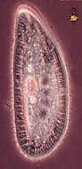
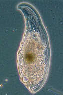
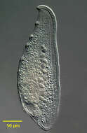
Portrait of Loxophyllum helus (Stokes, 1884, Penard, 1922) a rhabdophorine ciliate. The body is elongate and laterally compressed. The left side is sparsely ciliated and slightly domed the flat right side is more densely ciliated. Very flexible. Prominent trichocyst warts occur at intervals along the dorsal edge (seen well here). A narrow flattened band traversed by trichocysts runs along the entire ventral edge. The slit-like oral aperture is located on the anterior ventral edge. There is one posterior contractile vacuole. The macronucleus is bipartite flanking a small micronucleus. Preys on rotifers and other ciliates. Collected from freshwater pond near Boise, Idaho in June 2003. DIC optics.
-
Portrait of Loxophyllyum, large pleurostomatid ciliate, which is highly laterally, compressed. Glides with ribbon-like movement over substrate. Oral region is slit-like and oriented to the right in this image. Wart-like aggregates of extrusomes are seen at intervals along the dorsal (left) surface. Macronucleus is multinodal in this species. Many species. This species, from standing fresh water near Boise, Idaho, has been preying on rotifers. Oblique illumination.
-
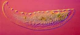
Portrait of the pleurostomatid ciliate, Loxophyllyum meleagris (Mueller,1773) Dujardin, 1841. Glides with ribbon-like movement over substrate. Oral region is slit-like and oriented to the right in this image. Wart-like aggregates of extrusomes are seen at intervals along the dorsal (left) surface. Macronucleus is multinodal in this species. From standing temporary fresh water puddlenear Boise, Idaho. Phase contrast illumination.
-

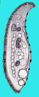
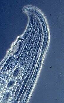
Detail view (dorsal surface) of the large pleurostomatid ciliate, Loxophyllum meleagris (Mueller,1773) Dujardin, 1841. The strongly laterally compressed cell is scimitar-shaped in outline. . The cell is slightly contractile and highly flexible. The rounded anterior end is curved dorsally. The posterior is bluntly tapered. The right side is more densely ciliated than the left. Somatic kineties are longitudinal. The dorsal edge bears characteristic nodular protrusions called extrusome warts (seen well here). The slit-like cytostome is located along the anteroventral edge. There is one posterior contractile vacuole which has a long collecting canal extending anteriorly along the dorsal edge of the cell. The macronucleus (part of which is seen well here) is moniliform. There are multiple inconspicuous micronuclei (not seen here). L.meleagris swims slowly, gliding gracefully over the substrate. L.meleagris feeds on other ciliates and even metazoans such as rotifers. Differentiated from the similar L. helus by its much larger size. Collected from a freshwater agricultural irrigation ditch near McCall, Idaho 9/21/03. DIC.
-
Portrait (right side) of the large pleurostomatid ciliate, Loxophyllum meleagris (Mueller,1773) Dujardin, 1841. The strongly laterally compressed cell is scimitar-shaped in outline. . The cell is slightly contractile and highly flexible. The rounded anterior end is curved dorsally. The posterior is bluntly tapered. The right side is more densely ciliated than the left. Somatic kineties are longitudinal. The dorsal edge bears characteristic nodular protrusions called extrusome warts. The slit-like cytostome is located along the anteroventral edge. There is one posterior contractile vacuole which has a long collecting canal extending anteriorly along the dorsal edge of the cell. The macronucleus is moniliform. There are multiple inconspicuous micronuclei (not seen here). L.meleagris swims slowly, gliding gracefully over the substrate. L.meleagris feeds on other ciliates and even metazoans such as rotifers. Differentiated from the similar L. helus by its much larger size. Collected from a freshwater agricultural irrigation ditch near McCall, Idaho 9/21/03. DIC.
-
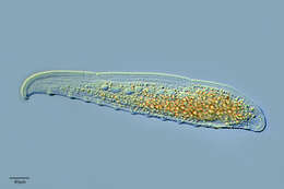
This image of the anterior end shows the curved oral region with faintly visible extrusomes that are used to capture protists as food. The surface is folded along the lines of the kineties. The contractile vacuole, upper left, has a long feeding canal, and the macronucleus is in the form of a series of linked beads. Phase contrast.
-
Interference contrast image of a single living cell. the warts on the oral face are distinctive.
-
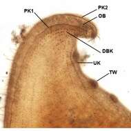
Infraciliature (left side) of Loxophyllum meleagris (Mueller,1773) Dujardin, 1841. The oral bulge (OB is bordered by one left perioral kinety (PK1) and two right perioral kineties (only PK2 visible here). The dorsal edge bears characteristic nodular protrusions called extrusome or trichocyst warts (TW). A row of unciliated kinetids is seen at the base of each trichocyst wart (UK)The slit-like cytostome is located in the center of the oral bulge. DK= dorsal brush kinetids. Collected from a freshwater canal in Boise,Idaho 10/27/08. Protargol.Brightfield.