-
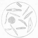
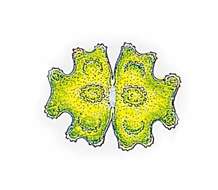
Micrasterias rotata (GREV.) RALFS Length: 200 â 300 µm, width: 200 â 270 µm. This specie is very tolerant concerning living conditions. Therefore the species is widely spread in all altitudes, in forestal ditches and lowland fens sometimes abundant. The cells are 1.08 to 1.15 times longer than wide, the shape seems almost circular or wide elliptical. The cell is devided into lobes due to deep cuts, the terminations of lobes are denticulated. The central lobe is broadened evenly at the end. The termination is formed concavely and is lightly arched upwards at both sides. The lateral angles of the central lobe are little denticulated. The cut in the middle of the cell (sinus) is very deep and peripherally a little widened. The cellwall is densly punctuated by tiny pores. The Chromatophores have several scattered pyrenoids with varying sizes.
-
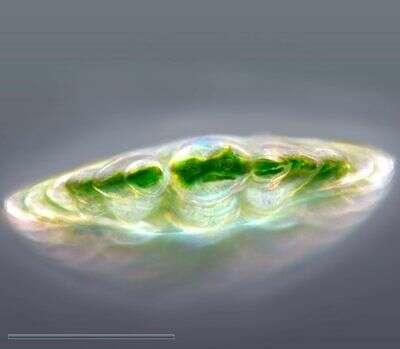
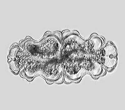
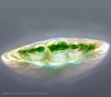
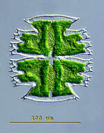
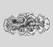
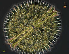
A very special view of Micrasterias rotata using an inverted microscope. The cell ist standing perpenticular upon its apical lobe. Like all the desmids Micrasterias can move using mucilage for backstroke. They move towards the light. When it's dark they erect with help of mucilage ejection. The scale bar indicates 100 µm. Sample from sphagnum pond situated in the northern alpine region of Austria near Salzburg. Images were taken using Zeiss IM35 with Olympus C7070 CCD camera.
-
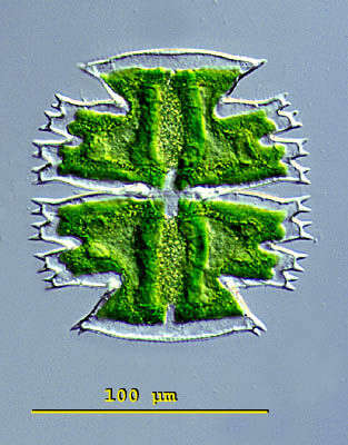
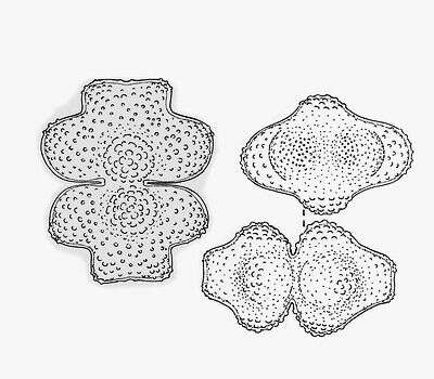
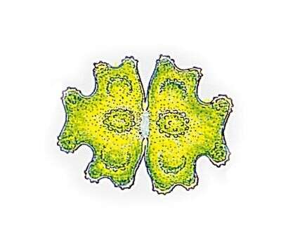
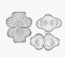
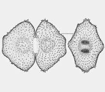
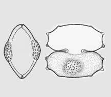
Micrasterias(mike-raz-tear-ee-ass) is a genus of unicellular algae in the family Desmidiaceae. The cells are flattened and disc-like. The cells of the genus Micrasterias are organized in two semi-cells that are mirror images of each other. The semicells have a distinctive shape with an intricate lobes and indentation. At the end of the lobes the cell wall may sometimes form notches or short spines. The nucleus is located in the centrfe between the semicells. Each semicell has a chloroplast with some pyrenoids. Usually found in oligotrophic, acid waters. This is a specimen of Micrasterias truncata collected in a moor pond located in the vicinity of Konstanz, Germany. Differential interference contrast.
-
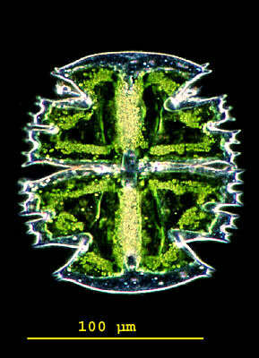
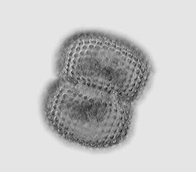
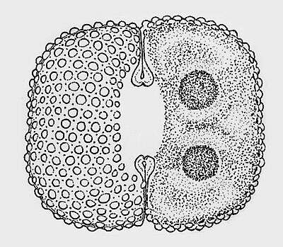
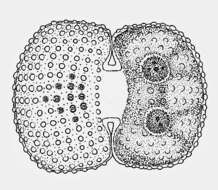
Micrasterias(mike-raz-tear-ee-ass) is a genus of unicellular algae in the family Desmidiaceae. The cells are flattened and disc-like. The cells of the genus Micrasterias are organized in two semi-cells that are mirror images of each other. The semicells have a distinctive shape with an intricate lobes and indentation. At the end of the lobes the cell wall may sometimes form notches or short spines. The nucleus is located in the centrfe between the semicells. Each semicell has a chloroplast with some pyrenoids. Usually found in oligotrophic, acid waters. This is a specimen of Micrasterias truncata collected in a moor pond located in the vicinity of Konstanz, Germany. Dark ground illumination.
-
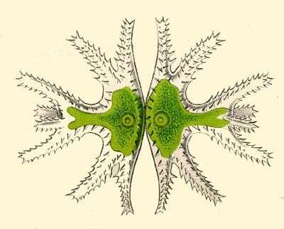
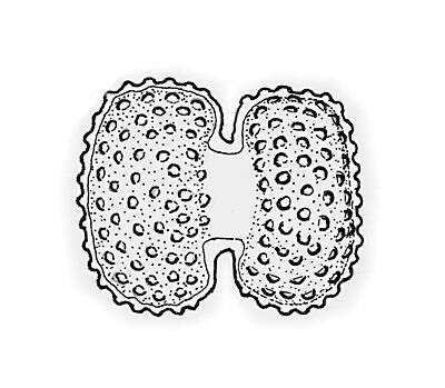
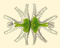
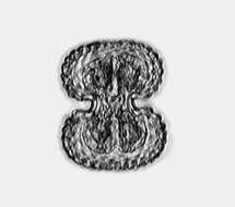
Micrasterias trigemina.
-
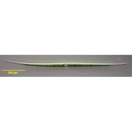
Closterium aciculare (T. West,1860) collected from a freshwater pond near Boise, Idaho. September 2007. DIC.
-
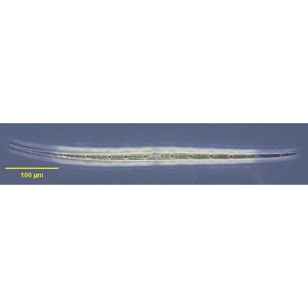
Closterium aciculare (T. West,1860) collected from a freshwater pond near Boise, Idaho. September 2007. Phase contrast.
-
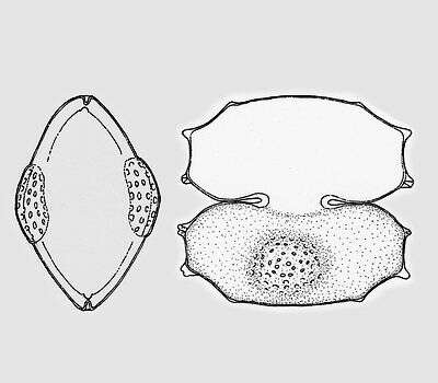
Cosmarium conspersum RALFS var. latum (BREB.) WEST & G. S. WEST Tle cells are appr. 1.3 times longer than wide. The cell halves are somewhat broader toward the ends than at the basis and thus trapezoid. The cell ends are rounded off flat, the central cuts are deep and peripherally extended. The cell surface is covered with hemispheric warts, arranged in longitudinal rows between them are small pores. Length 90 - 100 µm, width 70 - 85 µm. Occurrence: Widely spread in moderate acidic waters of Central Europe.
-
Cosmarium conspersum RALFS var. latum (BREB.) WEST & G. S. WEST The cells are appr. 1.3 times longer than wide. The cell halves are somewhat broader toward the ends than at the basis and thus trapezoid. The cell ends are rounded off flat, the central cuts are deep and peripherally extended. The cell surface is covered with hemispheric warts, arranged in longitudinal rows between them are small pores. Length 90 - 100 µm, width 70 - 85 µm. Occurrence: Widely spread in moderate acidic waters of Central Europe
-
Cosmarium pachydermum P. LUNDELL The cells are up to 1.4 times longer than wide and coarsely elliptical in shape. The cell halves are semicircular. The central cuts are narrowly rounded on the inside and extend strongly to the outside. The cell wall is thick. Some areas are covered closely, others are covered loosely with standing pores. The vertex view is broadly elliptical. Length 100 - 120 µm, width 70 - 90 µm. Occurrence: acidophilic alga, widely spread in sphagnum bogs
-
Cosmarium protractum (NÃG.) DE BARY var. procerum LENZENW. The cells are only little longer than wide. The cell halves form three lobes, the lateral lobes are unequally rounded off, whereby they appear gently rolling. The vertex lobes are slightly widened outwards, rounded off laterally and slightly concave in the center. The central cuts are deep and outward extended. At the basis of both cell halves there is a hump, which is covered with larger verrucae. The remaining cell wall is covered with smaller warts. The vertex view is oblong oval with one distinct surface bulg each at the sides. Length 95 - 100 µm, width 55 - 65 µm. Occurrence: New description, well-known only from Greenland so far.
-
Cosmarium quadrum P. LUNDELL The cells are a little longer than wide, rounded off rectangular in shape. The sides are straight or flat convex. The central cuts are deep, linear, and extended outwards. The cell wall is covered with warts. They run in crossing rows (under an angle of appr. 45°). Around each of the warts are pores, which build symmetrical hexagon in their arrangement. Length 55 - 85 µm, width 50 - 80 µm. Occurrence: Common in littoral region and quaking bogs of moor ponds and in moderate acidic moorlands in Central Europe.
-
Cosmarium reniforme (RALFS) ARCHER var. alaskanum CROASDALE The cells and the cell halves are rectangular, the cell ends are weakly rounded off. The central cuts are opened widely along the entire length. The cell wall is covered with numbers of spherical warts, between these small pores. Length 50 µm, width 40 µm. Occurrence: The habitat is apparently limited to northern latitudes.
-
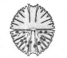
Euastrum gemmatum (BREB.) BREB. var. alatum KOSSINSAJA The cells are appr. 1.5 times longer than wide and.squarely shaped in the center section. Due to an emargination the lateral line of the shape shows two lobes. The vertex lobes are broadened towards the periphery, rounded on the lateral line and slightly concave in the center of the lobe. The cut in the middle of the cell is deep and peripherally widened. In the center of the cell halves there is a clear hump with small verrucae, on both sides of this hump there is a further, flat hump. This area together with the lateral lobes and the vertex lobes are clearly covered with tiny warts. Length 55 - 60 µm, width 45 - 50 µm. Occurrence: Not rare also in weakly acidic ponds in the alpine regions up to an altitude of 2000 m
-
Euastrum oblongum (GREV.) RALFS ex RALFS The cells are appr. 2 times longer than wide and slenderly elliptical in shape. The cell halves consists of two clearly distinguishable sets of lobes which are weakly concave in the center. The vertex lobes are clearly contrasted, widened wedge-shaped with a cut in the center. The cuts in the middle of the cell are not peripherally widened. There exists a big pore in the middle of the cell halves between the two humps. Further humps are visible at the lateral lobes. The cell wall is covered with densely packed pores. Length 150 - 170 µm, width 70 - 85 µm. Occurrence: Adaptable alga, ubiquitous
-
Euastrum verrucosum EHRENB. var. groenlandicum (LARSEN) WILLI KRIEG So far only two quite inaccurate illustrations of this alga exist: The original illustration of LARSEN (1904) and an illustration of GROENBLAD (1952). The cell halves are rounded off trapezoidal, both the lateral lobes and the vertex lobe drawn weakly into the center turn into one another, separated only by a shallow emargination. At the ends of the lateral lobes there are several short pricks. The central cuts are far opened. The hump near the isthmus range of the cell halves are covered with large warts, the remaining cell wall with concentric rows of small warts. Length 100 - 105 µm, width 80 - 85 µm. Occurrence: So far only known from Greenland
-
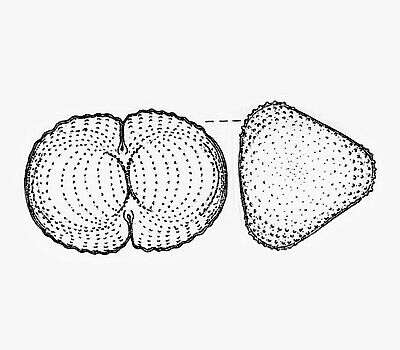
Staurastrum gratum WHELDEN The cells are 1/3 longer than wide coarsely oval in shape, tri-radial, but similar to a Cosmarium. Thr central cuts are shallow, the cell edges with small waves. The cell wall is covered with concentric rings of small warts. The vertex view is triangular with broadly rounded off ends. Length 40 µm, width 30 µm. Occurrence: Known only from northern areas (Baffin Island ans Labrador).
-
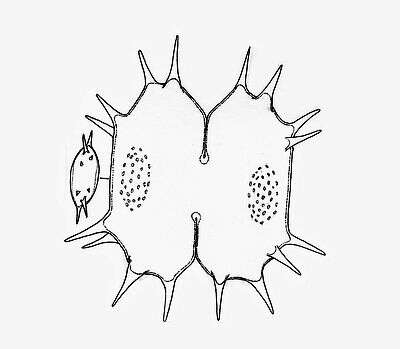
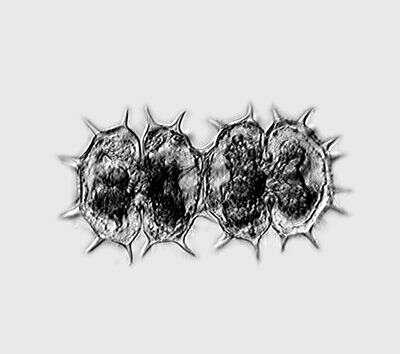
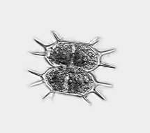
Xanthidium antilopaeum (BREB.) KÃTZ. var. crameri GRÃNBLAD The cells are a little wider than long and octagonal in coarse shape. The central cut is deep and extends strongly outwards. The cell halves are oblong hexagonal with straight or weakly concave sides, the vertices are broadly truncated. At each lateral and apical angle a pair of long pricks originate. Above the center is a flat, often slightly brown colored swelling of the cell wall occupies with small warts. Length without pricks 50 - 60 µm, width without pricks 58 - 63 µm. Occurrence: In littoral zones of mountain lakes in Central Europe rather rarely.
-
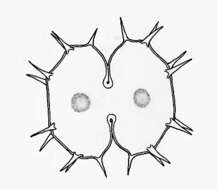
Xanthidium cristatum BREB. In RALFS The cells are little longer than wide, octagonal in coarse shape with straight or weakly concave sides. The central cut is strongly extended outwards. On each side of each cell half origins one prick . The lateral and apical angles likewise have one pair of pricks each. In the center of the cell halves is flat, hemispheric swelling. Length without pricks 50 - 55 µm, width without pricks 40 - 43 µm. Occurrence: In Central Europe sporadic in moderate acidic waters of fens, siltation zones et cetera.
-
Xanthidium groenlandicum BOLD fa. depauperata LARSEN The cells are little wider than long and almost square in shape. The central cut is deep and extends strongly outwards. The cell halves are stretched octagonally with broadly rounded ends. At the cell sides there are two broadly blunted, conical extensions and a hardly visible projection on both sides of the cell vertices. In the center of the cell halves is a flat swelling, which is covered with small warts or dimples. From this alga only three illustrations exist so far: BOLDT (1888), LARSEN (1907) und GRÃNBLAD (1952). Length 60 - 63 µm, width 65 - 68 µm. Occurrence: Known only from Greenland so far.
-
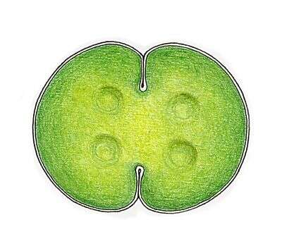
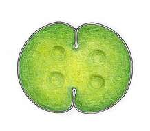
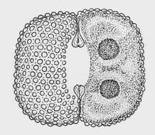
Micrasterias is one of the desmids, flattened green algae in which the organism has a central constriction which gives the organism the appearance of being two cells joined together. Phase contrast micrograph.