Description
provided by Journal of Hymenoptera Research (archived)
Description of the worker.
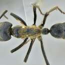
Abundant setae; black integument, ranges from smooth and shiny with no microsculpturing, to finely micropunctate or scaled depending on species (Fig. 12). Head: Mandibles long and curved posteriorly in side view; seven large teeth; erect setae on dorsum. Ventral surface of head with sparse decumbent and subdecumbent setae; may have fine striations depending on species; Papal formula 4, 4; large bilobed labrum. Clypeus with two laterally projecting teeth on anterior edge, clypeus bulging medially, extending posteriorly between frontal lobes, anterior edge with row of long setae; sparse appressed setae from distal edges to medial area of clypeus. Area posterior to clypeus with varying amounts of striation. Tentorial pits apparent. Frontal lobes raised and conspicuous, with striations at posterior constriction. Antennae: geniculate, 12 segments, all with flagellate setae; scape long, extending past posterior border of head; funiculus covered in minute appressed pubescence. Gena depressed medially of eye; dense appressed setae on the antero-lateral sides of the head; covered in conflected punctulate sculpturing. Eyes large, elliptical with slight depression (ocular ring) around circumference. Frons with large pads of long flagellate pubescence (lost in older or poorly curated specimens). Median furrow running from posterior termination of clypeus, between frontal lobes to center of frons, terminates in shallow pit in most specimens. Entire head covered in long flagellate subdecumbent setae (Fig. 1A). Mesosoma: in lateral view weakly convex; covered in long subdecumbent to erect flagellate pilosity and dense pubescence; pronotal disc with slight bulges; promesonotal suture distinct, suture between mesopleuron and propodeum distinct; mesonotum fused with propodeum and episternum, separated by slight furrows; basilar sclerite large, ovaloid; propodeum with broadly rounded dorsal outline, dorsal surface gradually curves into posterior face (Fig. 2); propodeal spiracle forms nearly vertical slit; sulcus running from center of propodeum along lower edge of propodeal spiracle to posterior edge of propodeum at dorsal edge of bulla, patches of short white pubescence at curved posterior border of pronotum and basilar sclerite. Legs long, covered in long setae with short, stiff pubescence. One well-developed, antennae cleaning, comb-like spur on foreleg; one spine-like appendage and one less developed denticular comb on mesothoracic tibia; one spine and one comb-like spur on hind tibia. Posterior side of fore leg basitarsus with dense pads of golden setae; tarsal claws bidentate. Petiole: node large and tabular in lateral view, narrow attachments at base to propodeum and gaster; in dorsal view largest width less than propodeum and gaster, varies from ovate rectangular to ovate triangular in outline; covered in long subdecumbent to erect flagellate pilosity; pubescence on anterior face and ridges of subpetiolar process; subpetiolar process reduced, slightly variable between species. Gaster: typical of ponerines; covered with flagellate setae with short pubescence; small protuberance at articulation of gastric sternite III and the petiole; stridulatory file of varying size on acrotergite of gastral tergum II; posterior edges of the pygidium and hypopygidium with characteristic rows of minute spines. Description of the male.
Integument: smooth and nitid; reddish to dark brown/black. Head: Mandibles greatly reduced, rounded, spoon shaped, lacking teeth; palps elongated, maxillary palps 4 segmented, labial palps 3 segmented; labrum reduced, rounded to truncate, emarginated distal margin in Dinoponera snellingi and Dinoponera longipes covered with setae. Clypeus large, triangular, bulging medially; anterior tentorial pits large; frontal lobes absent; antennal sockets almost touching, located at posterior apex of clypeus. Antennae: geniculate, 13–segmented, pilosity varies from fine pubescence to long setae in different species; scape shorter than second funicular segment, but shorter than 1st, 1st funicular segment reduced. Compound eyes large, along lateral side of head, deeply emarginated medially. Three ocelli at posterior margin of head, bulging beyond margin of head in all species except Dinoponera australis. Entire head immaculate, covered in fine pubescence and long erect setae (Fig. 3). Mesosoma: pronotum triangular, exposed narrowly dorsally anterior to scutum; scutum large, bulging antero-dorsally, with 3 longitudinal carina; small tegula over insertion of forewing; scutellum domed, side with vertical carina, dorsal surface smooth; basilar sclerite under hind wing reduced; fused mesopleuron, separated by furrow into anepisternum and katepisternum; metanotum exposed between scutellum and propodeum, reduced; dorsal face of propodeum shorter than posterior face, rounded into posterior face; coxa large, conical (Fig. 3). Wings: covered in minute pubescence, venation as shown in Figure 5. Legs: one well-developed, antennae cleaning, pectinate spur on foreleg; one spine-like and one less developed denticular comb on mesothoracic tibia; one spine and one comb-like spur on hind tibia. Posterior side of fore basitarsus with dense pads of golden setae; tarsal claws bidentate. Petiole: narrow attachments at base to propodeum and gaster; petiolar node humped dorsally, subpetiolar process anteriorly triangular. Gaster: large, cylindrical, covered in fine silvery pubescence; pygidium terminating in spine posteriorly, with short cerci; hypopygidium with long fine erect setae, tabular subgenital plate with posterior end truncated, often emarginated. Genitalia (Figs 6–11): basal ring with dorso-anterior loop structures; parameres long, rounded, with emarginated ventro-basal edge (Fig. 9); volsella articulated with basiparamere along ventral edge, lateral finger-like cuspis volsellaris, medial digitus volsellaris with distal wide toothed cusp, basal medial lobe with tooth-like structures varying with species (Fig. 10); penis valve of aedeagus roughly triangular and rounded, aedeagal apodeme curved horn-like antero-lateral arm structure arising from mid-valve ridge, terminating at interior surface of basiparamere (Fig. 11). Description of the larvae.
A basic description of the larva of Dinoponera quadriceps (cited as Dinoponera grandis mutica) is present in Mann (1916). A detailed description of the egg and all larval stages of Dinoponera gigantea are present in Wheeler and Wheeler (1985). The following generic description of Dinoponera larvae is from their work:
"Profile pogonomyrmecoid (i.e., diameter greatest near the middle of abdomen, decreasing gradually toward anterior end and more rapidly toward posterior end, which is rounded; thorax more slender than abdomen and forming a neck, which is curved ventrally). Body with numerous (114–160) mammiform tubercles, each with 2–25 short simple hairs; body hairs lacking elsewhere. Cranial hairs lacking. Mandible dinoponeroid (i.e. narrowly subtriangular in anterior view; anterior portion curved posteriorly; with or without medial teeth.)"
- license
- cc-by-3.0
- copyright
- Paul A. Lenhart, Shawn T. Dash, William P. Mackay
- bibliographic citation
- Lenhart P, Dash S, Mackay W (2013) A revision of the giant Amazonian ants of the genus Dinoponera (Hymenoptera, Formicidae) Journal of Hymenoptera Research 31: 119–164
- author
- Paul A. Lenhart
- author
- Shawn T. Dash
- author
- William P. Mackay