-
-
-
-
-
-
-
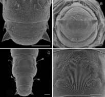
Michel P. Valim, Jason D. Weckstein
Zookeys
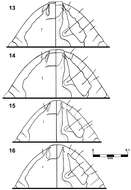
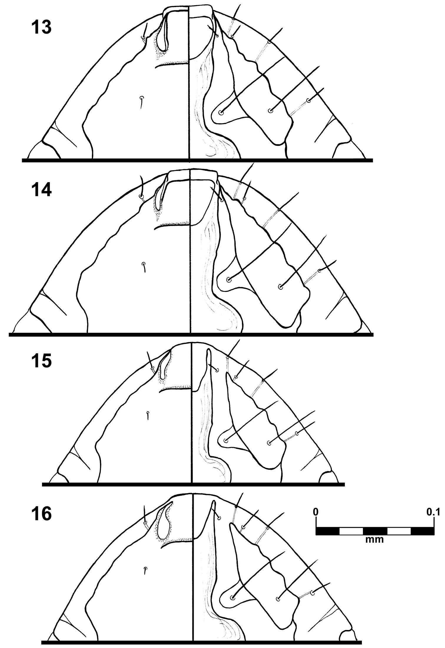
Figures 13–16.Brueelia sueta sp. n.: male preantenal region, dorso-ventral views (13); female preantenal region, dorso-ventral views (14); Brueelia cicchinoi sp. n.: male preantenal region, dorso-ventral views (15); female preantenal region, dorso-ventral views (16).
-
Juli Pujade-Villar, Paul Hanson, Claudia A. Medina, Miguel Torres, George Melika
Zookeys
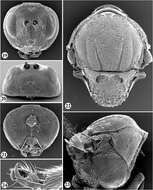
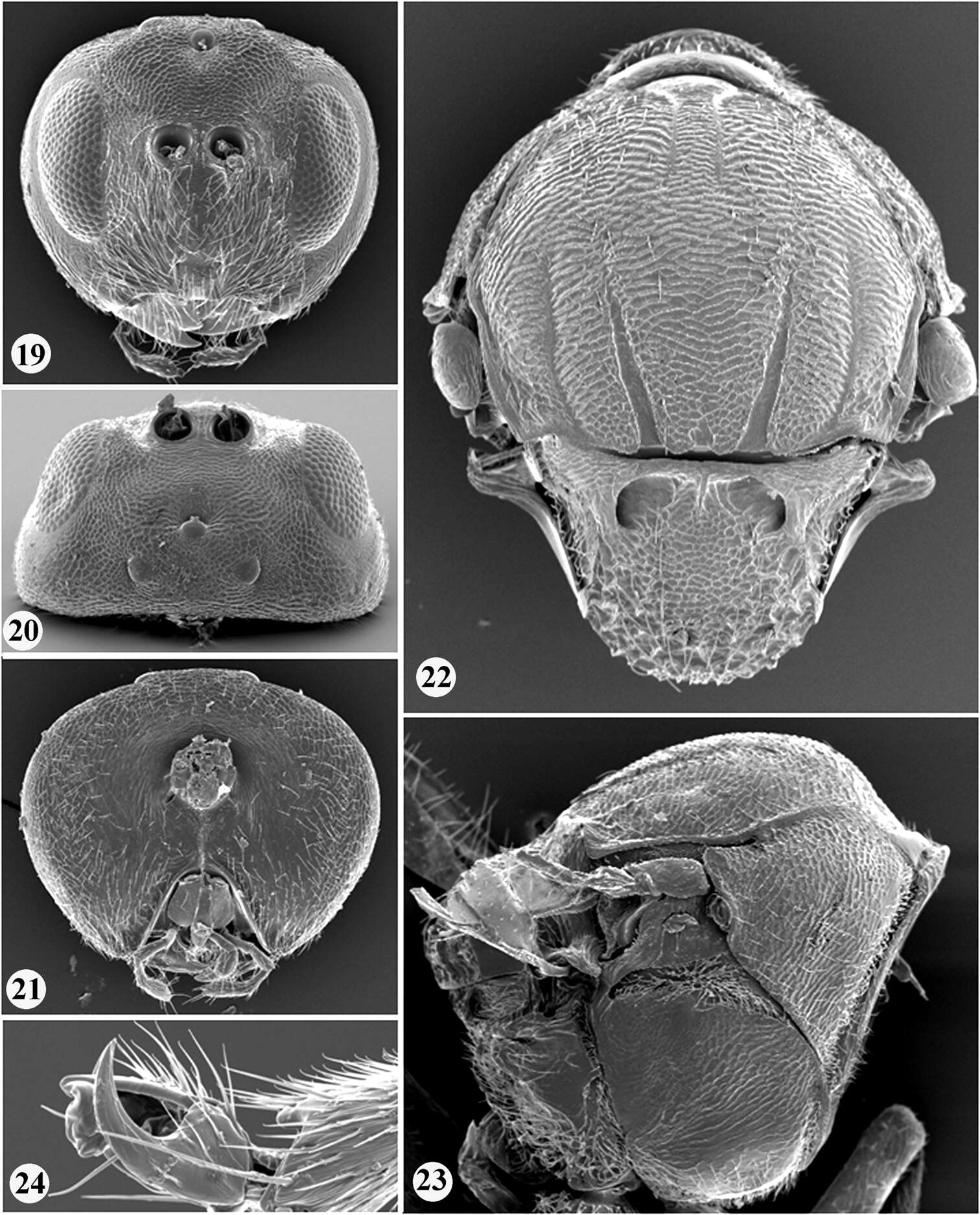
Figures 19–24.Zapatella nievesaldreyi, female 19 head (anterior view) 20 head (dorsal view) 21 head (posterior view) 22 mesosoma (dorsal view) 23 mesosoma (lateral view) 24 tarsal claw.
-
Pierfilippo Cerretti, D. Monty Wood, James E. O’Hara
Zookeys
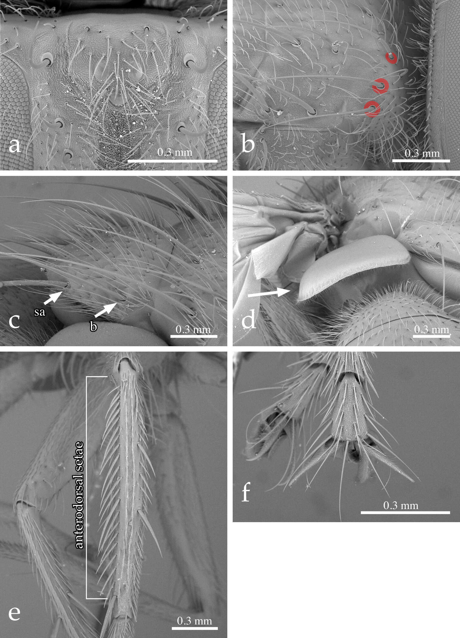
Figure 2.Neoethilla gen. n.ignobilis (male, New Mexico) a vertex in anterodorsal view b right postpronotum and part of presutural portion of scutum in laterodorsal view [circles indicate basal postpronotal saetae] c scutellum in laterodorsal view [b = basal scutellar seta; sa = subapical scutellar seta] d lower calypter in posterior view e left hind tibia in dorsal view f fore claws.
-
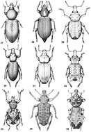
Figures 27–35.Habitus of representative Listroderini. 27 Lanteriella microphtalma 28 Telurus caudiculatus 29 Acrorius papallacta 30 Acrostomus bruchi 31 Antarctobius lacunosus 32 Germainiellus dentipennis 33 Lamiarhinus aelficus 34 Listroderes annulipes 35 Philippius superbus.
-
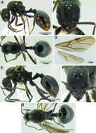
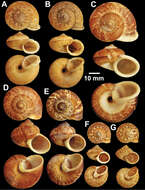
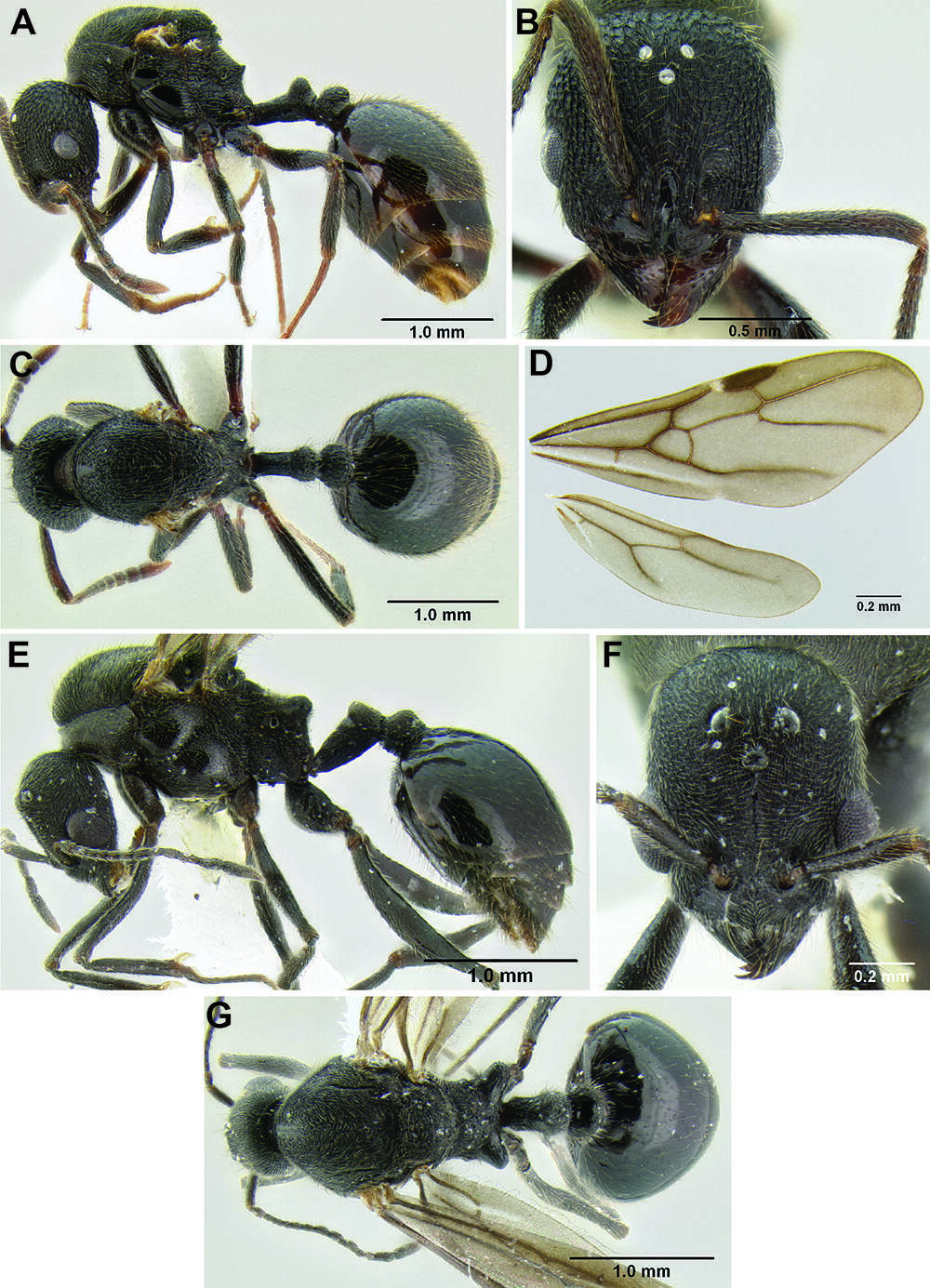
Figure 121.Stenamma megamanni A Paratype queen (CASENT0604840), profile B Same, face C Same, dorsum D Same, wings E Male (CASENT000007293), profile F Same, face G Same, dorsum.
-
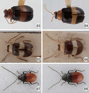
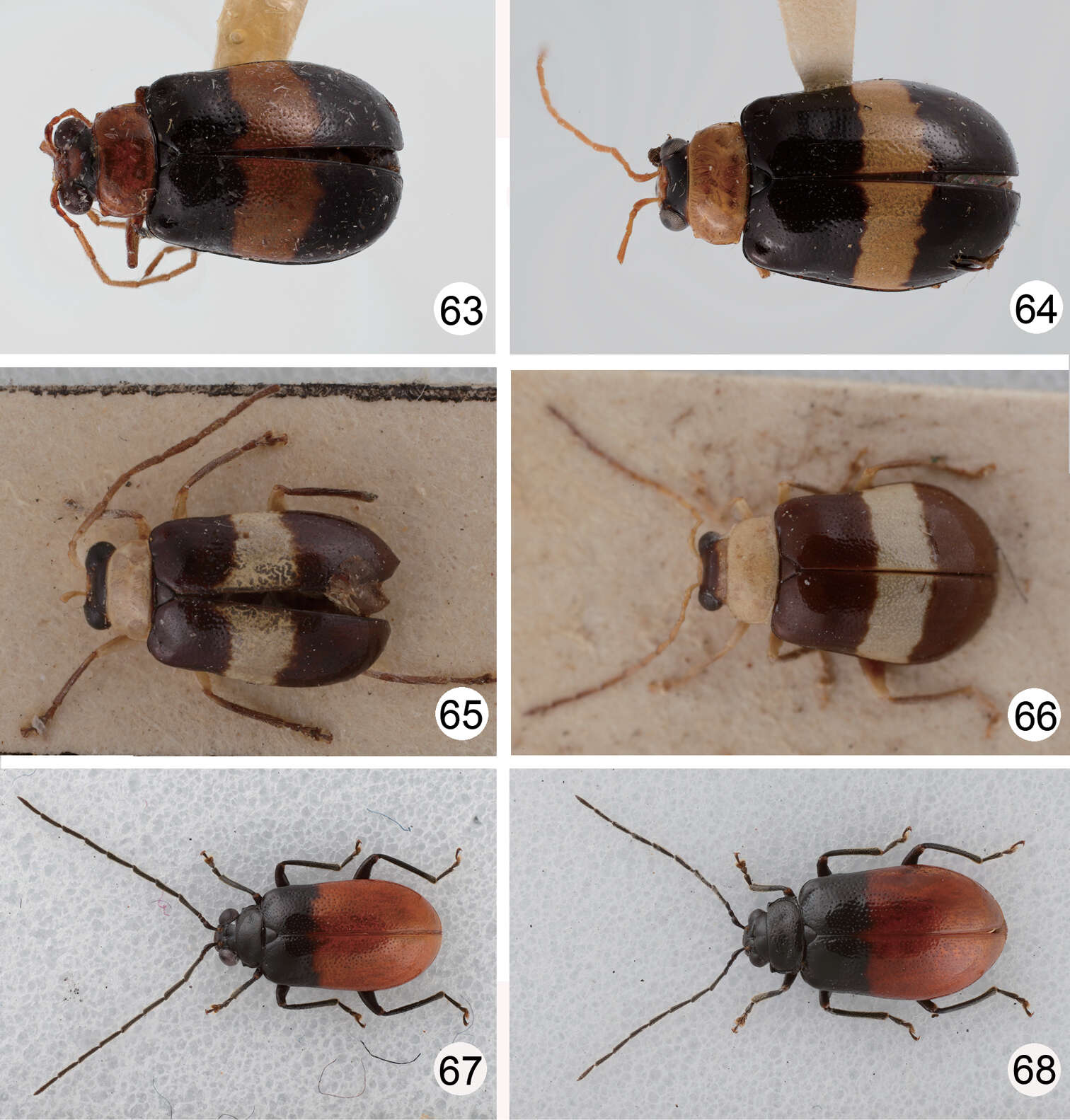
Figures 63–68.Dorsal habitus of Dercetina and Arthrotus species. 63 Dercetina unifasciata, male 64 Dercetina unifasciata, female 65 Arthrotus flavocincta, male 66 Arthrotus flavocincta, female 67 Arthrotus nakanei, male; 68. Arthrotus nakanei, female.
-
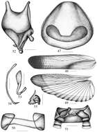
Figures 47–55.Symploce evidens sp. n. 47 pronotum 48 tegmen 49 hind wing 50 abdominal tergum 9 and lateral plates, ventral view 51 supra-anal plate and paraprocts, ventral view 52 subgenital plate, dorsal view 53 hook-like phallomere 54 median phallomere 55 right phallomere. Scale bars = 1.0 mm (Fig. 47), 2.0 mm (Figs 48–49), 0.5 mm (Figs 50–55).
-
Lizhi Huo, Xingmin Wang, Xiaosheng Chen, Shunxiang Ren
Zookeys
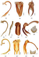
Figures 53–64.53–55 Aspidimerus guangxiensis Yu, male genitalia: 53 penis 54 tegmen, ventral view 55 tegmen, lateral view 56–60 Aspidimerus matsumurai Sasaji 56–59 male genitalia: 56 penis 57 apex of penis 58 tegmen, ventral view 59 tegmen, lateral view 60 female genitalia: ovipositor 61–64 Aspidimerus kabakovi Hoàng 61–63 male genitalia 61 penis 62 tegmen, ventral view 63 tegmen, lateral view 64 female genitalia: ovipositor. Scale bars: 0.1mm.
-
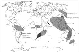
Map 12.A distribution of the tribe Ancyronini.
-
Nattawadee Nantarat, Chirasak Sutcharit, Piyoros Tongkerd, Jonathan Ablett, Fred Naggs, Somsak Panha
Zookeys
Figure 12.Types of Cyclophorus species. A, B Cyclophorus koboensis Godwin-Austen, 1915 A lectotype NHMUK 1903.7.1.3579/1, and B paralectotype NHMUK 1903.7.1.3579/2-4 C Cyclophorus labiosus (Pfeiffer, 1854), lectotype NHMUK 20130080 D, E Cyclophorus linguiferus (Sowerby I, 1843) D lectotype NHMUK 20110269/1, and E paralectotype NHMUK 20110269/2-3 F, G Cyclophorus lingulatus (Sowerby I, 1843) F lectotype NHMUK 20110272/1, and G paralectotype NHMUK 20110272/2-3.
-
Thomas Wesener, Daniel Minh-Tu Le, Stephanie F. Loria
Zookeys
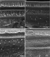
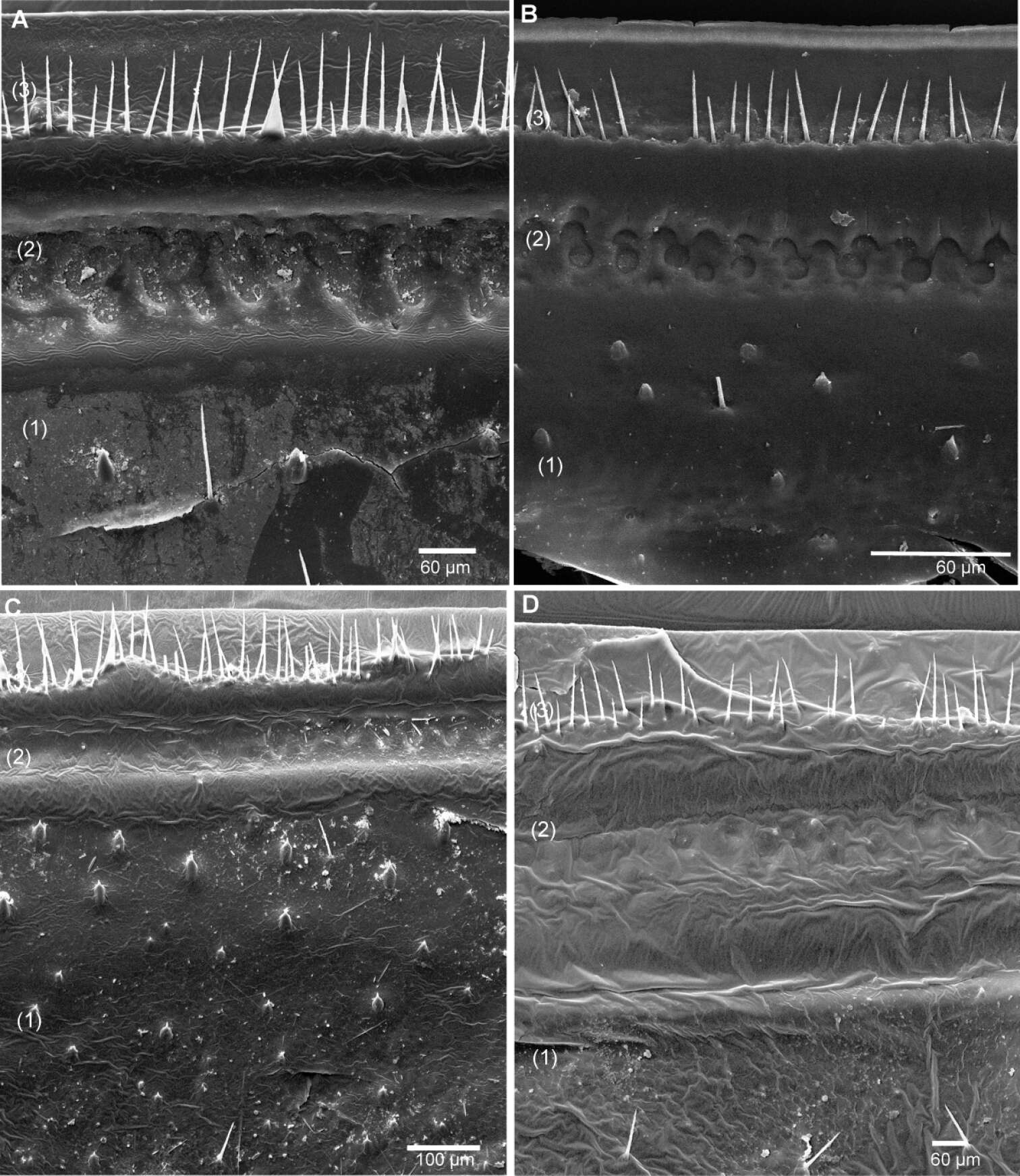
Figure 16.SEM, Endoterga of mid-body tergite. A Sphaeromimus ivohibe sp. n., paratype B Sphaeromimus saintelucei sp. n., holotype from Isaka-Ivondro C Sphaeromimus andrahomana sp. n., holotype D Sphaeromimus andrahomana cave specimen. Abbreviations: (1) = inner area with large spines and long setae; (2) = area with cuticular patterns; (3) = outer area with row(s) of marginal bristles and tergite margin.
-
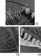
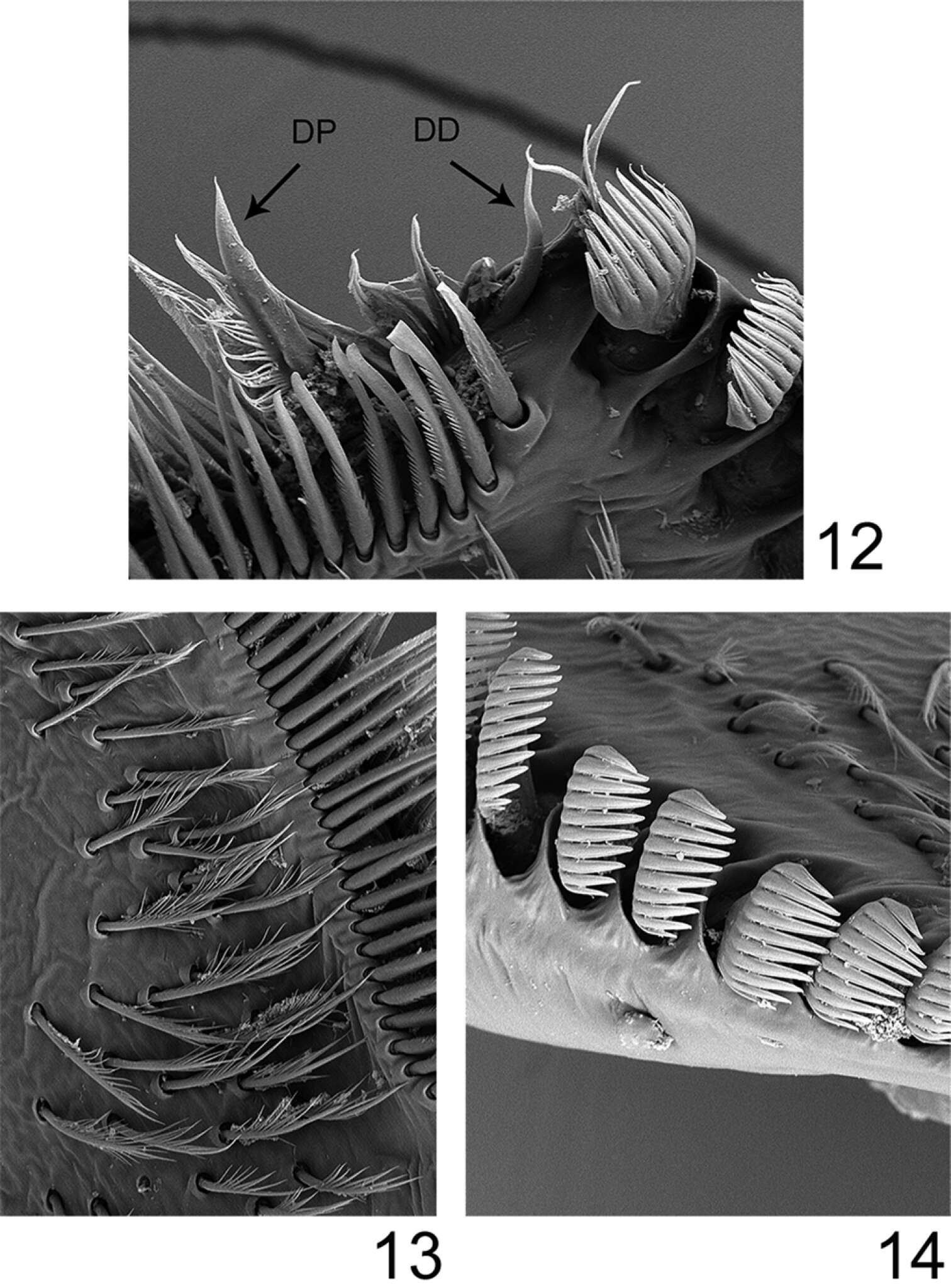
Figures 12–14.Rhithrogeniella ornata Ulmer, 1939, SEM pictures of the maxilla. 12 Dentisetae (DP: proximal dentiseta, DD: distal dentiseta) 13 Fimbriate setae on the ventral surface 14 Comb-shape setae on the crown of the galea-lacinia.
-
Donald R. Davis, David L. Wagner
Zookeys
Figure 15.Phyllocnistis perseafolia sp. n. pupa. A Abdominal terga 7-10 (100 µm) B Caudal end of abdomen (100 µm) C Abdominal sterna 6-10 (100 µm) D Spinules of sternum 6 in longitudinal rows (100 µm). (Length of bar scales shown in parentheses.)
-
Georgia, United States
-
-
Keekorok, Narok, Kenya
-
Bournemouth, England, United Kingdom
-
Løvenholm Skov