-
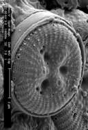
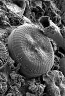
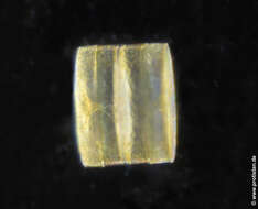
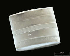
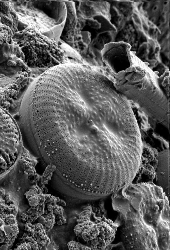
Cyclotella spec. Frustules mounted on a lorica from ciliate Codonella crater gathered in the lake Schierensee near Kiel, Germany5/2004). Use of SEM equipment courtesy of Lab Dr. Karl-Heinz Schäffner, Solingen, Germany.Image under Creative Commons License V 3.0 (CC BY-NC-SA). Place name: Lake Schierensee near Kiel (Schleswig-Holstein, Germany) Latitude: 54.26256721 Longitude: 9.98369694 Kieselalgenschalen in einer Lorica des Ciliaten Codonella crater ; Probe aus dem Schierensee bei Kiel. Datum der Aufsammlung: 5/2004. Aufgenommen mithilfe eines Rasterelektronenmikroskops im Labor Dr. Schäffner, Solingen. Creative Commons License V 3.0 (CC BY-NC-SA). For permission to use of (high-resolution) images please contact postmaster@protisten.de.
-
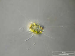
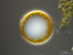
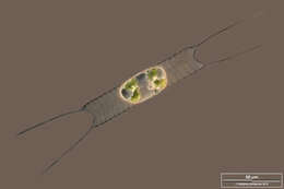
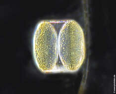
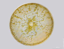
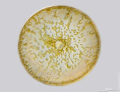
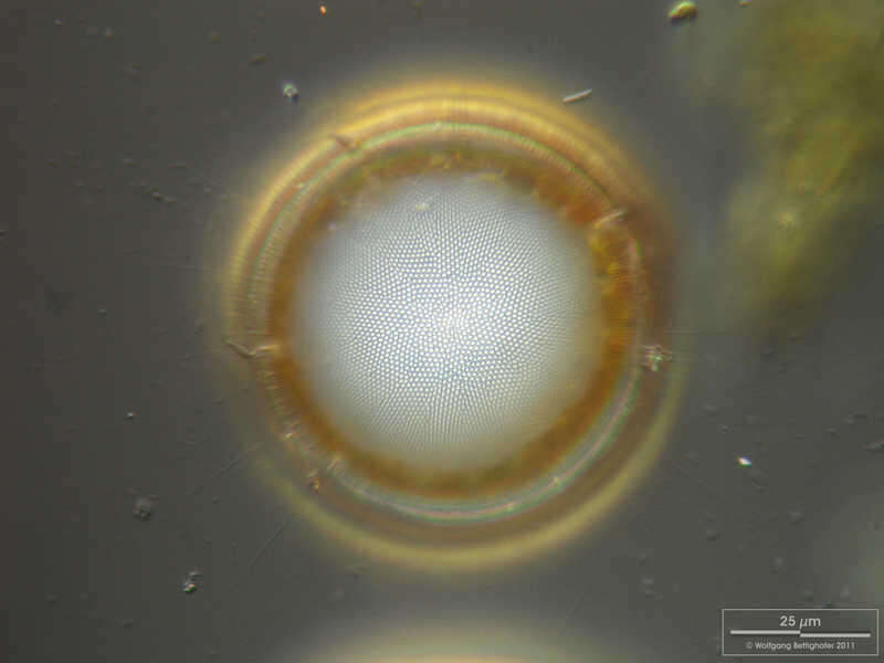
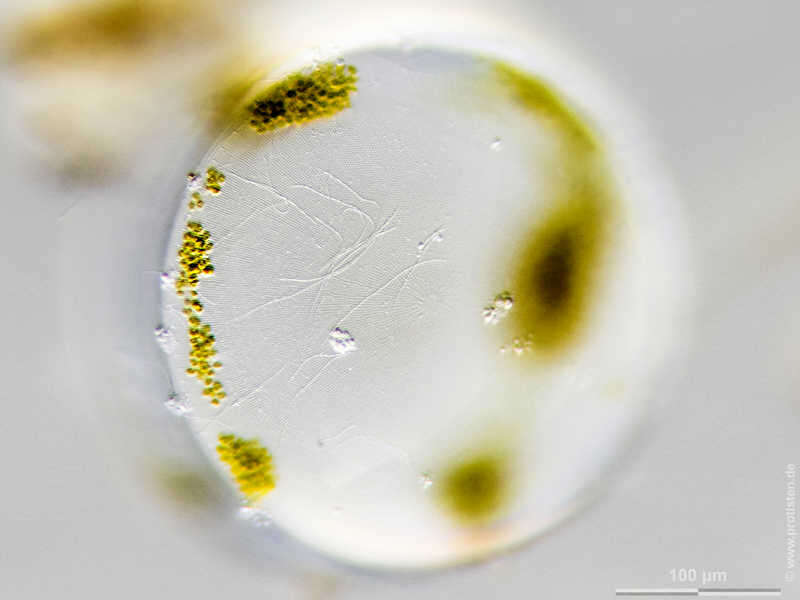
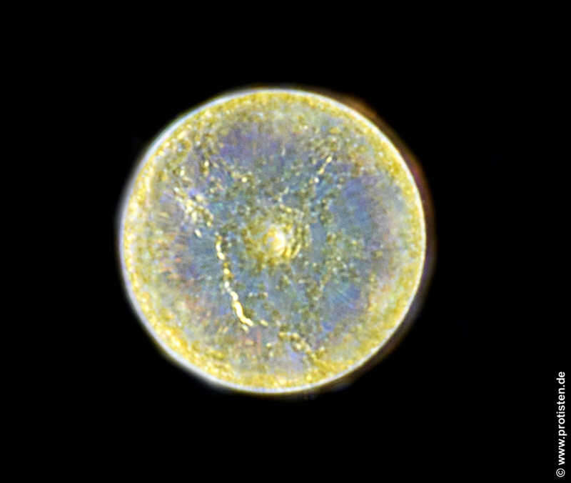
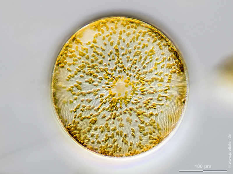
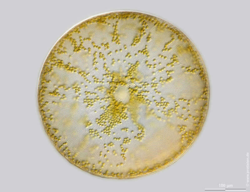
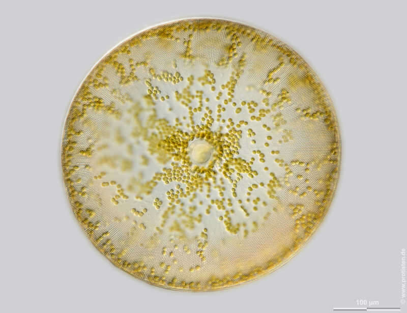
Cyclotella spec. Optical tranversal section, showing the nucleus. The thin, barely visible floating extensions are made of chitin. Furthermore, filamentous bacteria colonies are attached. Scale bar indicates 50 µm. Sample from the Lake Constance (vicinity of Bodman). The image was built up using several photomicrographic frames with manual stacking technique. Images were taken using Zeiss Universal with Olympus C7070 CCD camera.Image under Creative Commons License V 3.0 (CC BY-NC-SA). Place name: Lake Constance vicinity of Bodman (Germany) Latitude: 47.796494 Longitude: 9.047656 Optischer Querschnitt mit Darstellung des Zellkerns. Die dünnen, kaum erkennbaren Schwebefortsätze sind aus Chitin. Des weiteren sitzen fadenförmige Bakterienkolonien auf. Multiebenen-Abbildung, manuell gestapelt. Der Messbalken markiert eine Länge von 50 µm. Probe aus dem Bodensee in der Nähe von Bodman. Mikrotechnik: Zeiss Universal, Kamera: Olympus C7070. Creative Commons License V 3.0 (CC BY-NC-SA). For permission to use of (high-resolution) images please contact postmaster@protisten.de.
-
Cyclotella spec. Frustules mounted on a lorica from ciliate Codonella crater gathered in the lake Schierensee near Kiel, Germany5/2004). Use of SEM equipment courtesy of Lab Dr. Karl-Heinz Schäffner, Solingen, Germany.Image under Creative Commons License V 3.0 (CC BY-NC-SA). Place name: Lake Schierensee near Kiel (Schleswig-Holstein, Germany) Latitude: 54.26256721 Longitude: 9.98369694 Kieselalgenschalen in einer Lorica des Ciliaten Codonella crater ; Probe aus dem Schierensee bei Kiel. Datum der Aufsammlung: 5/2004. Aufgenommen mithilfe eines Rasterelektronenmikroskops im Labor Dr. Schäffner, Solingen. Creative Commons License V 3.0 (CC BY-NC-SA). For permission to use of (high-resolution) images please contact postmaster@protisten.de.
-
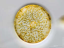
Thalassiosira lundiana Silicious processes (the labiate and the occluded ones) are visible. Scale bar indicates 25 µm. The image was built up using several photomicrographic frames with manual stacking technique. Sample from North Sea near Heligoland (spring diatom bloom). Images were taken using Zeiss Universal with Olympus C7070 CCD camera.Image under Creative Commons License V 3.0 (CC BY-NC-SA). Place name: North Sea around Heligoland Latitude: 54.186311 Longitude: 7.895034 Silikatfortsätze der Typen labiate und occluded sind sichtbar. Multiebenen-Abbildung, manuell gestapelt. Der Messbalken markiert eine Länge von 25 µm. Probe aus der Nordsee vor Helgoland in der Zeit der Frühjahrsblüte. Mikrotechnik: Zeiss Universal, Kamera: Olympus C7070.Creative Commons License V 3.0 (CC BY-NC-SA). For permission to use of (high-resolution) images please contact postmaster@protisten.de.
-
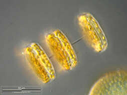
Thalassiosira lundiana Labiate processes and chitinous spines are visible. Scale bar indicates 25 µm. The image was built up using several photomicrographic frames with manual stacking technique. Sample from North Sea near Heligoland (spring diatom bloom). Images were taken using Zeiss Universal with Olympus C7070 CCD camera.Image under Creative Commons License V 3.0 (CC BY-NC-SA). Place name: North Sea around Heligoland Latitude: 54.186311 Longitude: 7.895034 Silikatfortsätze des Typs labiate zusammen mit chitinösen Schwebefortsätzen sind sichtbar. Multiebenen-Abbildung, manuell gestapelt. Der Messbalken markiert eine Länge von 25 µm. Probe aus der Nordsee vor Helgoland in der Zeit der Frühjahrsblüte. Mikrotechnik: Zeiss Universal, Kamera: Olympus C7070.Creative Commons License V 3.0 (CC BY-NC-SA). For permission to use of (high-resolution) images please contact postmaster@protisten.de.
-
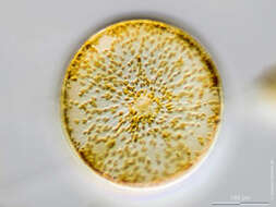
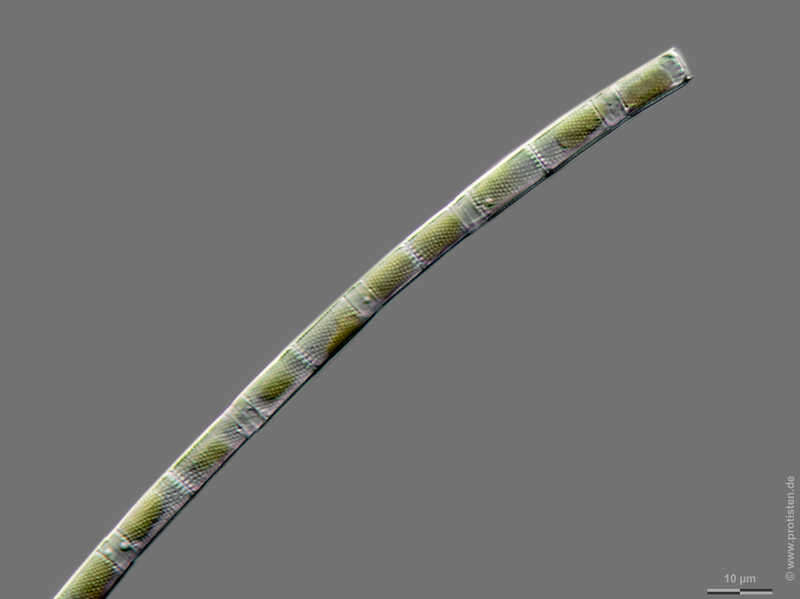
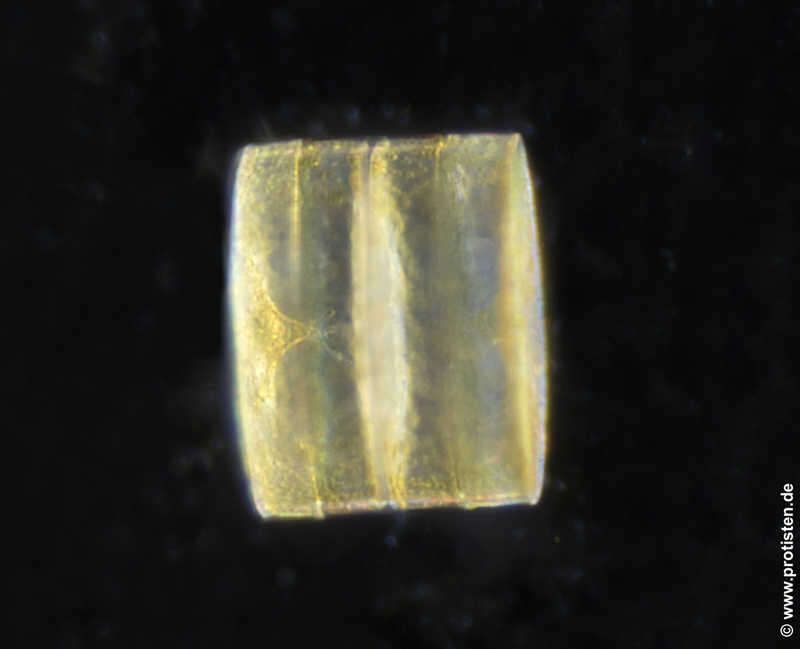
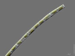
Aulacoseira spec. Scale bar indicates 10 µm. Sample from the Lake Vollstedter See near Kiel, Germany. Sampling date 9/2019. The image was built up using several photomicrographic frames with manual stacking technique. Images were taken using Zeiss Axioplan with Olympus OM-D M5 MKII. Image under Creative Commons License V 3.0 (CC BY-NC-SA). Place name: Lake Vollstedter See near Kiel (Germany) Latitude: 54.24105528 Longitude: 9.859339 Multiebenen-Abbildung, manuell gestapelt. Der Messbalken markiert eine Länge von 10 µm. Probe aus dem Vollstedter See bei Groß Vollstedt. Datum der Aufsammlung: 9/2019. Mikrotechnik: Zeiss Axioplan, Kamera: Olympus OM-D M5 MKII. Creative Commons License V 3.0 (CC BY-NC-SA). For permission to use of (high-resolution) images please contact postmaster@protisten.de.
-
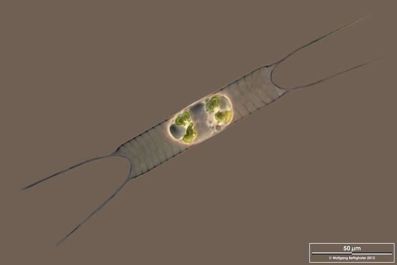
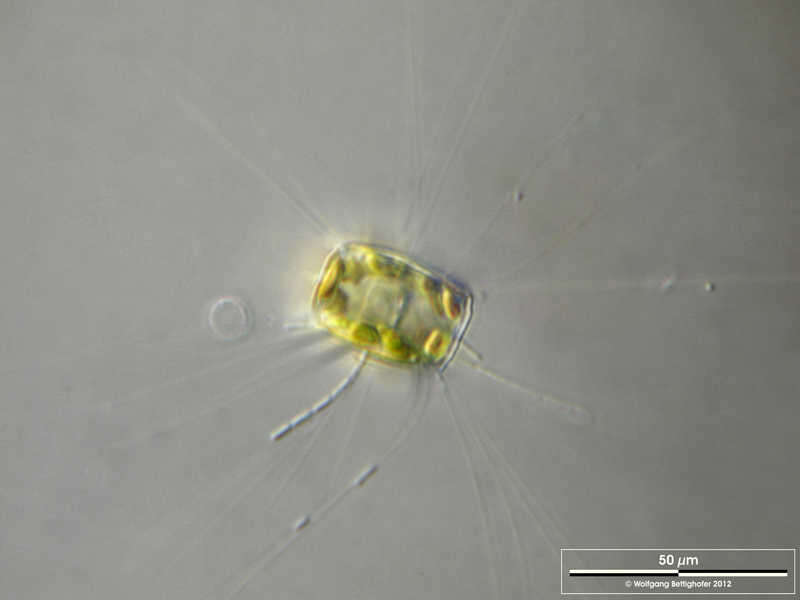
Acanthoceras spec. Scale bar indicates 50 µm. The specimen was gathered in the wetlands of national park Unteres Odertal (100 km north east of Berlin). The image was built up using several photomicrographic frames with manual stacking technique. Images were taken using Zeiss Universal with Olympus C7070 CCD camera. Image under Creative Commons License V 3.0 (CC BY-NC-SA). Place name: Creek in Oder valley 100 km north east of Berlin (Germany) Latitude: 53.135032 Longitude: 14.348738 Der Messbalken markiert eine Länge von 50 µm. Die Probe wurde in den Feuchtgebieten des Nationalpark Unteres Odertal (100 km nordöstlich von Berlin) gesammelt. Mikrotechnik: Zeiss Universal, Kamera: Olympus C7070. Creative Commons License V 3.0 (CC BY-NC-SA). For permission to use of (high-resolution) images please contact postmaster@protisten.de.
-
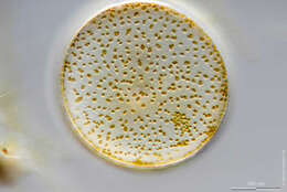
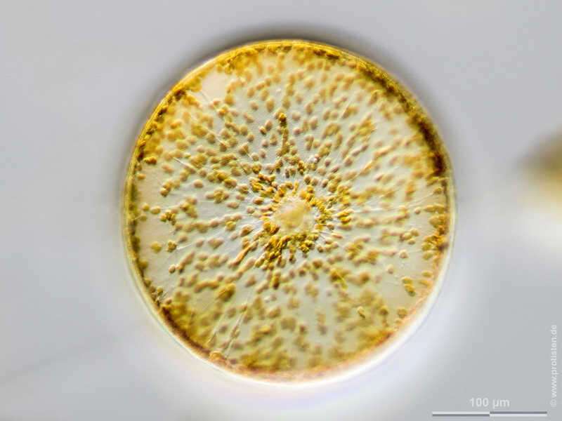
Coscinodiscus concinnus Scale bar indicates 100 µm. The specimen was gathered in the Kieler Förde (Baltic Sea). Sampling date 1/2022. The image was built up using several photomicrographic frames with manual stacking technique. Images were taken using Olympus stereo microscope SZX16/Planapo 2.0x with Olympus OM-D M5 MKII. Image under Creative Commons License V 3.0 (CC BY-NC-SA). Place name: Baltic Sea, Kieler Förde, Kiel Fjord (Germany) Latitude: 54.3894126 Longitude: 10.1749055 Multiebenen-Abbildung, manuell gestapelt. Der Messbalken markiert eine Länge von 100 µm. Probe aus der Kieler Förde. Datum der Aufsammlung: 1/2022. Mikrotechnik: Olympus stereo microscope SZX16/Planapo 2.0x, Kamera: Olympus OM-D M5 MKII. Creative Commons License V 3.0 (CC BY-NC-SA). For permission to use of (high-resolution) images please contact postmaster@protisten.de.
-
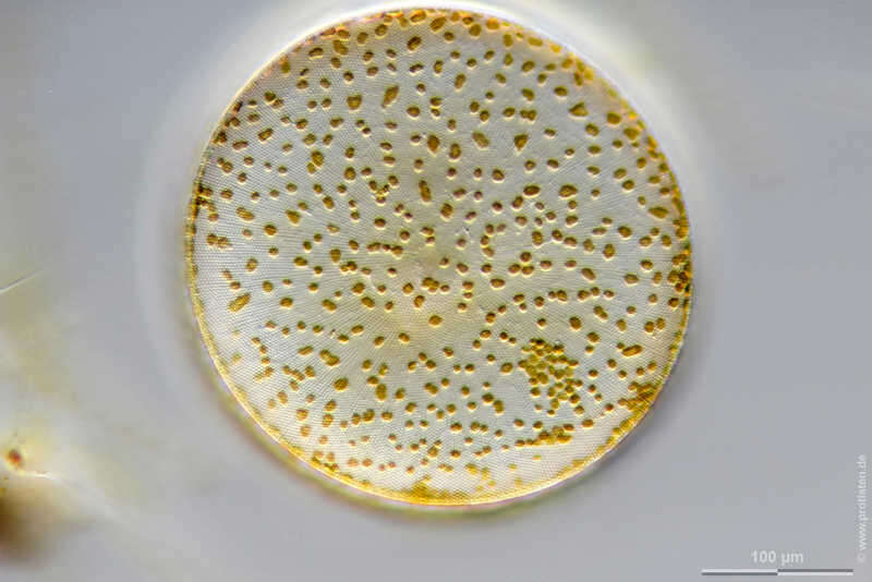
Coscinodiscus concinnus Sample from North Sea near Heligoland (spring diatom bloom). The image was built up using several photomicrographic frames with manual stacking technique. Images were taken using Leica dissecting microscope with MFT camera Olympus OM-D E-M5 II.Image under Creative Commons License V 3.0 (CC BY-NC-SA). Place name: North Sea around Heligoland Latitude: 54.186311 Longitude: 7.895034 Probe aus der Nordsee vor Helgoland in der Zeit der Frühjahrsblüte. Mikrotechnik: Leica Stereomikroskop, Kamera: Olympus OM-D E-M5 II.Creative Commons License V 3.0 (CC BY-NC-SA). For permission to use of (high-resolution) images please contact postmaster@protisten.de.
-
Coscinodiscus concinnus Scale bar indicates 100 µm. The specimen was gathered in the Kieler Förde (Baltic Sea). Sampling date 1/2022. The image was built up using several photomicrographic frames with manual stacking technique. Images were taken using Olympus stereo microscope SZX16/Planapo 2.0x with Olympus OM-D M5 MKII. Image under Creative Commons License V 3.0 (CC BY-NC-SA). Place name: Baltic Sea, Kieler Förde, Kiel Fjord (Germany) Latitude: 54.3894126 Longitude: 10.1749055 Multiebenen-Abbildung, manuell gestapelt. Der Messbalken markiert eine Länge von 100 µm. Probe aus der Kieler Förde. Datum der Aufsammlung: 1/2022. Mikrotechnik: Olympus stereo microscope SZX16/Planapo 2.0x, Kamera: Olympus OM-D M5 MKII. Creative Commons License V 3.0 (CC BY-NC-SA). For permission to use of (high-resolution) images please contact postmaster@protisten.de.
-
Thalassiosira rotula Scale bar indicates 100 µm. The specimen was gathered in the Kieler Förde (German Baltic Sea). Sampling date 3/2018. The image was built up using several photomicrographic frames with manual stacking technique. Images were taken using Zeiss Axioplan with Olympus OM-D M5 MKII. Image under Creative Commons License V 3.0 (CC BY-NC-SA). Place name: Baltic Sea, Kieler Förde, Kiel Fjord (Germany) Latitude: 54.3894126 Longitude: 10.1749055 Multiebenen-Abbildung, manuell gestapelt. Der Messbalken markiert eine Länge von 100 µm. Probe aus der Kieler Förde. Datum der Aufsammlung: 3/2018. Mikrotechnik: Zeiss Axioplan, Kamera: Olympus OM-D M5 MKII. Creative Commons License V 3.0 (CC BY-NC-SA). For permission to use of (high-resolution) images please contact postmaster@protisten.de.
-
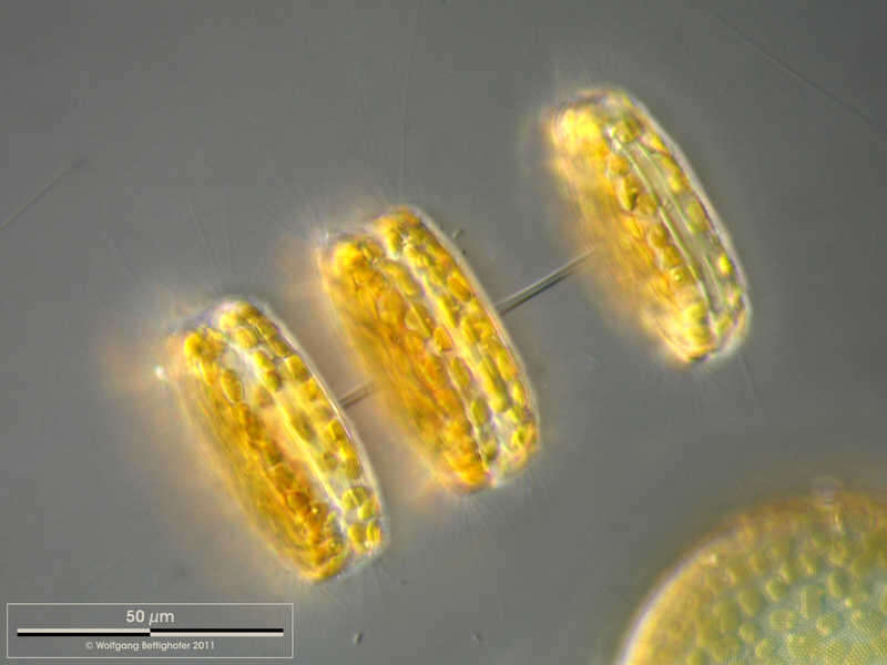
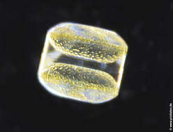
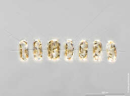
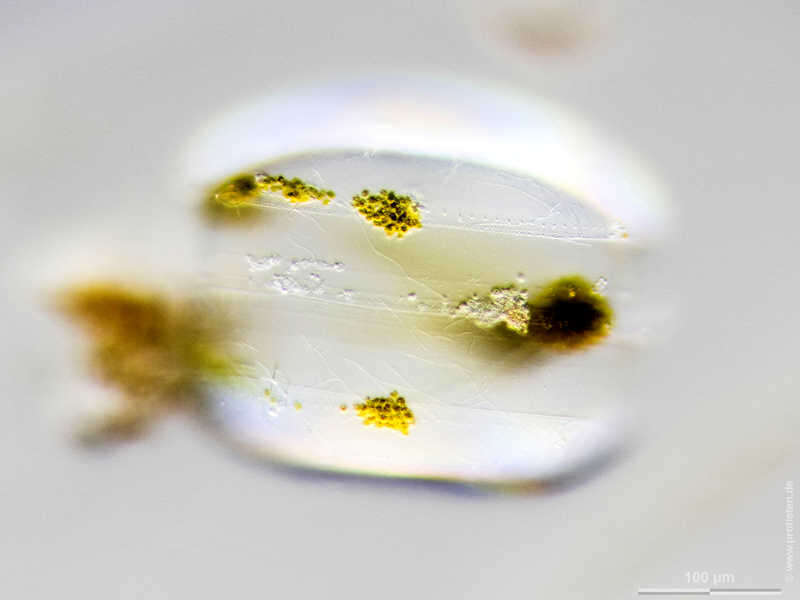
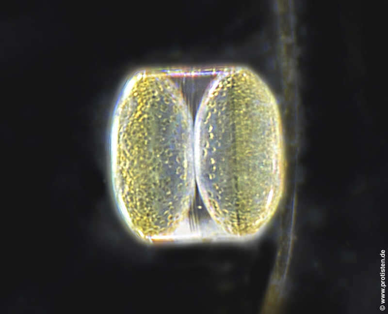
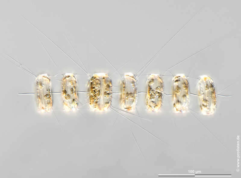
Thalassiosira rotula The members of the colony are interconnected with a bundle of threads. Numerous delicate spines protruding from the valve's margin are visible. Scale bar indicates 50 µm. The image was built up using several photomicrographic frames with manual stacking technique. Sample from North Sea near Heligoland (spring diatom bloom). Images were taken using Zeiss Universal with Olympus C7070 CCD camera.Image under Creative Commons License V 3.0 (CC BY-NC-SA). Place name: North Sea around Heligoland Latitude: 54.186311 Longitude: 7.895034 Die Zellen der Kolonie sind mit Bündeln aus dünnen Fortsätzen verbunden. Zahlreiche dünne Schwebefortsätze aus Chitin entspringen an den Schalenrändern. Multiebenen-Abbildung, manuell gestapelt. Der Messbalken markiert eine Länge von 50 µm. Probe aus der Nordsee vor Helgoland in der Zeit der Frühjahrsblüte. Mikrotechnik: Zeiss Universal, Kamera: Olympus C7070.Creative Commons License V 3.0 (CC BY-NC-SA). For permission to use of (high-resolution) images please contact postmaster@protisten.de.
-
Thalassiosira rotula Scale bar indicates 100 µm. The specimen was gathered in the Kieler Förde (German Baltic Sea). Sampling date 3/2018. The image was built up using several photomicrographic frames with manual stacking technique. Images were taken using Zeiss Axioplan with Olympus OM-D M5 MKII. Image under Creative Commons License V 3.0 (CC BY-NC-SA). Place name: Baltic Sea, Kieler Förde, Kiel Fjord (Germany) Latitude: 54.3894126 Longitude: 10.1749055 Multiebenen-Abbildung, manuell gestapelt. Der Messbalken markiert eine Länge von 100 µm. Probe aus der Kieler Förde. Datum der Aufsammlung: 3/2018. Mikrotechnik: Zeiss Axioplan, Kamera: Olympus OM-D M5 MKII. Creative Commons License V 3.0 (CC BY-NC-SA). For permission to use of (high-resolution) images please contact postmaster@protisten.de.
-
Coscinodiscus wailesii Sample from North Sea near Heligoland (spring diatom bloom). The image was built up using several photomicrographic frames with manual stacking technique. Images were taken using Leica dissecting microscope with MFT camera Olympus OM-D E-M5 II.Image under Creative Commons License V 3.0 (CC BY-NC-SA). Place name: North Sea around Heligoland Latitude: 54.186311 Longitude: 7.895034 Probe aus der Nordsee vor Helgoland in der Zeit der Frühjahrsblüte. Mikrotechnik: Leica Stereomikroskop, Kamera: Olympus OM-D E-M5 II.Creative Commons License V 3.0 (CC BY-NC-SA). For permission to use of (high-resolution) images please contact postmaster@protisten.de.
-
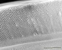
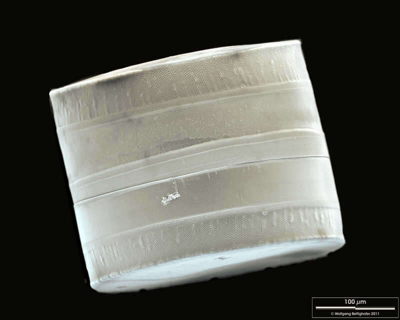
Coscinodiscus wailesii SEM of girdle view. The ligulae which fit in the open girdle bands are weakly visible. Scale bar indicates 100 µm. Sample from North Sea near Heligoland (spring diatom bloom). The image was built up using several photomicrographic frames with manual stacking technique. Use of SEM equipment courtesy of Lab Dr. Karl-Heinz Schäffner, Solingen, Germany. Place name: North Sea around Heligoland Latitude: 54.186311 Longitude: 7.895034 REM-Aufnahme des Gürtelbandes. Man erkennt schwach die sogenannten Ligulae, die in den Lücken der offenen Gürtelbänder liegen. Der Messbalken markiert eine Länge von 100 µm. Probe aus der Nordsee vor Helgoland in der Zeit der Frühjahrsblüte. Die Aufnahme entstand im Labor Dr. Karl-Heinz Schäffner, Solingen.Creative Commons License V 3.0 (CC BY-NC-SA). For permission to use of (high-resolution) images please contact postmaster@protisten.de.
-
Coscinodiscus wailesii Sample from North Sea near Heligoland (spring diatom bloom). Scale bar indicates 100 µm.The image was built up using several photomicrographic frames with manual stacking technique. Images were taken using Zeiss Axioplan with MFT camera Olympus OM-D E-M5 II.Image under Creative Commons License V 3.0 (CC BY-NC-SA). Place name: North Sea around Heligoland Latitude: 54.186311 Longitude: 7.895034 Der Messbalken markiert eine Länge von 100 µm. Probe aus der Nordsee vor Helgoland in der Zeit der Frühjahrsblüte. Mikrotechnik: Zeiss Axioplan, Kamera: Olympus OM-D E-M5 II.Creative Commons License V 3.0 (CC BY-NC-SA). For permission to use of (high-resolution) images please contact postmaster@protisten.de.
-
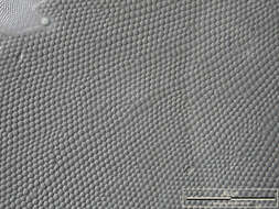
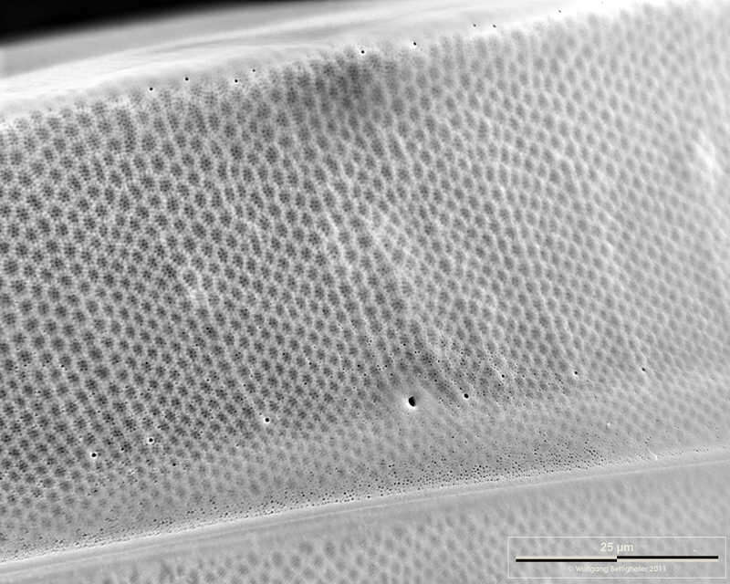
Coscinodiscus wailesii Closeup of the lateral side of the valve. Scale bar indicates 25 µm. Sample from North Sea near Heligoland (spring diatom bloom). The image was built up using several photomicrographic frames with manual stacking technique. Use of SEM equipment courtesy of Lab Dr. Karl-Heinz Schäffner, Solingen, Germany. Place name: North Sea around Heligoland Latitude: 54.186311 Longitude: 7.895034 Ausschnitt aus der Seitenansicht. Der Messbalken markiert eine Länge von 25 µm. Probe aus der Nordsee vor Helgoland in der Zeit der Frühjahrsblüte. Die Aufnahme entstand im Labor Dr. Karl-Heinz Schäffner, Solingen.Creative Commons License V 3.0 (CC BY-NC-SA). For permission to use of (high-resolution) images please contact postmaster@protisten.de.
-
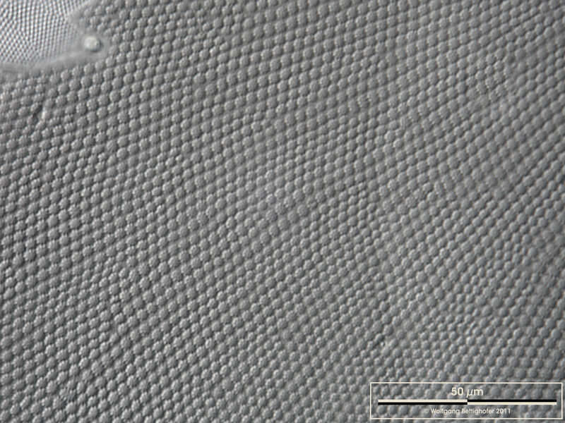
Coscinodiscus wailesii DIC closeup of valve of a living specimen. Scale bar indicates 50 µm. Sample from North Sea near Heligoland (spring diatom bloom). The image was built up using several photomicrographic frames with manual stacking technique. Images were taken using Zeiss Universal with Olympus C7070 CCD camera.Image under Creative Commons License V 3.0 (CC BY-NC-SA). Place name: North Sea around Heligoland Latitude: 54.186311 Longitude: 7.895034 Ausschnitt einer Schale, aufgenommen von einer lebenden Zelle. Multiebenen-Abbildung, manuell gestapelt. Der Messbalken markiert eine Länge von 50 µm. Probe aus der Nordsee vor Helgoland in der Zeit der Frühjahrsblüte. Mikrotechnik: Zeiss Universal, Kamera: Olympus C7070.Creative Commons License V 3.0 (CC BY-NC-SA). For permission to use of (high-resolution) images please contact postmaster@protisten.de.