-
Giacinta Angela Stocchino, Ronald Sluys, Paolo Deri, Renata Manconi
Zookeys
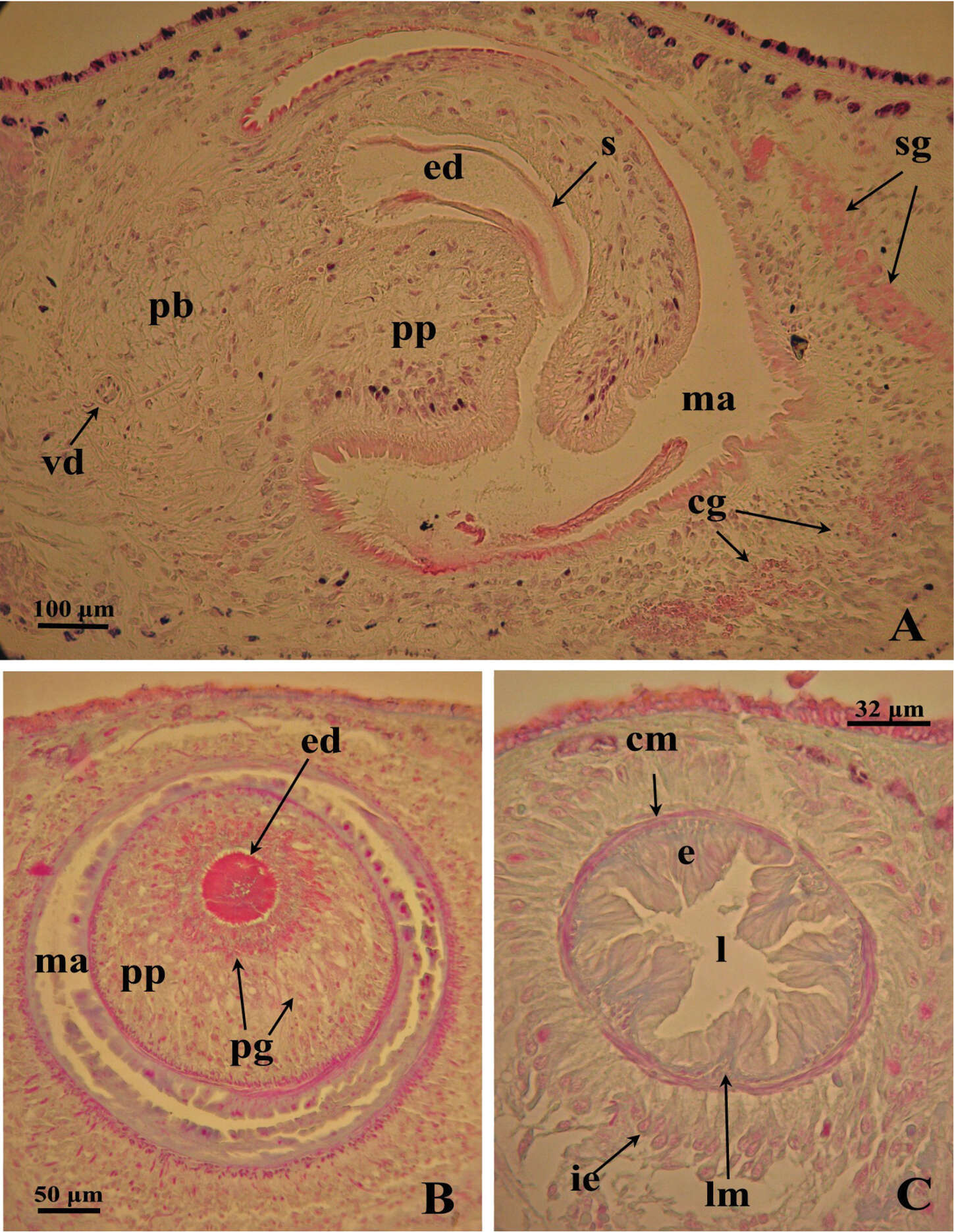
Figure 4.Dugesia superioris. Photomicrographs of the copulatory apparatus. A Holotype ZMA V.Pl. 7153.1, sagittal section showing the penis bulb and the penis papilla with the ejaculatory duct B Paratype CGAS Pla 6. 3, transverse section of the penis papilla and the ejaculatory duct surrounded by numerous glands C Paratype CGAS Pla 6. 3, transverse section of the bursal canal.
-
Carolina Noreña, Daniel Marquina, Jacinto Perez, Bruno Almon
Zookeys
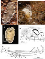
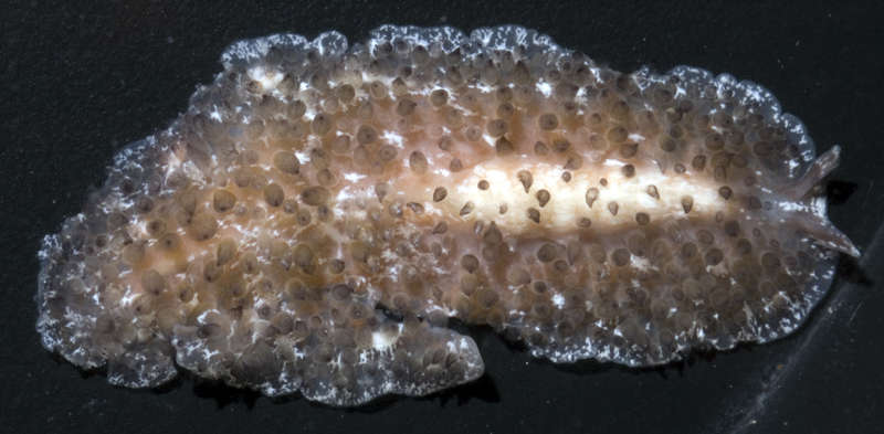
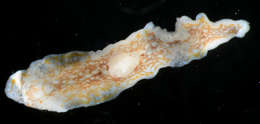
Figure 3.Euryleptodes galikias. A, B dorsal view of a living animal C dorsal and F, ventral views of a fixed specimen E dorsal and D ventral details of the eyes G sagittal reconstruction of a whole specimen H sagittal reconstruction of the reproductive system H. Anterior to the left in A, F and G.
-
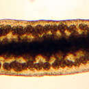
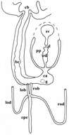
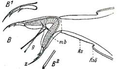
Giacinta Angela Stocchino, Ronald Sluys, Renata Manconi
Zookeys
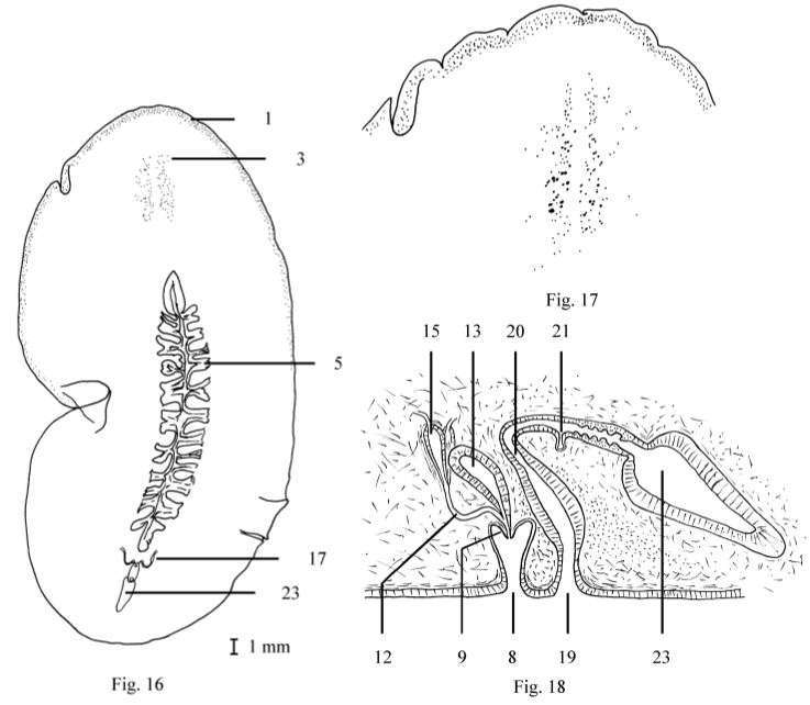
Figure 4.Dugesia bifida. Schematic horizontal reconstruction of the copulatory apparatus.
-
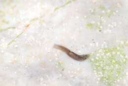
All Biocode files are based on field identifications to the best of the researcher’s ability at the time.
-
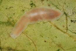
All Biocode files are based on field identifications to the best of the researcher’s ability at the time.
-
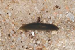
All Biocode files are based on field identifications to the best of the researcher’s ability at the time.
-
-
-
-
-
Coral Sea, Duration 28 seconds
-
Coral Sea, Duration 13 seconds, Shot includes Torquigener pleurogramma (Weeping toado)
-
Coral Sea, Duration 15 seconds
-
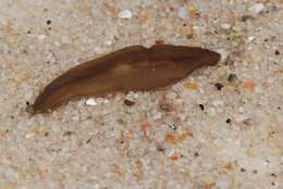
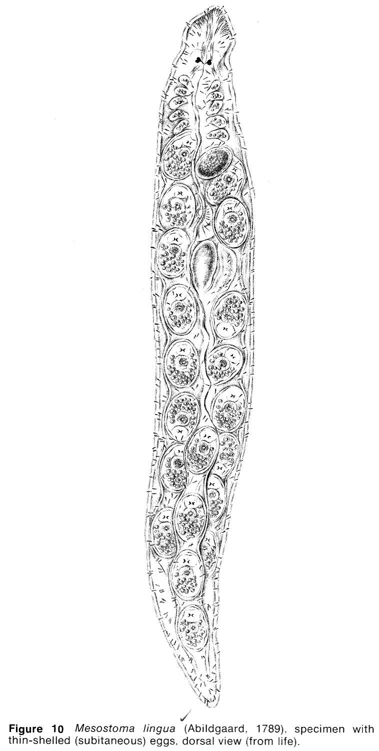
Hampen Sø, Midtjylland, Danmark
-
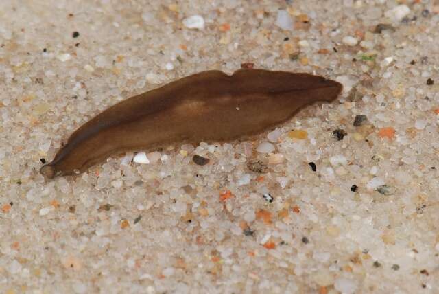
Roskilde
-
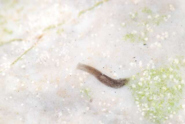
Kastbjerg Å
-
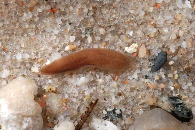
Gudenå opstrøms Klostermølle, Mattrup Å ved Stidsmølle
-
Ørn Sø, Silkeborg, Danmark
-
Dollerup Kilde ved Hald Sø, Jylland, Danmark
-
Centers for Disease Control/Division of Parasitic Diseases and Malaria
EOL staff
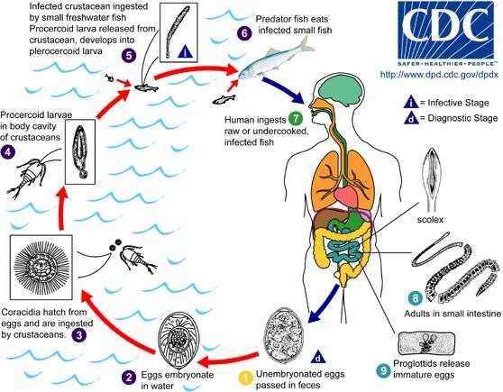
Life cycle of the Broad Tapeworm (Diphyllobothrium latum) Immature eggs of the Broad Tapeworm (Diphyllobothrium latum) are passed in feces (1) from the definitive host. Under appropriate conditions, the eggs mature (after approximately 18 to 20 days) (2) and yield oncospheres, which develop into a coracidia (3). After ingestion by a suitable freshwater crustacean (the copepod first intermediate host), the coracidia develop into procercoid larvae (4). Following ingestion of the copepod by a suitable second intermediate host, typically minnows and other small freshwater fish, the procercoid larvae are released from the crustacean and migrate into the fish flesh, where they develop into a plerocercoid larva (sparganum) (5). The plerocercoid larvae are the infective stage for humans. Because humans do not generally eat undercooked minnows ans similar freshwater fish, these do not represent an important source of infection. Nevertheless, these small second intermediate hosts can be eaten by larger predator species such as trout, perch, and walleyed pike (6). In this case, the sparganum can migrate to the musculature of the larger predator fish and humans can acquire the disease by eating these later intermediate infected host fish raw or undercooked (7). After ingestion of the infected fish, the plerocercoid develop into immature adults and then into mature adult tapeworms, which will reside in the human host's small intestine. The adults of D. latum attach to the intestinal mucosa by means of the two bilateral grooves (bothria) of the scolex (8). The adult tapeworms can exceed 10 m in length, with more than 3,000 proglottids. Immature eggs are discharged from the proglottids (up to 1,000,000 eggs per day per worm) (9) and are passed in the feces (1). Eggs appear in the feces 5 to 6 weeks after infection. In addition to humans, many other mammals can also serve as definitive hosts for D. latum. From
Centers for Disease Control Parasites and Health website.
-
Carolina Noreña, Daniel Marquina, Jacinto Perez, Bruno Almon
Zookeys
Figure 4.Prostheceraeus vittatus. A dorsal view of a living animal B living animal feeding on ascidians C dorsal view of a fixed specimen D sagittal reconstruction of the copulatory apparatus E sagittal reconstruction of a whole specimen. Anterior to the left in D, E.
-
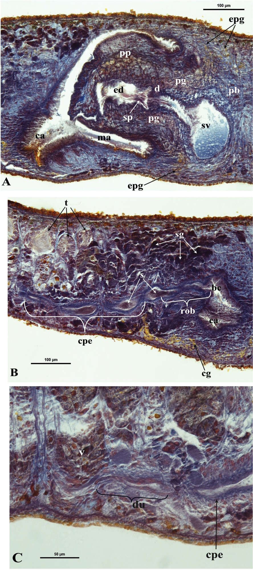
Giacinta Angela Stocchino, Ronald Sluys, Renata Manconi
Zookeys
Figure 5.Dugesia bifida. Microphotographs of the copulatory apparatus. A Holotype ZMA V.Pl. 7189.1, sagittal section showing the penis bulb (pb) and the penis papilla (pp) with the seminal vesicle (sv) and the ejaculatory duct (ed) B Holotype ZMA V.Pl. 7189.1, sagittal section showing the opening of the right oviducal branch (rob) through the posterior wall of the bursal canal (bc), and the common posterior oviducal extension (cpe) full of sperm (s) C Paratype CGAS Pla 7.1, sagittal section showing the caudal part of the common posterior oviducal extension (cpe) and the ductule (du) communicating with the ventral part of an adjacent vitellarium (v).
-
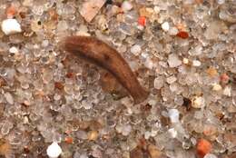
All Biocode files are based on field identifications to the best of the researcher’s ability at the time.
-
All Biocode files are based on field identifications to the best of the researcher’s ability at the time.