-
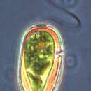
Euglena mutabilis. Cell observed in freshwater habitats in the vicinity of Broome, Western Australia in September 2003. This image was taken using differential interference contrast optics. This work was supported by the Australian Biological Resources Study.
-
Moving Euglena. By Bob Moore and Dan Lahr.
-
Euglena mutabilis. Cell observed in freshwater habitats in the vicinity of Broome, Western Australia in September 2003. This image was taken using differential interference contrast optics. This work was supported by the Australian Biological Resources Study.
-
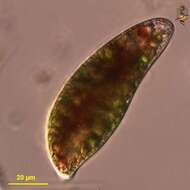
Euglena sanguinea. Cell observed in freshwater habitats in the vicinity of Broome, Western Australia in September 2003. This work was supported by the Australian Biological Resources Study.
-
Euglena mutabilis. Cell observed in freshwater habitats in the vicinity of Broome, Western Australia in September 2003. This image was taken using differential interference contrast optics. This work was supported by the Australian Biological Resources Study.
-
Euglena sanguinea. Detail showing the red granules which give this species its distinctive colour. Cell observed in freshwater habitats in the vicinity of Broome, Western Australia in September 2003. This work was supported by the Australian Biological Resources Study.
-
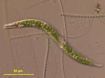
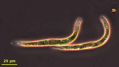
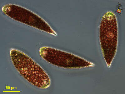
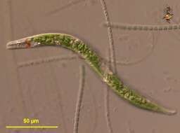
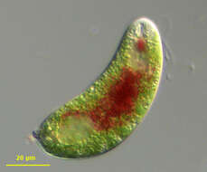
Two cells of this worm-like species of euglena. These cells have no emerging flagella, and they move by squirming and gliding. The cell contains numerous chloroplasts, and these provide the cell with their green colour. The red dots are the eye-spots.
-
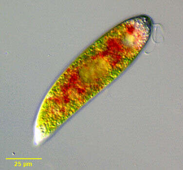
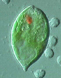
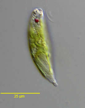
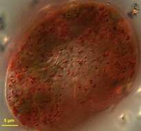
Portrait of Euglena sanguinea (probably synonymous with E. rubra and some other red euglenae). This species has numerous red granules containing astaxanthin among other carotenoids scattered in the cytoplasm. The granules, which may provide protection of cell structures from UV-B radiation, concentrate in a central mass under low light conditions and disperse in bright light (as seen in this image). The cell is has a blunt point posteriorly and a broadly rounded anterior. The large stigma (left anterior in this image) has the same coloration as the red cytoplasmic granules. The plastids are elongate, thin and tend to parallel the fine pellicular striations. The flagellum is about body length. Cells are flexible but not highly metabolic. The central nucleus is seen in this image. Small paramylon grains of various shapes are scattered through the cytoplasm. Swimming is slow with rotation around the long axis. Cells often appear to glide on the substrate. From stagnant fresh water with abundant rotting vegetation near Boise, Idaho. DIC optics
-
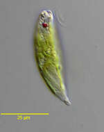
Euglena velata. Cell observed in freshwater habitats in the vicinity of Broome, Western Australia in September 2003. Animations by Rosemary Arbur of flagellar beat patterns are available
here.This image was taken using differential interference contrast optics. This work was supported by the Australian Biological Resources Study.
-
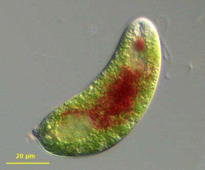
Portrait of Euglena sanguinea (probably synonymous with E. rubra and some other red euglenae). This species has numerous red granules containing astaxanthin among other carotenoids scattered in the cytoplasm. The granules, which may provide protection of cell structures from UV-B radiation, concentrate in a central mass under low light conditions (as seen in this image) and disperse in bright light. The cell is has a blunt point posteriorly and a broadly rounded anterior. The large stigma (mid-anterior in this image) has the same coloration as the red cytoplasmic granules. The plastids are elongate, thin and tend to parallel the fine pellicular striations. The flagellum is about body length. Cells are flexible but not highly metabolic. The nucleus is seen in the posterior 1/3 in this image. Small paramylon grains of various shapes are scattered through the cytoplasm. Swimming is slow with rotation around the long axis. Cells often appear to glide on the substrate. From stagnant fresh water with abundant rotting vegetation near Boise, Idaho. DIC optics.
-
Euglena velata. Plastids with pyrenoids. Cell observed in freshwater habitats in the vicinity of Broome, Western Australia in September 2003. This image was taken using differential interference contrast optics. This work was supported by the Australian Biological Resources Study.
-
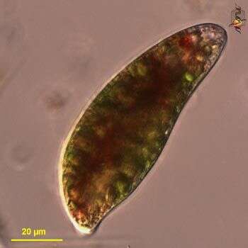
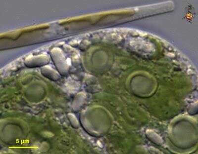
"Palmelloid" or resting phase of Euglena sanguinea. This species has numerous red granules containing astaxanthin among other carotenoids scattered in the cytoplasm. The granules, which may provide protection of cell structures from UV-B radiation, concentrate in a central mass under low light conditions and disperse in bright light. Under inhospitable conditions cells enter the palmelloid phase. The normally elongate cells shed their flagella and encyst within a clear mucous coat (seen in this image) and reduce metabolic activity. Cell division within the mucous cysts may occur with the aggregate of cells spreading to form large sheets. From stagnant fresh water with abundant rotting vegetation near Boise, Idaho. DIC optics.
-
Euglena velata. Cell observed in freshwater habitats in the vicinity of Broome, Western Australia in September 2003. This image was taken using differential interference contrast optics. This work was supported by the Australian Biological Resources Study.
-
Euglena sanguinea is one of several nominal species in the genus that are capable of producing dark red granules. This species can withdraw and extend the granules in the cell, and the species may occur in sufficient numbers to color water green or red (from which they have got the ne 'traffic-light euglena'). The anterior flagellum is often short or does not emerge at all.
-
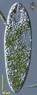
Euglena velata. Cell observed in freshwater habitats in the vicinity of Broome, Western Australia in September 2003. This image was taken using differential interference contrast optics. This work was supported by the Australian Biological Resources Study.
-
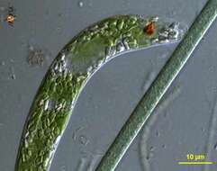
Euglena sanguinea is one of several nominal species in the genus that are capable of producing dark red granules. This species can withdraw and extend the granules in the cell. The anterior flagellum is often short or does not emerge at all. In this cell the flagellum is well developed. The larger clear inclusions are paramylon deposits.
-
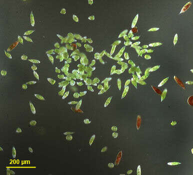
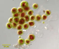
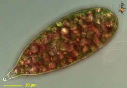
This image shows two species of Euglena, Euglena velata (the green cells) and Euglena sanguinea (the red cells). Low magnification image.
-
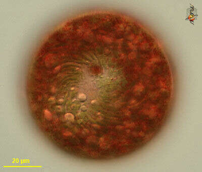
Euglena sanguinea is one of several nominal species in the genus that are capable of producing dark red granules. This image focussed on the surface shows the strand-like margins of the green platids, various spherical paramylon granules, and the red color. Differential interference contrast image.
-
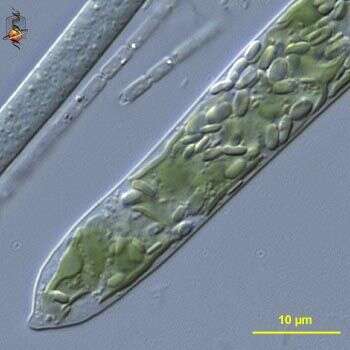
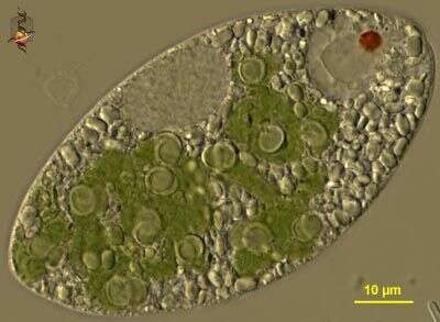
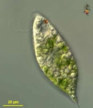
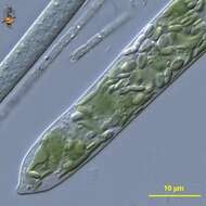
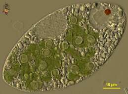
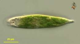
Euglena velata is a medium-sized Euglena with large flat plastids with central pyrenoid regions. The cytoplasm often has many fine granules. Differential interference contrast optics.
-
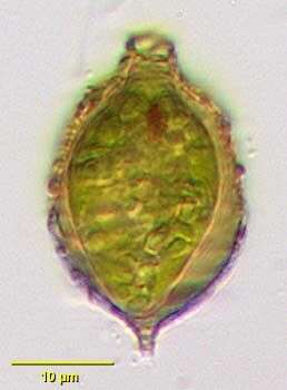
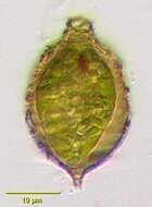
Portrait of Strombomonas acuminata a loricate euglenoid flagellate. Lorica in this species is brownish with a tapering neck and the posterior drawn out to a tapering point. Single flagellum emerges from aperture in neck. Debris adheres to the lorica in this species. Cell body does not fill lorica.Prominent stigma and discoid plastids.From freshwater pond near Boise, Idaho. Oblique illumination.
-
Euglena velata is a medium-sized Euglena with large flat plastids with central pyrenoid regions. The cytoplasm often has many fine granules. Eyespot and paramylon polysaccharide by-product are visible. Animations by Rosemary Arbur of flagellar beat patterns are available
here.Differential interference contrast optics.
-
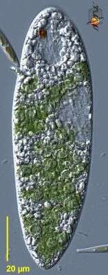
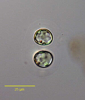
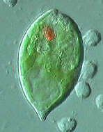
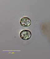
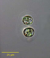
Portrait of the eustigmatophyte, Chlorobotrys regularis West, 1903 (Bohlin, 1901). Spherical paired cells enclosed in a faintly laminated mucilagenous sheath. Multiple parietal chloroplasts. The red spot (visible in the upper cell here) is an oil globule. Collected from a freshwater pond near Idaho City, idaho. June 2005. DIC.
-
Portrait of the euglenoid flagellate (Ehrenberg,1830).Collectedfrom a slow-flowing freshwaterstream near Boise, Idaho. November,2005.DIC.
-
Portrait of the eustigmatophyte, Chlorobotrys regularis West, 1903 (Bohlin, 1901). Spherical paired cells enclosed in a faintly laminated mucilagenous sheath (visible in this high contrast image). Multiple parietal chloroplasts. The red spot (best seen in the upper cell here) is an oil globule. Collected from a freshwater pond near Idaho City, Idaho. June 2005. DIC.