-
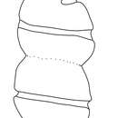
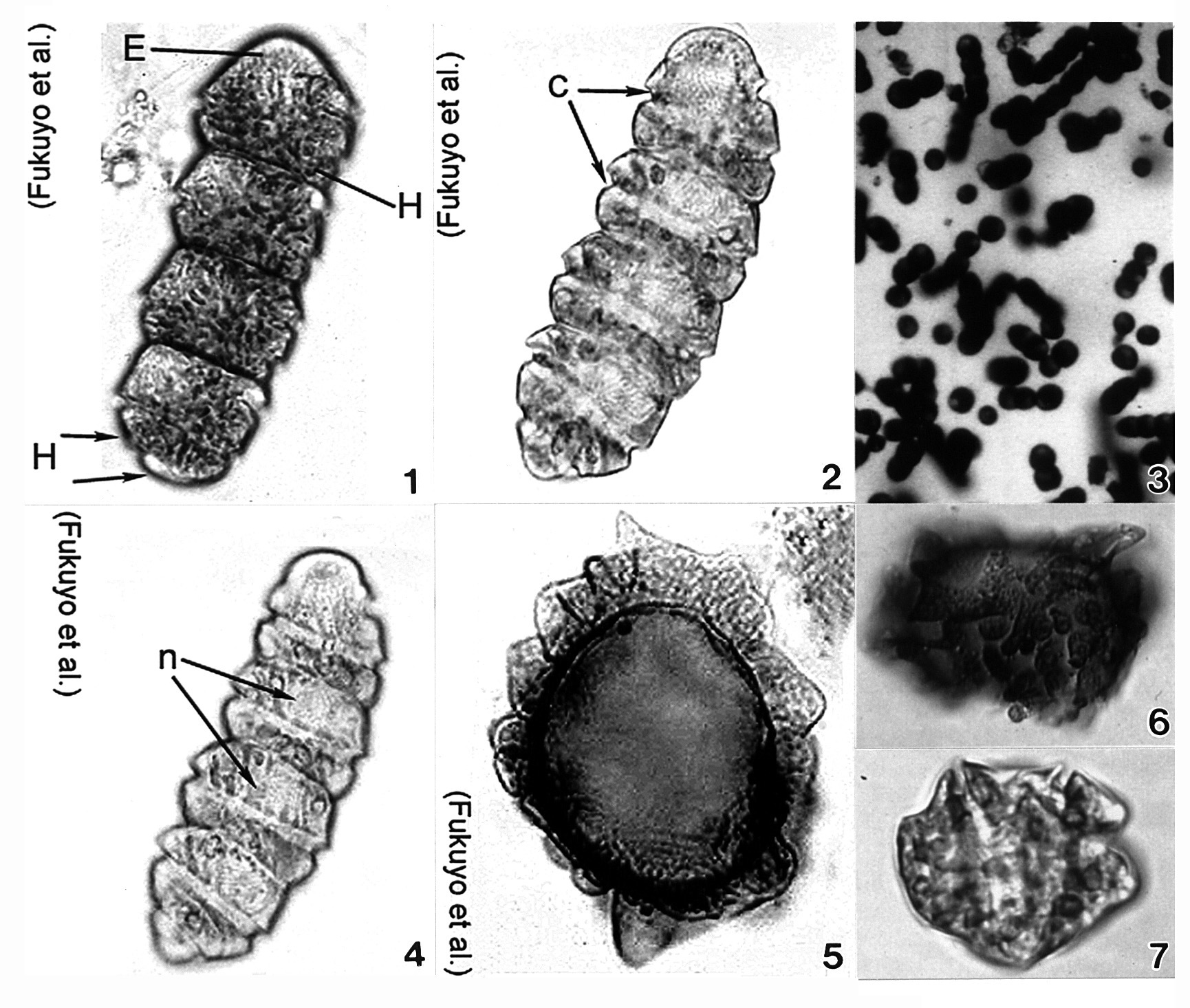
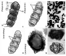
Plate 9. Cochlodinium polykrikoides. Figs. 1-7. LM. Fig. 1. Four cell chain. Single cell small and ellipsoid. Epitheca (E) rounded and conical. Hypotheca (H) divided into two posterior lobes (arrows). Numerous rod-shaped chloroplasts. Fig. 2. Cingulum (c) deeply excavated; circles cell 1.8-1.9 times. Fig. 3. Colony of single and chained cells. Fig. 4. Large nucleus (n) in epitheca. Figs. 5-7. Cysts. (Figs. 3,6,7 by Matsuoka & Fukuyo)
-
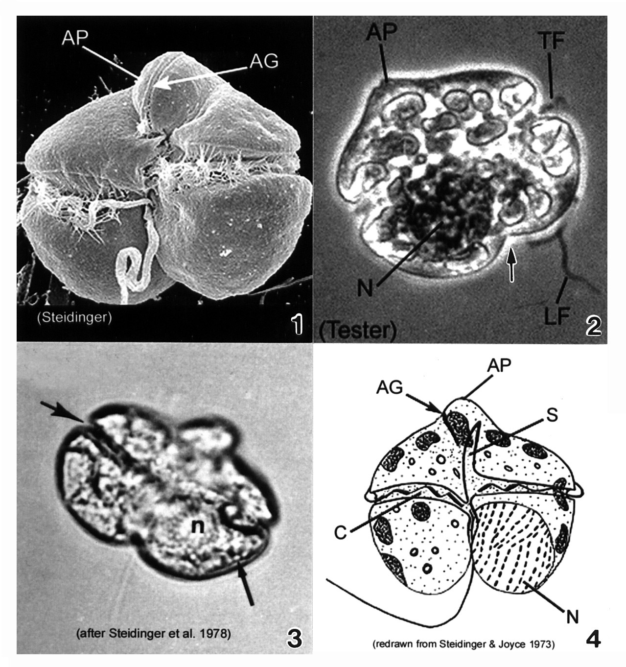
Plate 22. Gymnodinium breve. Fig. 1. SEM: ventral view. Cell small, wider than long, dorso-ventrally flattened. Cell nearly square in outline; prominent apical process (AP) directed ventrally. Apical groove (AG) present on apical process, adjacent to sulcus. Figs. 2-3. LM. Fig. 2. Dorsal view: large nucleus (N) in hypotheca. Transverse (TF) and longitudinal (LF) flagella present. Hypotheca bilobed (arrow). Fig. 3. Ventral view: displaced cingulum (large arrow) and lipid globule (small arrow). Fig. 4. Line drawing. Cingulum (C) displaced, descending. Long sulcus (S) extends to apex of cell.
-
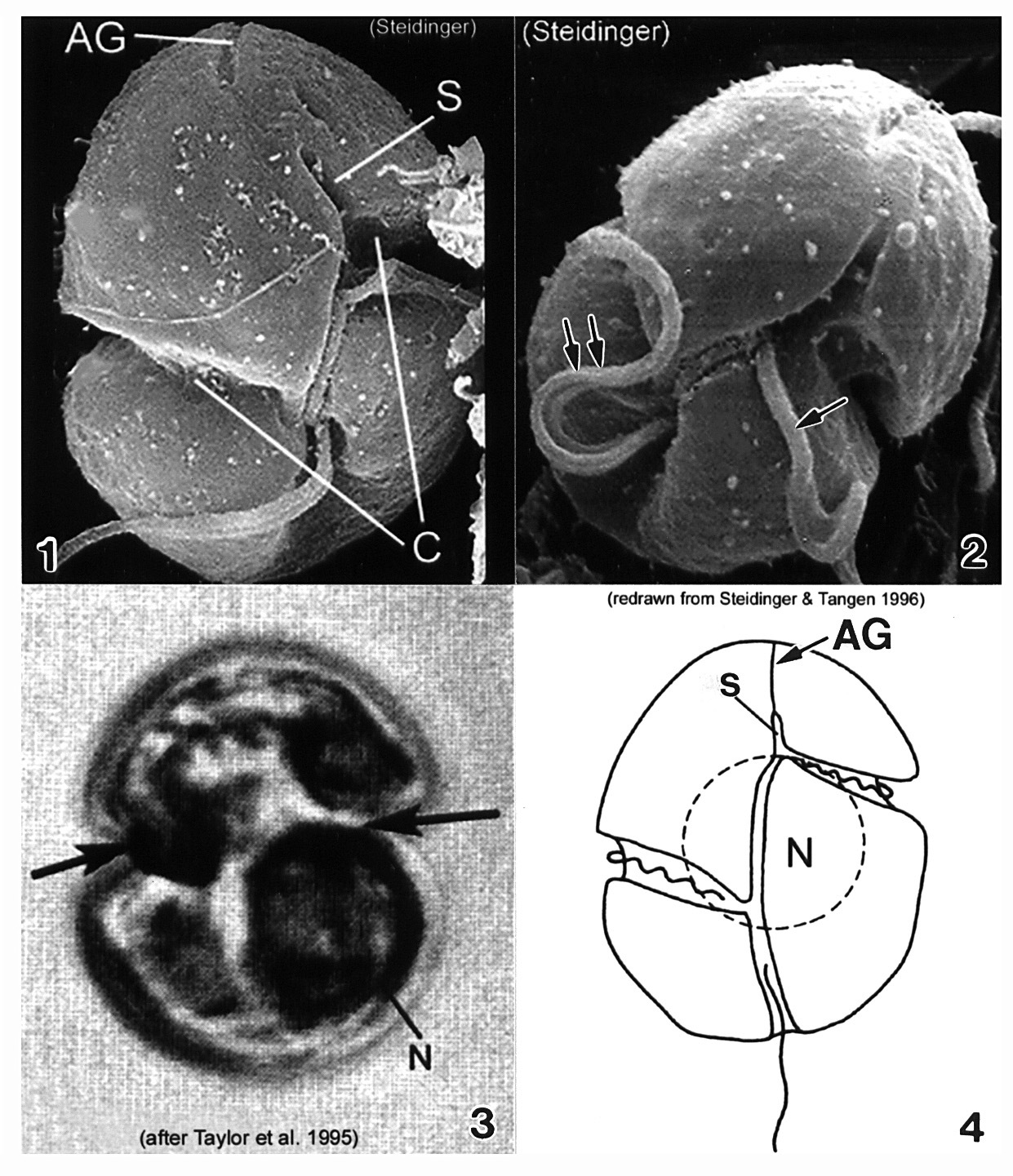
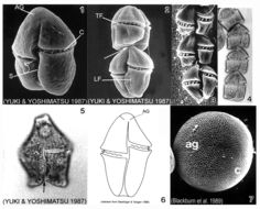
Plate 23. Gymnodinium catenatum. Figs. 1-3. SEM: ventral view. Fig. 1. Cell small, elongate-ovoid with slight dorso-ventral compression. Conical apex; rounded and notched antapex. Cingulum (C) excavated; sulcus (S) long. Distinctive horse-shoe shaped apical groove (AG) encircles apex. Fig. 2. Two cell chain; attachment point visible (arrow). Premedian cingulum displaced 2X its width. Longitudinal (LF) and transverse (TF) flagella visible. Fig. 3. Chain cells with anterior-posterior compression. Terminal cell slightly longer. Thecal surface rugose to smooth (Blackburn et al. 1989). Figs. 4-5. LM. Fig. 4. Chain-formation (Yuki and Yoshimatsu 1987). Fig. 5. Single cell. Conical epitheca with concave to flat apex. Bilobed hypotheca (arrow). Fig. 6. Line drawing. Fig. 7. SEM: cyst with microreticulations. ag=apical groove; c=cingulum
-
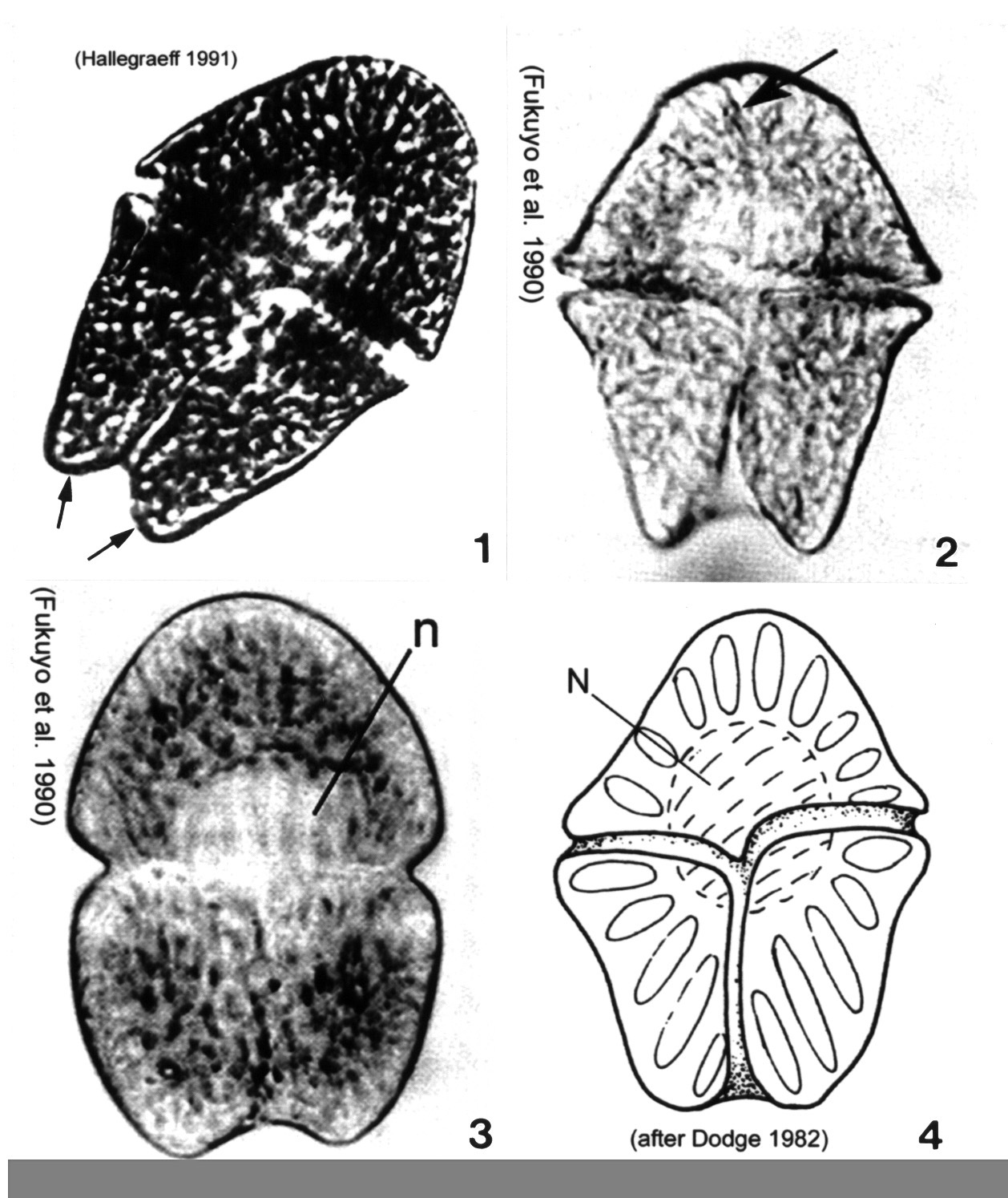
Plate 24. Gymnodinium mikimotoi. Figs. 1-4. SEM. Fig. 1. Ventral view: cell small, broadly oval to almost round. Epitheca slightly smaller than hypotheca. Characteristic straight apical groove (AG). Cingulum (C) deep, displaced 2 times its width. Sulcus (S) slightly invades epitheca (arrowheads). Hypotheca notched by widening sulcus (arrow). Fig. 2. Dorsal view: apical groove extends to dorsal side of epitheca creating slight indentation at the apex (arrowhead). Hypotheca bilobed (arrow). Fig. 3. Apical view of apical groove (arrow)(after Fukuyo et al.). Fig. 4. Cell compressed dorso-ventrally (after Fukuyo et al.). Figs. 5-7. LM. Fig. 5. Cingulum displaced 2 times its width (arrows)(from Larsen & Moestrup 1989: fig. 16g). Fig. 6. Large nucleus (N) in left lobe of hypotheca. Fig. 7. Vegetative division. Division plane oblique.
-
Plate 26. Gymnodinium sanguineum. Figs. 1-3. LM. Cell large, pentagonal, and slightly dorso-ventrally flattened. Cells vary in shape and size. Fig. 1. Ventral view. Epitheca and hypotheca nearly equal in size: epitheca conical, hypotheca bilobed (arrows). Fig. 2. Ventral view. Deep cingulum median, displaced 1-2 times its width. Sulcus deeply notches hypotheca. Apical groove present (arrow). Fig. 3. Cell deeply pigmented; central nucleus (n). Fig. 4. Line drawing. Spindle-shaped chloroplasts radially arranged.
-
Plate 27. Gymnodinium veneficum. Figs. 1-3. Line drawings. Fig. 1. Ventral view: cell small and ovoid. Epitheca slightly pointed, without apical groove. Cingulum deep and displaced 1-2 times its width. Fig. 2. Dorsal view: large central nucleus (N). Two to eight irregular chloroplasts present (C). Fig. 3. Sigmoid sulcus slightly invades epitheca (arrow).
-
Plate 28. Gyrodinium galatheanum. Figs. 1-2. SEM: ventral view. Fig. 1. Cell small, oval to round, with distinct apical groove (AG). Cingulum (C) displaced 3 times its width. Short and narrow sulcus (S) slightly invades epitheca. Fig. 2. Epitheca and hypotheca round. Cingulum wide, houses transverse flagellum (single arrow). Longitudinal flagella present (double arrow). Fig. 3. LM: ventral view. Cingulum deeply excavated (arrows). Nucleus (N) large and central. Fig. 4. Line drawing.