-
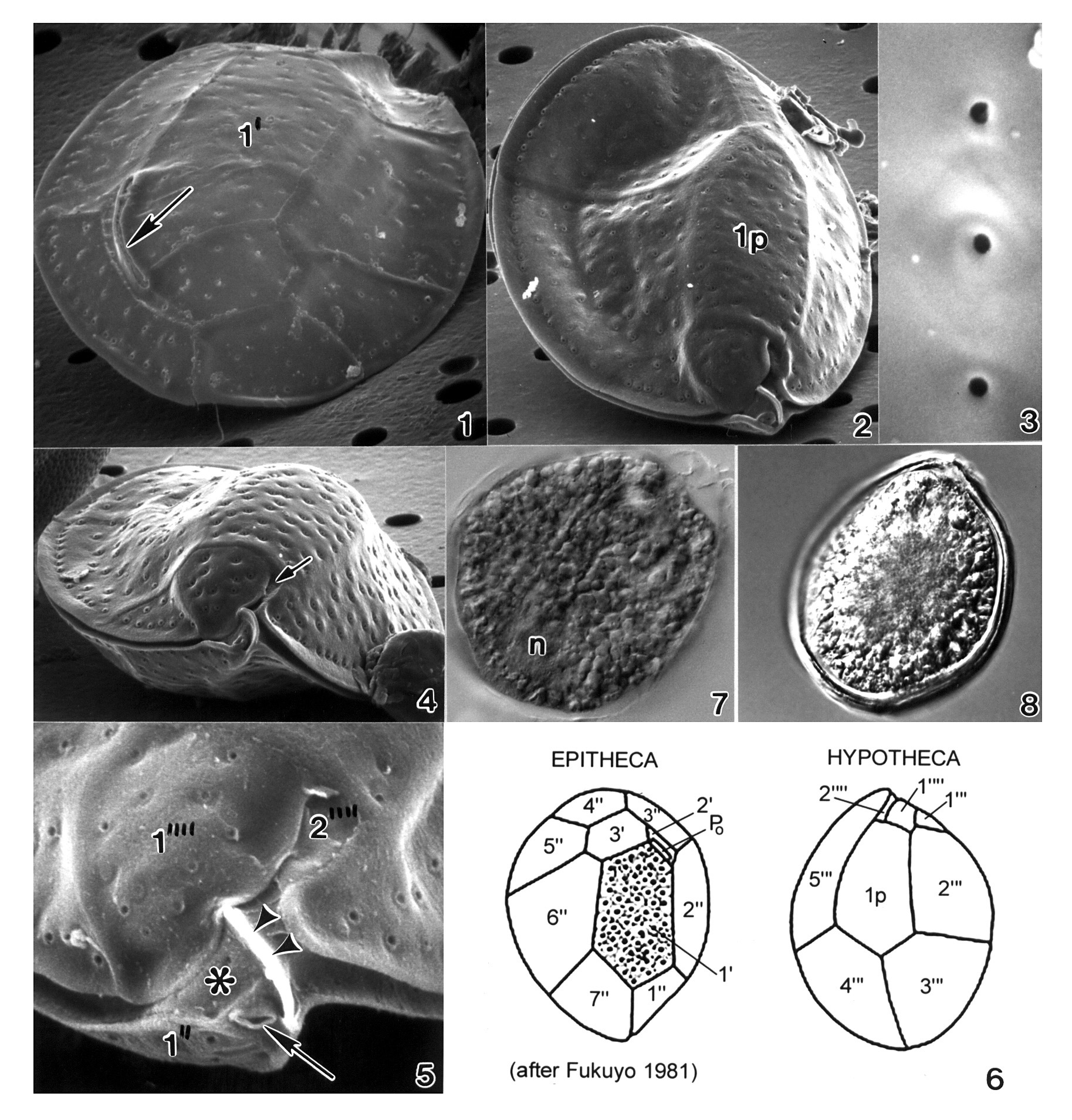
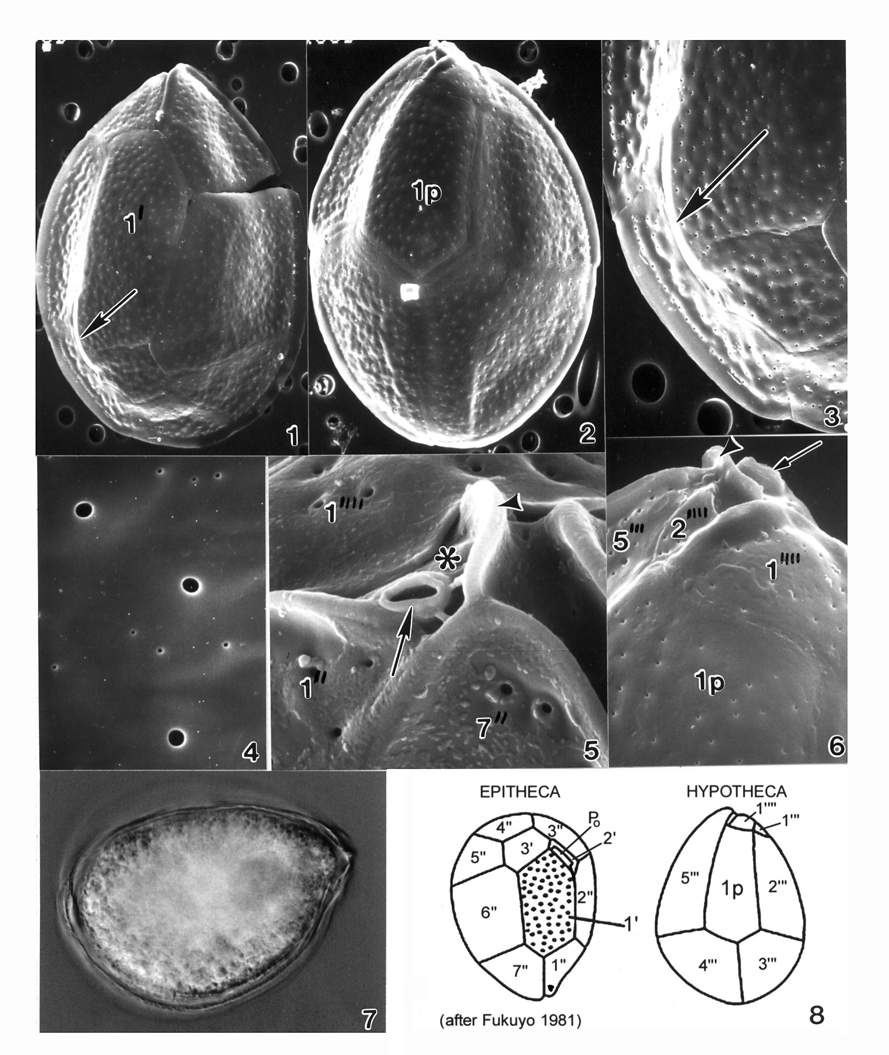
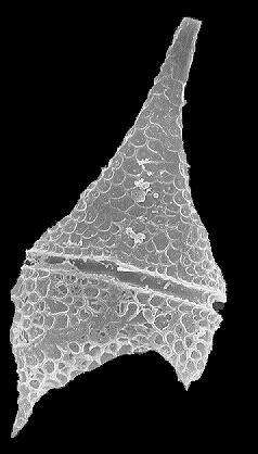
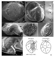
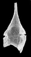
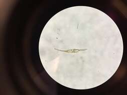
Plate 32. Ostreopsis lenticularis. Figs. 1-5. SEM. Fig. 1. Epithecal view: cell lenticulate to broadly oval. Curved off-center apical pore plate with a slit-like apical pore (arrow). Plate 1' irregularly pentagonal. Fig. 2. Hypothecal view: plate 1p central and pentagonal. Fig. 3. Smooth thecal surface. Round pores with smooth raised edges. Fig. 4. Hypothecal ventral view: cell anterio-posteriorly compressed. Shallow cingulum with smooth edge. Small sulcus hidden (arrow). Fig. 5. Location of ventral opening (arrow), ventral plate (asterisk), and rigid plate (arrowheads) within cingulum. Fig. 6. Line drawing: thecal plate arrangement. Figs. 7,8. LM. Fig. 7. Cytoplasma granulated; posterior nucleus (n). Fig. 8. Distinct cingular list.
-
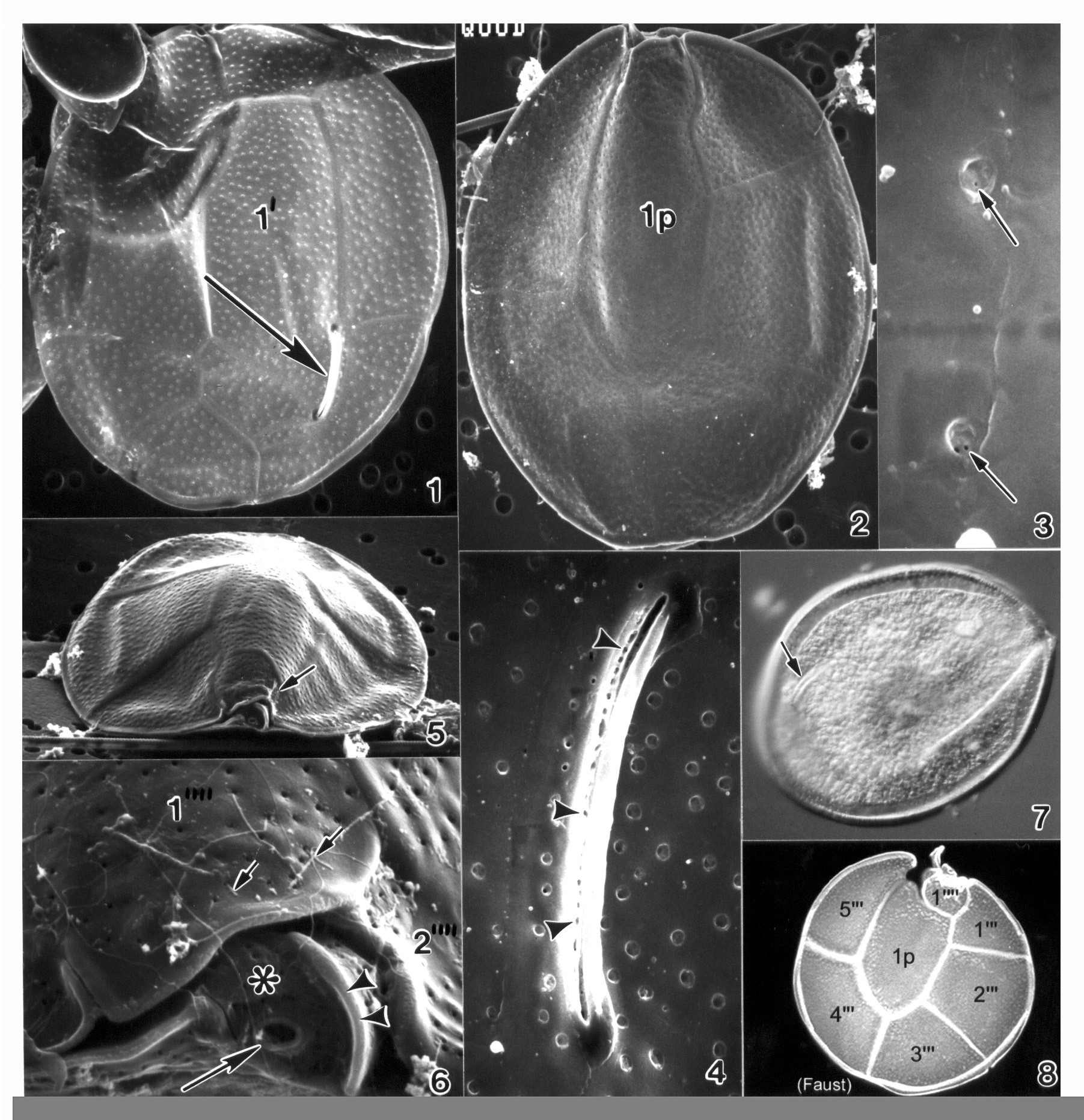
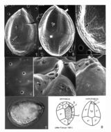
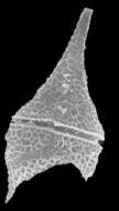
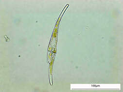
Plate 33. Ostreopsis mascarenensis. Figs. 1-5. SEM. Fig. 1. Epitheca: inner thecal surface. Cell very large, broadly ovate, large plates. Plate 1' elongate and hexagonal. Apical pore plate (Po) nearly straight. Fig. 2. Hypotheca: plate 1p long and wide. Fig. 3. Smooth cell surface with round pores; pores with two small openings (arrows). Fig. 4. Po with long narrow apical pore; small pores line the opening (arrowheads). Figs. 5-6. Ventral view of epitheca. Fig. 5. Cell compressed anterio-posteriorly; cingulum narrow with smooth edge. Small sulcus hidden (arrow). Fig. 6. Location of ventral opening (large arrow), ventral plate (asterisk), and rigid plate (arrowheads) within cingulum. Pores with ejected trichocysts (small arrows). Fig. 7. LM. Epitheca: Po (arrow) and cingulum in focus. Fig. 8. Line drawing: hypotheca plate arrangement.
-
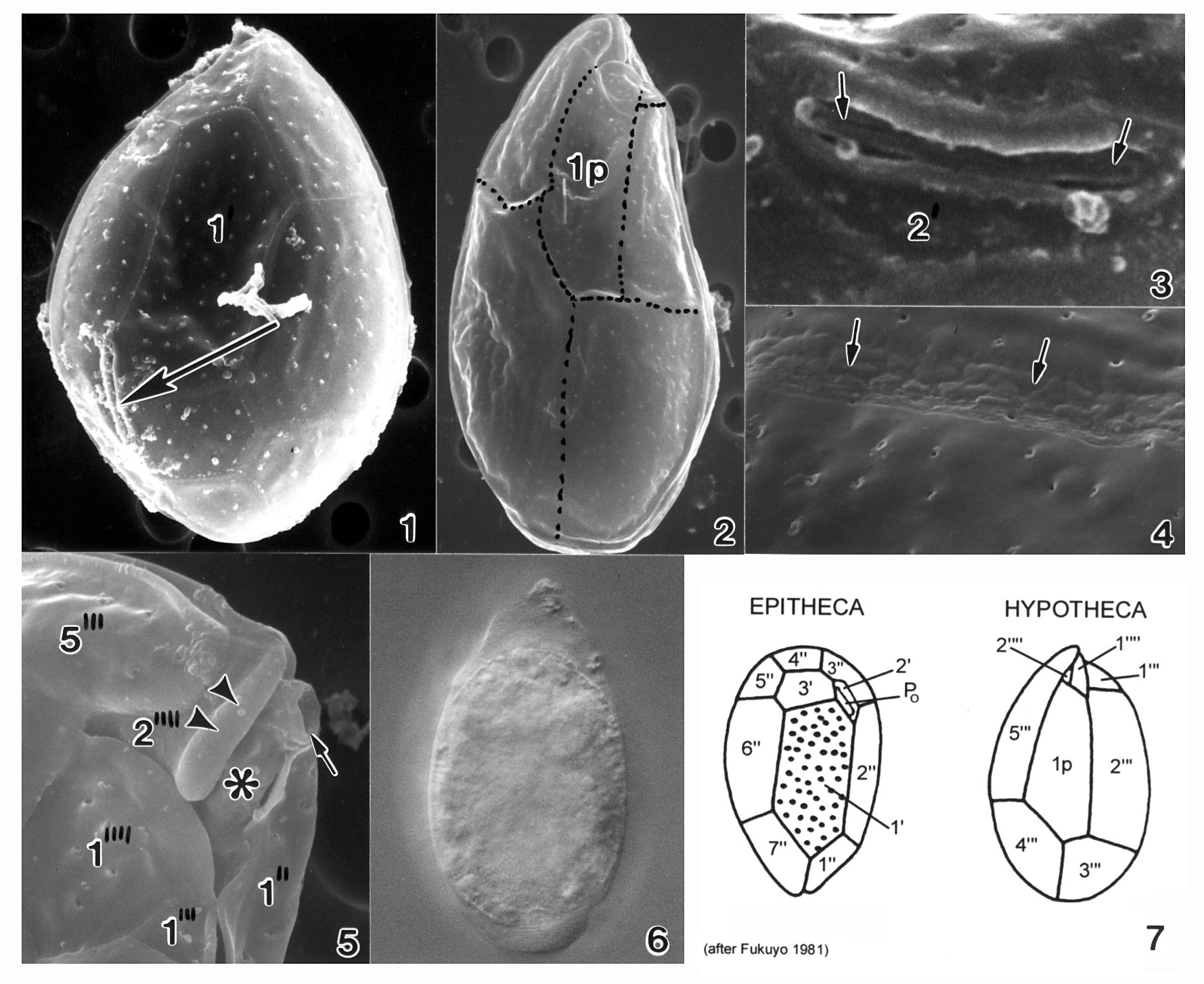
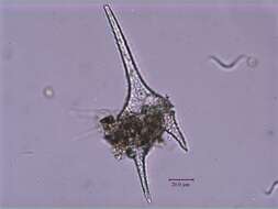
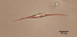
Plate 34. Ostreopsis ovata. Figs. 1-5. SEM. Fig. 1. Epithecal view: cell slender and tear-shaped. Apical pore plate (Po) off-center (arrow). Plate 1' large and hexagonal. Cingulum wide with narrow lists. Fig. 2. Hypothecal view: plates delicate. Plate 1p long and narrow. Fig. 3. Po: short and straight, adjacent to plate 2'. Fig. 4. Thecal surface smooth with scattered small pores. Suture line uneven and bumpy (arrows). Fig. 5. Hypothecal view: ventral opening (arrow), ventral plate (asterisk), and rigid plate (arrowhead) on cingulum. Fig. 6. LM. Large posterior nucleus. Fig. 7. Line drawing: thecal plate arrangement.
-
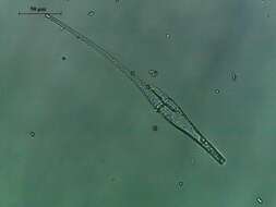
Plate 35. Ostreopsis siamensis. Figs. 1-6. SEM. Fig. 1. Epithecal view: cell broad and tear-shaped. Thecal surface smooth with scattered pores. Apical pore plate (Po) off-center (arrow). Narrow cingulum with smooth edge. Plate 1' narrow and pentagonal. Fig. 2. Hypothecal view: plate 1p long and pentagonal. Fig. 3. Po: long, curved and narrow. Fig. 4. Large and small pores on thecal surface. Fig. 5. Ventral view: location of ventral opening (arrow), ventral plate (asterisk), and rigid plate (arrowhead) on cingulum. Fig. 6. Hypothecal view: Vo (arrow) and Rp (arrowhead). Fig. 7. LM. Hypotheca. Fig. 8. Line drawing: thecal plate arrangement.
-
-
-
-
-
-
-
-
-
-
-
-
-
-
-
-
-
-
-
-