-
-
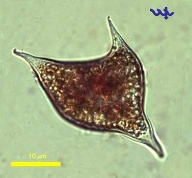
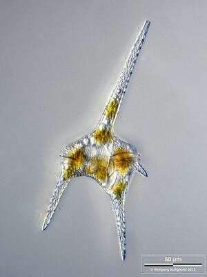
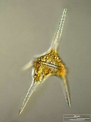
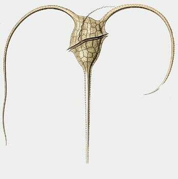
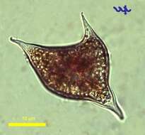
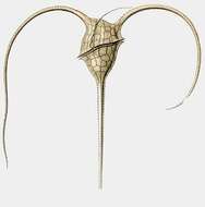
A distinctive species with a long apical horn and a long left antapical horn. The left anatapical horn is very short. This species is found in oceanic coastal and estuarine sites. It is known to form blooms in autumn in the North and Irish Sea.
-
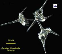
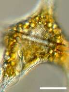
Portrait of the dinoflagellate, Ceratium hirundinella (O.F. Müller) Schrank, 1882. The body is drawn out into a long anterior horn and three subequal posterior horns at angles to one another. The transverse girdle or cingulum bears a flagellum (not seen in this image), which differs structurally from the trailing flagellum (seen here between the right and central posterior horns). Complex faceted cellulose plates cover the body. Small discoid plastids contain chlorophyll a and c along with other pigments, which may give a yellow-brown or brownish-red color. Composite image. Collected from freshwater pond near Boise, Idaho September 2003. DIC optics.
-
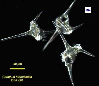
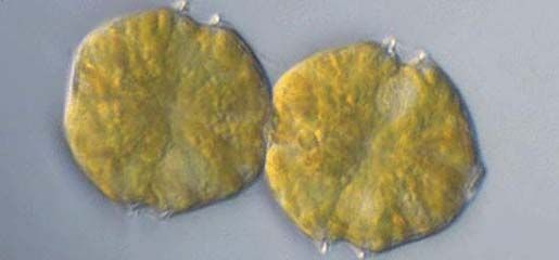
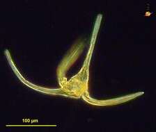
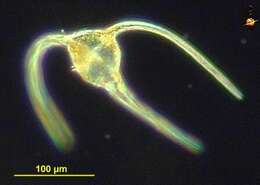
Ceratium hirundinella is one of the larger dinoflagellates (length ~ 400 μm) occurring in Lake Kinneret. It is easily identified by its typical 3 or 4 horns of varying length and overall shape reminiscent of the Eiffel tower. It is abundant in spring, when it accompanies the more abundant dinoflagellate Peridinium gatunense at its bloom decline phase. At this time of year other, smaller dinoflagellates are also present in the water column (Peridiniopsis elpatiewsky, Ps. cunningtonii, Ps. borgei, Ps. polonicum). While usually less abundant than the other dinoflagellates, it produces a large number of cysts (resting spores) that sink to the sediments. An exceptional bloom of Ceratium occurred in spring 1993 when cell densities in the upper 0-2 m layer reached 150/mL.
-
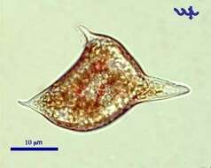
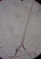
Cysts of Ceratium are found in the water column during the exponential growth phase and sink to the sediments. Their shape is typical, with 3 horns and at least one red spot. The specimen was sampled from shallow water near the Kinneret Limnological Laboratory in April 2006.
-
Uit: www.nies.go.jp/biology/ mcc/strainlist_a.htm
Ecomare
Alexandrium; Alexandrium.
-
Ceratium hirundinella is one of the larger dinoflagellates (length ~ 400 μm) occurring in Lake Kinneret. It is easily identified by its typical 3 or 4 horns of varying length and overall shape reminiscent of the Eifel tower. It is abundant in spring, when it accompanies the more abundant dinoflagellate Peridinium gatunense at its bloom decline phase. At this time of year other, smaller dinoflagellates are also present in the water column (Peridiniopsis elpatiewsky, Ps. cunningtonii, Ps. borgei, Ps. polonicum). While usually less abundant than the other dinoflagellates, it produces a large number of cysts (resting spores) that sink to the sediments. An exceptional bloom of Ceratium occurred in spring 1993 when cell densities in the upper 0-2 m layer reached 150/mL.
-
Cysts of Ceratium hirundinella are found in the water column during the exponential growth phase and sink to the sediments. Their shape is typical, with 3 horns and at least one red spot. The specimen was sampled from shallow water near the Kinneret Limnological Laboratory in April 2006.
-
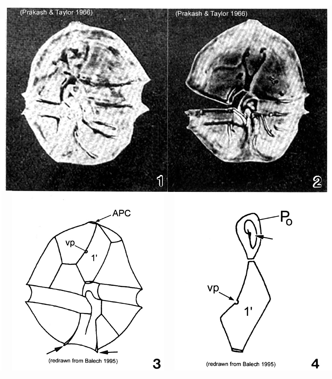
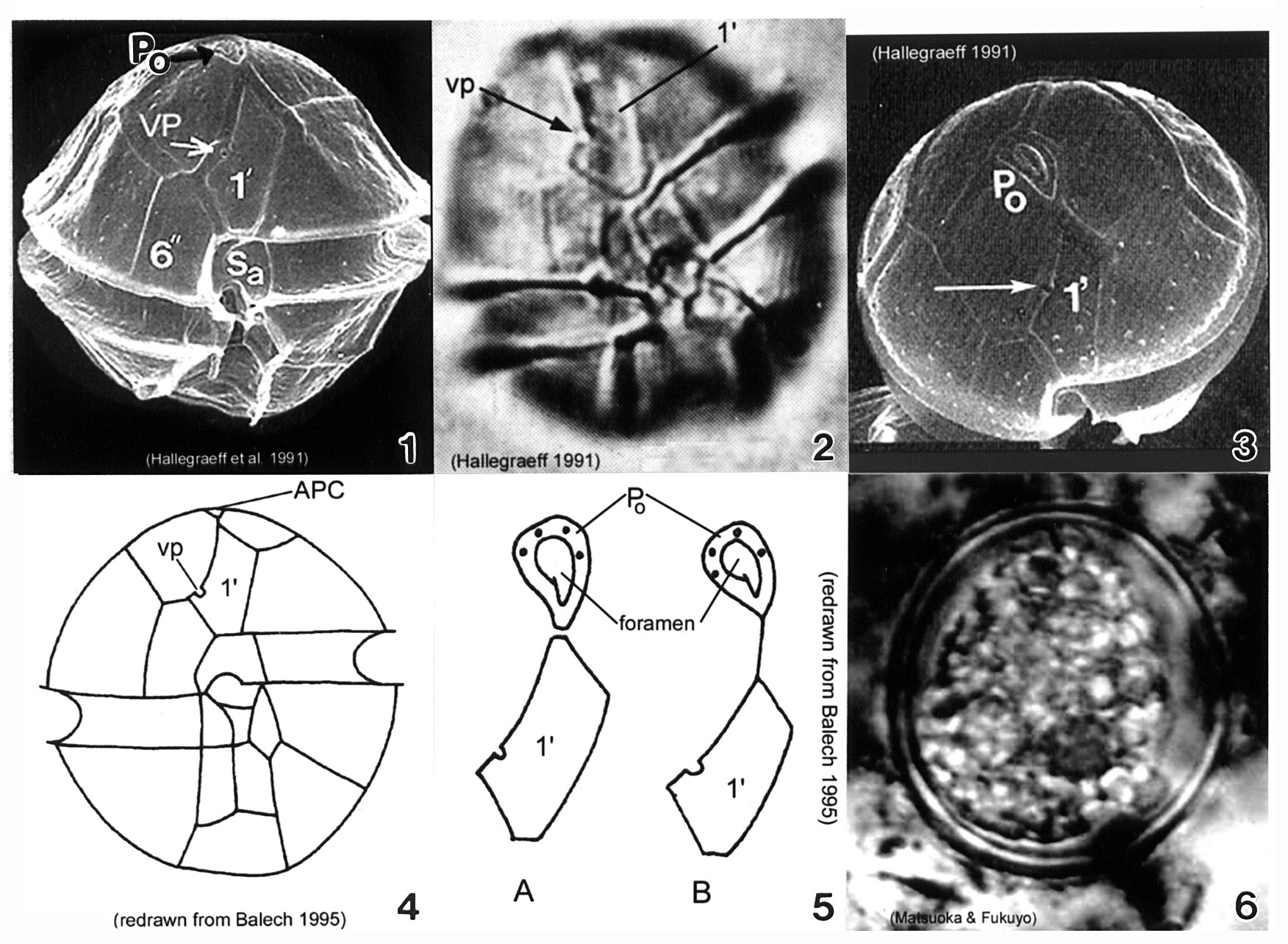
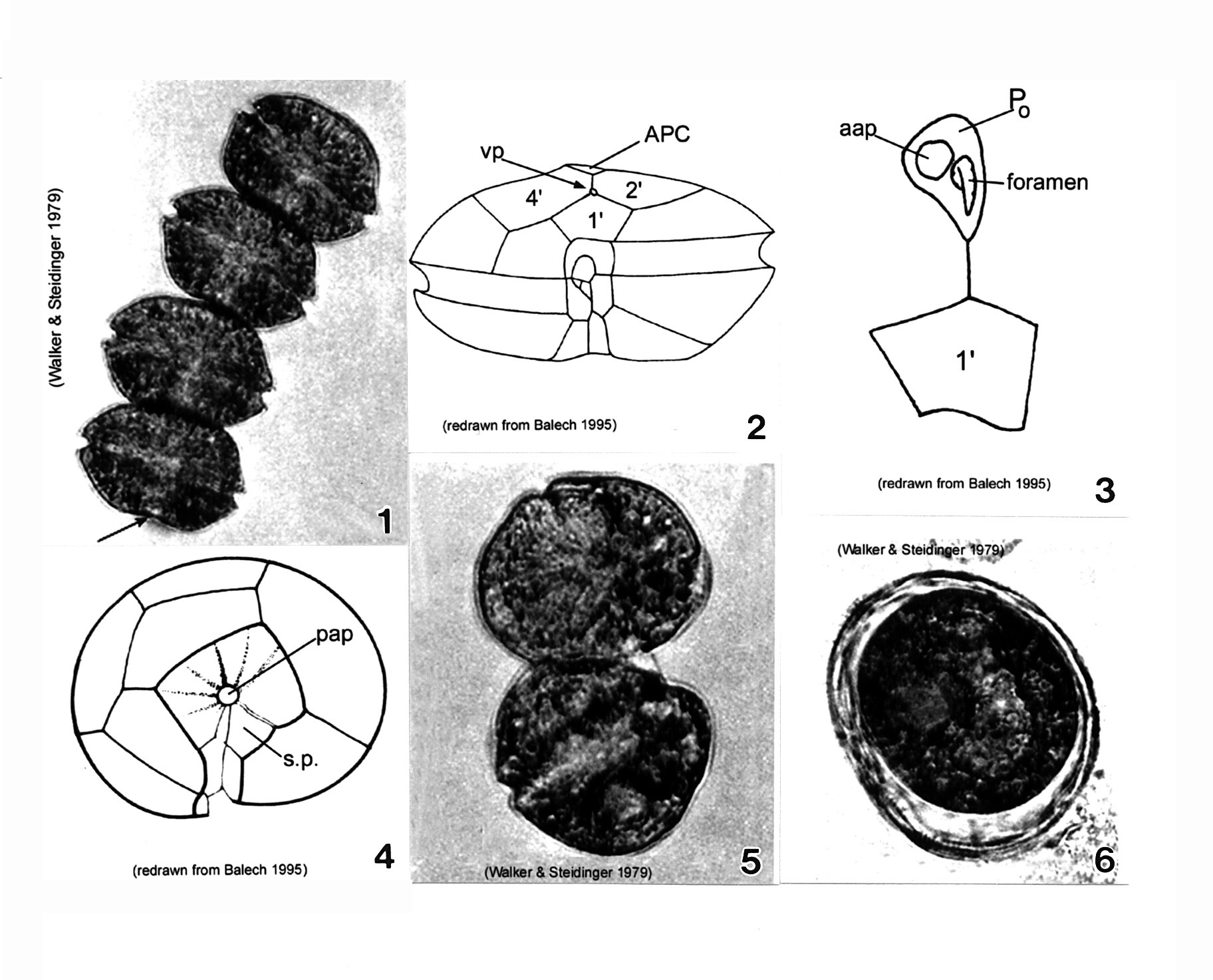
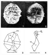
Plate 1. Alexandrium acatenella. Figs. 1-2. LM: ventral view of empty thecae. Cell small to medium, longer than wide, angular to round. Conical epitheca with shoulders; larger than hypotheca. Figs. 3-4. Line drawings. Fig. 3. Ventral view: 1' plate bears ventral pore (vp). Hypotheca with two antapical spines (arrows). Fig. 4. Po comes in direct contact with 1' plate. APC: comma-shaped foramen (arrow).
-
Dividing cell of Ceratium hirundinella (Dinoflagellata), and a second cell with a abnormal horn, pointed in the "wrong" direction
-
Scale bar indicates 50 µm. Sample from the Lake Constance (vicinity of Bodman). The image was built up using several photomicrographic frames with manual stacking technique. Images were taken using Zeiss Universal with Olympus C7070 CCD camera.Image under Creative Commons License V 3.0 (CC BY-NC-SA).
-
Transverse groove is shown, which contains the transverse flagellum. One can also see a part of the longitudinal groove and the whole longitudinal flagellum. Scale bar indicates 50 µm. Sample from the Lake Constance (vicinity of Bodman). The image was built up using several photomicrographic frames with manual stacking technique. Images were taken using Zeiss Universal with Olympus C7070 CCD camera.Image under Creative Commons License V 3.0 (CC BY-NC-SA).
-
Zoom-in with nucleus, part of the longitudinal groove and flagellum. Scale bar indicates 25 µm. Sample from the Lake Constance (vicinity of Bodman). The image was built up using several photomicrographic frames with manual stacking technique. Images were taken using Zeiss Universal with Olympus C7070 CCD camera.Image under Creative Commons License V 3.0 (CC BY-NC-SA).
-
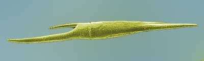
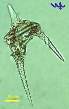
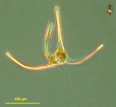
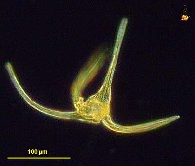
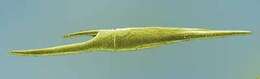
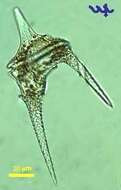
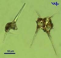
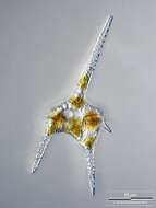
Ceratium (serr-at-ee-um) longipes, a representative of a large and distinctive genus of marine autotrophic dinoflagellates - made distinctive by having one anterior and two or as in this case three, posterior horns. Phase contrast microscopy.
data on this strain.
-
Ceratium (serr-at-ee-um) longipes, a representative of a large and distinctive genus of marine autotrophic dinoflagellates - made distinctive by having one anterior and two or as in this case three, posterior horns. This may be a cell in early division. The posterior trailing flagellum is also visible. Dark ground illumination.
data on this strain.
-
This species is closely related to Ceratium horridum. A distinguishing feature is the the apical horn which is bent to the right in C. longipes. It is a cold water species
-
Plate 3. Alexandrium minutum. Fig. 1. SEM: ventral view. Cell small and ellipsoidal. Epitheca conical, larger than hypotheca. Hypotheca short and wide; antapex obliquely flattened. Intercalary bands present. Cingulum deep, lipped; displaced 1X its width. Sulcus shallow (sa=anterior sulcal plate). Apical pore plate (Po) in direct contact with 1' plate. Fig. 2. LM: ventral view. Ventral pore (vp) present on 1' plate. Fig. 3. SEM: apical view. Po large, narrow and oval; indirectly connected to 1' plate. Vp present (arrow). Figs. 4-5. Line drawing. Fig. 4. Ventral view. 1' plate slender and rhomboidal. Fig. 5. Po connection to 1' plate: a. direct; b. indirect via thin suture. Fig. 6. LM: cyst circular in apical view.
-
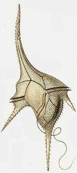
Ceratium macroceros.
-
-
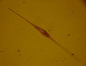
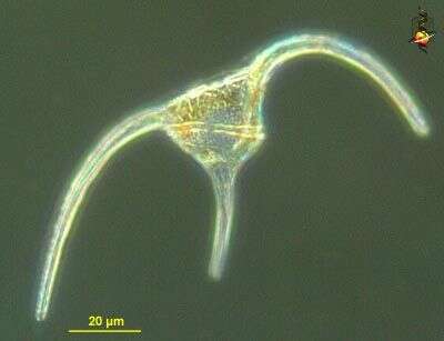
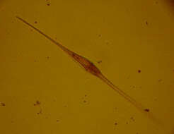
Ceratium bipes (serrate-ee-um try-poss), a common dinoflagellate encountered in marine waters, with both cones of the cell drawn into long horns, the circumferential flagellum lying in a groove in the expanded central region. With plastids. Dark ground illumination.
-
Ceratium bipes (serrate-ee-umtry-poss), a common dinoflagellate encountered in marine waters, with both cones of the cell drawn into long horns. Differential interference contrast
-
Ceratium tripos.
-
Its morphology is highly variable. It has a large dorsoventrally flattened cell body. The left antapical horn is more strongly developed than the right one. It is found in many areas of the Irish Sea but is never as abundant as C. furca.
-
Plate 4. Alexandrium monilatum. Fig. 1. LM: four-cell chain. Cells large, wider than long, flattened anterio-posteriorly. Antapex slightly concave (arrow). Figs. 2-4. Line drawings. Fig. 2. Ventral pore (vp) depicted (Florida specimens) at anterior margin of 1' plate where it comes in contact with plates 2' and 4'. Cingulum (C) deeply excavated, wide, descending; displaced one time its width. Fig. 3. Apical pore plate (Po) does not come in contact with 1' plate. Anterior attachment pore (aap) large, round and dorsally situated in the APC. Foramen comma-shaped. Fig. 4. Antapical view: posterior sulcal plate (sp) large, rhomboid and concave with radial markings. Posterior attachment pore (pap) large and centrally located. Figs. 5-6. LM. Fig. 5. Two isogamous gametes fusing at oblique angles. Fig. 6. Mature resting cysts: dark and round, with a triple layered wall.