-
Stained by the silver carbonate technique (see Foissner, W.Europ. J. Protistol.27:313-330;1991).Brightfield.
-
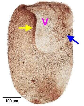
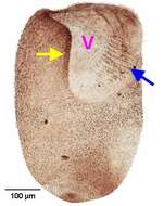
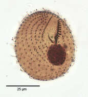
Closely spaced rows of dikinetids (paraoral rows) follow the anterior and lateral margins of the vestibulum (V). On the left side of the vestibulum there is an adoral zone of membranelles (blue arrow). Stained by the silver carbonate technique (see Foissner, W.Europ. J. Protistol.27:313-330;1991).Brightfield.
-
Stained by the silver carbonate technique (see Foissner, W.Europ. J. Protistol.27:313-330;1991).Brightfield.
-
-
-
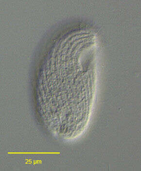
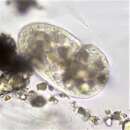
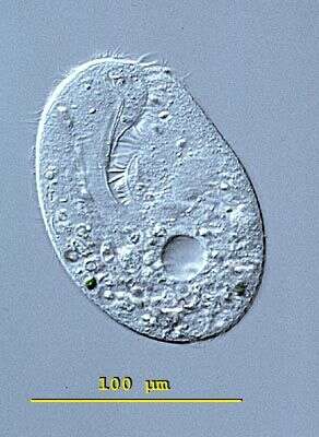
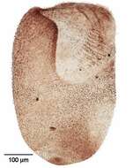
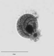
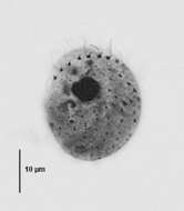
Right ventrolateral view of the colpodid ciliate, Bryometopus atypicus (Foissner,1980). Similar in overall shape to Colpoda maupasi. The dorsum of the cell is convex and the ventral surface straight. The subapical cytostome occupies the anterior 1/4 of the cell length.It is slightly oblique to the long axis of the cell. The somatic kineties (composed of dikinetids) are moderately spiralled curving around the cytostome to end on a short preoral suture.Approximately 7 postoral kineties terminate on the left border of the cytostome.There is a slightly curved right paraoral membrane and an adoral zone of membranelles on the left border of the cytostome.Rows of mucocysts occur between somatic kineties.The spherical macronucleus and adjacent single micronucleus is in the cell center.The posterior contractile vacuole has a distinctive large cylindrical excretory pore.Zoochlorellae are absent.Collected from an organically enriched rainwater pool with abundant decaying grass in Boise, Idaho. January 2006.DIC.
-

Ventral view of the colpodid ciliate, Bryometopus atypicus (Foissner,1980). Similar in overall shape to Colpoda maupasi. The dorsum of the cell is convex and the ventral surface straight. The subapical cytostome occupies the anterior 1/4 of the cell length.It is slightly oblique to the long axis of the cell. The somatic kineties (composed of dikinetids) are moderately spiralled curving around the cytostome to end on a short preoral suture.Approximately 7 postoral kineties terminate on the left border of the cytostome.There is a slightly curved right paraoral membrane and an adoral zone of membranelles on the left border of the cytostome.Rows of mucocysts occur between somatic kineties.The spherical macronucleus and adjacent single micronucleus is in the cell center.The posterior contractile vacuole has a distinctive large cylindrical excretory pore.Zoochlorellae are absent.Collected from an organically enriched rainwater pool with abundant decaying grass in Boise, Idaho. January 2006.DIC.
-

Right ventrolateral view of the colpodid ciliate, Bryometopus atypicus (Foissner,1980). Similar in overall shape to Colpoda maupasi. The dorsum of the cell is convex and the ventral surface straight. The subapical cytostome occupies the anterior 1/4 of the cell length.It is slightly oblique to the long axis of the cell. The somatic kineties (composed of dikinetids) are moderately spiralled curving around the cytostome to end on a short preoral suture.Approximately 7 postoral kineties terminate on the left border of the cytostome.There is a slightly curved right paraoral membrane and an adoral zone of membranelles on the left border of the cytostome.Rows of mucocysts occur between somatic kineties.The spherical macronucleus and adjacent single micronucleus is in the cell center.The posterior contractile vacuole has a distinctive large cylindrical excretory pore (yellow arrow).Zoochlorellae are absent.Collected from an organically enriched rainwater pool with abundant decaying grass in Boise, Idaho. January 2006.DIC.
-
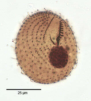
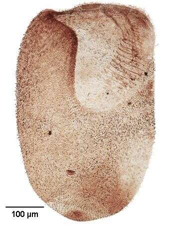
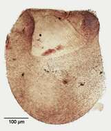
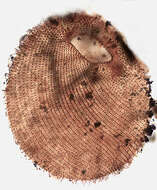
Ventral view of the infraciliature of the colpodid ciliate, Bryometopus atypicus (Foissner,1980). Similar in overall shape to Colpoda maupasi. The dorsum of the cell is convex and the ventral surface straight. The subapical cytostome occupies the anterior 1/4 of the cell length.It is slightly oblique to the long axis of the cell. The somatic kineties (composed of dikinetids) are moderately spiralled curving around the cytostome to end on a short preoral suture.Approximately 7 postoral kineties terminate on the left border of the cytostome.There is a slightly curved right paraoral membrane and an adoral zone of membranelles on the left border of the cytostome.Rows of mucocysts occur between somatic kineties.The spherical macronucleus and adjacent single micronucleus is in the cell center.The posterior contractile vacuole has a distinctive large cylindrical excretory pore.Zoochlorellae are absent.Collected from an organically enriched rainwater pool with abundant decaying grass in Boise, Idaho. January 2006.Stained by the silver carbonate technique (see Foissner, W. Europ. J. Protistol., 27:313-330;1991).Brightfield.
-

Dorsal view of the infraciliature of the colpodid ciliate, Bryometopus atypicus (Foissner,1980). Similar in overall shape to Colpoda maupasi. The dorsum of the cell is convex and the ventral surface straight. The subapical cytostome occupies the anterior 1/4 of the cell length.It is slightly oblique to the long axis of the cell. The somatic kineties (composed of dikinetids) are moderately spiralled curving around the cytostome to end on a short preoral suture.Approximately 7 postoral kineties terminate on the left border of the cytostome.There is a slightly curved right paraoral membrane and an adoral zone of membranelles on the left border of the cytostome.Rows of mucocysts occur between somatic kineties.The spherical macronucleus and adjacent single micronucleus is in the cell center(both seen here).The posterior contractile vacuole has a distinctive large cylindrical excretory pore.Zoochlorellae are absent.Collected from an organically enriched rainwater pool with abundant decaying grass in Boise, Idaho. January 2006.Stained by the silver carbonate technique (see Foissner, W. Europ. J. Protistol., 27:313-330;1991).Brightfield.
-
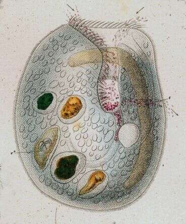
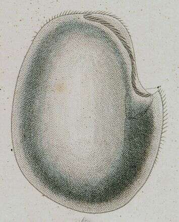
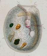
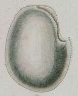
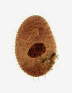
Bryometopus (bry-o-me-toe-puss) is a uniformly ciliated and the cell is rounded at both poles and is slightly reniform. The conspicuous peristome lies obliquely across the ventral surface. The single contractile vacuole is located approximately in the middle of the cell on the ventral side. The macronucleus may be oval or elongate with several micronuclei. Bryometopus can be confused with Balantidioides which does not have an undulating membrane and with Condylostoma which has a wide triangular peristome and a highly conspicuous undulating membrane. This specimen was collected in freshwater ponds near Konstanz, Germany. Differencial interference contrast.
-
Right side view of the colpodid ciliate, Bryometopus pseudochilodon (Kahl, 1932). The cell shape and size is quite variable. This population has a shape like Colpoda maupasi. The anterior end is pointed and the posterior end broadly rounded.The ventral surface is relatively straight and the dorsal surface is convex. There are 20-25 slightly spiraled somatic kineties consisting of doubly ciliated dikinetids. The kineties lie in shallow pellicular grooves. Collected from a temporary rainwater pool with decaying grass near Boise, Idaho march 2005. DIC.
-
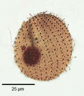
Right ventrolateral view of the infraciliature of the colpodid ciliate, Bryometopus pseudochilodon (Kahl, 1932). The cell shape and size is quite variable. This population has a shape like Colpoda maupasi. The anterior end is pointed and the posterior end broadly rounded. The elliptical oral vestibulum is in the anterior 1/3. There is a curved line of adoral membranelles along the left side of the vestibulum and an undulating membrane on the right. There are 20-25 slightly spiraled somatic kineties consisting of doubly ciliated dikinetids. The ellipsoid macronucleus contains many small micronuclei. There are two relatively large micronuclei adhering to the macronucleus. Collected from a temporary rainwater pool with decaying grass near Boise, Idaho march 2005. Silver carbonate stain (see Foissner, W. Europ. J. Protistol., 27:313-330;1991). Brightfield.
-
Left side of the colpodid ciliate, Bryometopus pseudochilodon (Kahl, 1932). The cell shape and size is quite variable. This population has a shape like Colpoda maupasi. The anterior end is pointed and the posterior end broadly rounded.The ventral surface is relatively straight and the dorsal surface is convex. There are 20-25 slightly spiraled somatic kineties consisting of doubly ciliated dikinetids. The kineties lie in shallow pellicular grooves. Collected from a temporary rainwater pool with decaying grass near Boise, Idaho march 2005. DIC.
-
Macronucleus and micronuclei of the colpodid ciliate, Bryometopus pseudochilodon (Kahl, 1932). The central ellipsoid macronucleus contains many small micronuclei (seen here). There are two relatively large micronuclei adhering to the macronucleus (seen here at 3 o'clock). Collected from a temporary rainwater pool with decaying grass near Boise, Idaho march 2005. Silver carbonate stain (see Foissner, W. Europ. J. Protistol., 27:313-330;1991). Brightfield.
-

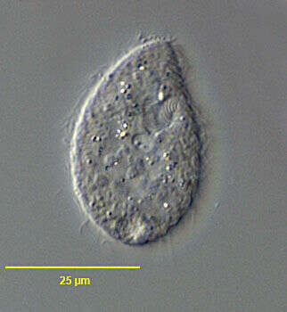
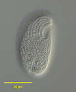
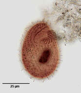
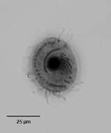
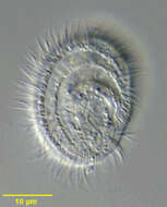
Ventral (right) view of Kreyella minuta (Foissner, 1979), one of the smallest known ciliates.Probably synonymous with K. muscicola. The body outline is obovoid to reniform. Flattened ventrally and distinctly convex dorsally. Somatic ciliature is markedly reduced on the left (dorsal) side to one row of anterior dikinetids, which lie in a pellicular notch. There are three concentric semicircular kineties on the right (ventral surface). Cilia are relatively long (about 6 microns). The ventrolateral oral aperture is relatively large extending from the posterior 1/3 to almost the posterior end. There is no cytopharyngeal basket of fibers or trichites. There is a paraoral mebrane on the right margin of the vestibulum and a group of about 7 adoral membranelles on the left margin (seen here). There is a single spherical macronucleus and one micronucleus. The contractile vacuole is located in the posterior half. Scurries rapidly back and forth over the substratum. Although described as being rare, Kreyella is found frequently in freshwater ponds and irrigation ditches in Idaho. Collected from freshwater pond near Boise, Idaho in June 2003.Silver carbonate stain (see Foissner, W.Europ. J. Protistol.27,313-330;1991). Black and white.Brightfield optics.
-

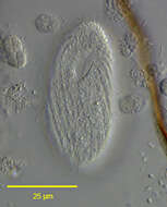
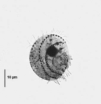
Infraciliature (right side) of the small colpodid ciliate, Kreyella minuta (Foissner,1979). The body outline is obovoid to reniform. The right side is flattened and the left distinctly convex. Somatic ciliature is markedly reduced on the left side to two rows of anterior dikinetids (two of the dikinetids are visible anteriorly here). There are two concentric semicircular kineties on the right surface. Cilia are relatively long (about 6 microns). The ventrolateral oral aperture in the posterior 1/3 is relatively large. There is no cytopharyngeal basket of fibers or trichites. A prominent curved paraoral membrane is located on the dorsal margin of the vestibulum. The adoral zone of membranelles is visible here as a series of short parallel lines opposite the paraoral membrane. The dark dots posterior to the oral aperture represent the short postoral kineties. There is a single spherical macronucleus and one micronucleus. The contractile vacuole is located in the posterior ½ (not seen here). This species may be synonymous with Kreyella musicale (Kahl, 1931). Collected from freshwater pond near Boise, Idaho in July 2004. Silver carbonate stain (see Foissner, W.Europ. J. Protistol.27, 313-330;1991). Brightfield.
-

Dorsal (left) view of Kreyella minuta (Foissner, 1979), one of the smallest known ciliates.Probably synonymous with K. muscicola. The body outline is obovoid to reniform. Flattened ventrally and distinctly convex dorsally. Somatic ciliature is markedly reduced on the left (dorsal) side to one row of anterior dikinetids, which lie in a pellicular notch. There are three concentric semicircular kineties on the right (ventral surface). Cilia are relatively long (about 6 microns). The ventrolateral oral aperture is relatively large extending from the posterior 1/3 to almost the posterior end. There is no cytopharyngeal basket of fibers or trichites. There is a paraoral mebrane on the right margin of the vestibulum and a group of about 7 adoral membranelles on the left margin (seen here). There is a single spherical macronucleus and one micronucleus. The contractile vacuole is located in the posterior half. Scurries rapidly back and forth over the substratum. Although described as being rare, Kreyella is found frequently in freshwater ponds and irrigation ditches in Idaho. Collected from freshwater pond near Boise, Idaho in June 2003.Silver carbonate stain (see Foissner, W.Europ. J. Protistol.27,313-330;1991). Black and white.Brightfield optics.
-
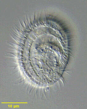
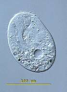
Ventral view of Kreyella minuta (Foissner, 1979), one of the smallest known ciliates.Probably synonymous with K. muscicola. The body outline is obovoid to reniform. Flattened ventrally and distinctly convex dorsally. Somatic ciliature is markedly reduced on the left (dorsal) side to one row of anterior dikinetids, which lie in a pellicular notch. There are three concentric semicircular kineties on the right (ventral surface). Cilia are relatively long (about 6 microns). The ventrolateral oral aperture is relatively large extending from the posterior 1/3 to almost the posterior end. There is no cytopharyngeal basket of fibers or trichites. Two rows of cilia are located on the margin of the vestibulum. There is a single spherical macronucleus and one micronucleus. The contractile vacuole is located in the posterior half. Scurries rapidly back and forth over the substratum. Although described as being rare, Kreyella is found frequently in freshwater ponds and irrigation ditches in Idaho. Collected from freshwater pond near Boise, Idaho in June 2003. DIC optics.
-
Ventral (right) view of Kreyella minuta (Foissner, 1979), one of the smallest known ciliates.Probably synonymous with K. muscicola. The body outline is obovoid to reniform. Flattened ventrally and distinctly convex dorsally. Somatic ciliature is markedly reduced on the left (dorsal) side to one row of anterior dikinetids, which lie in a pellicular notch. There are three concentric semicircular kineties on the right (ventral surface). Cilia are relatively long (about 6 microns). The ventrolateral oral aperture is relatively large extending from the posterior 1/3 to almost the posterior end. There is no cytopharyngeal basket of fibers or trichites. There is a paraoral mebrane on the right margin of the vestibulum and a group of about 7 adoral membranelles on the left margin (seen here). There is a single spherical macronucleus and one micronucleus. The contractile vacuole is located in the posterior half. Scurries rapidly back and forth over the substratum. Although described as being rare, Kreyella is found frequently in freshwater ponds and irrigation ditches in Idaho. Collected from freshwater pond near Boise, Idaho in June 2003.Silver carbonate stain (see Foissner, W.Europ. J. Protistol.27,313-330;1991). Black and white.Brightfield optics.
-
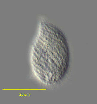
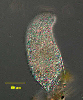
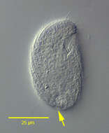
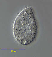
In vivo portrait of the colpodid ciliate, Rostrophrya camerounensis (Njine, 1979) Foissner, 1993 (ventral view). This population was feeding on diatoms. Collected from standing organically enriched roadside ditchwater near Boise, Idaho march 2005. DIC.
-
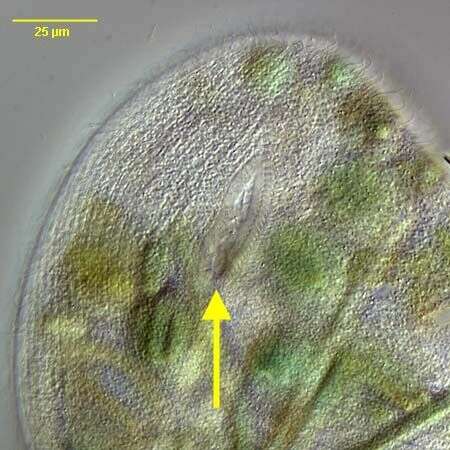
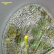
Detail of the oral aperture of the colpodid ciliate, Rostrophrya camerounensis (Njine, 1979) Foissner, 1993 (yellow arrow indicates posterior end of oral aperture).This population has been feeding on filamentous cyanobacteria (Oscillatoria sp.)Collected from an ephemeral puddle near Boise, Idaho March 2005. DIC.
-
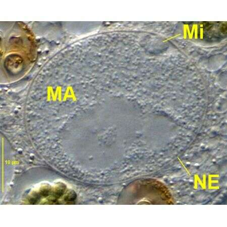
Detail view of the macronucleus (MA) and small elliptical micronucleus (Mi) within the macronuclear envelope (NE) in the large colpodid ciliate, Rostrophrya camerounensis (Njine, 1979) Foissner, 1993.The specimen is moderately compressed by coverglas pressure. Collected from an ephemeral puddle on an intermittently flood-irrigated grass lawn in Boise, Idaho. July 2007 DIC.
-
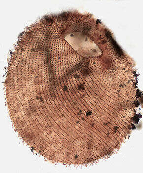
Ventrolateral view of the infraciliature of the colpodid ciliate, Rostrophrya camerounensis (Njine, 1979) Foissner, 1993. The shape of this specimen has been distorted by fixation and coverglass pressure. The relatively large obliquely oriented elliptical oral aperture (distorted in this specimen) is located at the junction of the rostrum with the main body. aperture. Collected from standing organically enriched roadside ditchwater near Boise, Idaho, March 2005.Stained by the silver carbonate technic (see Foissner, W. Europ. J. Protistol., 27:313-330;1991). Brightfield.