-
San Martn de Castaeda, Castilla y Len, Espaa
-
Ribadelago de Franco, Castilla y Len, Espaa
-
Santa Coloma, La Rioja, Spain
-
-
Floscularia (floss-cue-lair-ee-a) an illoricate rotifer (metazoa), lives within a test, the walls of which are made up of pellets constructed by the animal in a specialized organ on the head. Feeds with an array of cilia (called a corona) which is made up of four lobes. With lateral projections called antennae. Phase contrast.
-
Rattulus cylindricus Imhof. Left side of specimen from East Harbor, Lake Erie.
-
Phuripong Meksuwan, Pornsilp Pholpunthin, Hendrik Segers
Zookeys
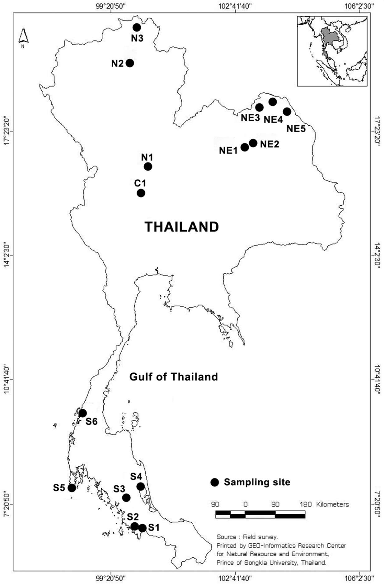
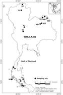
Figure 1.Sampling sites in Thailand. S, C, N and NE represent sampling sites in the Southern, Central, North and Northeast part of Thailand, respectively. Map from GIS center, PSU.
-
San Martin De Castaneda, Castille and Leon, Spain
-
-
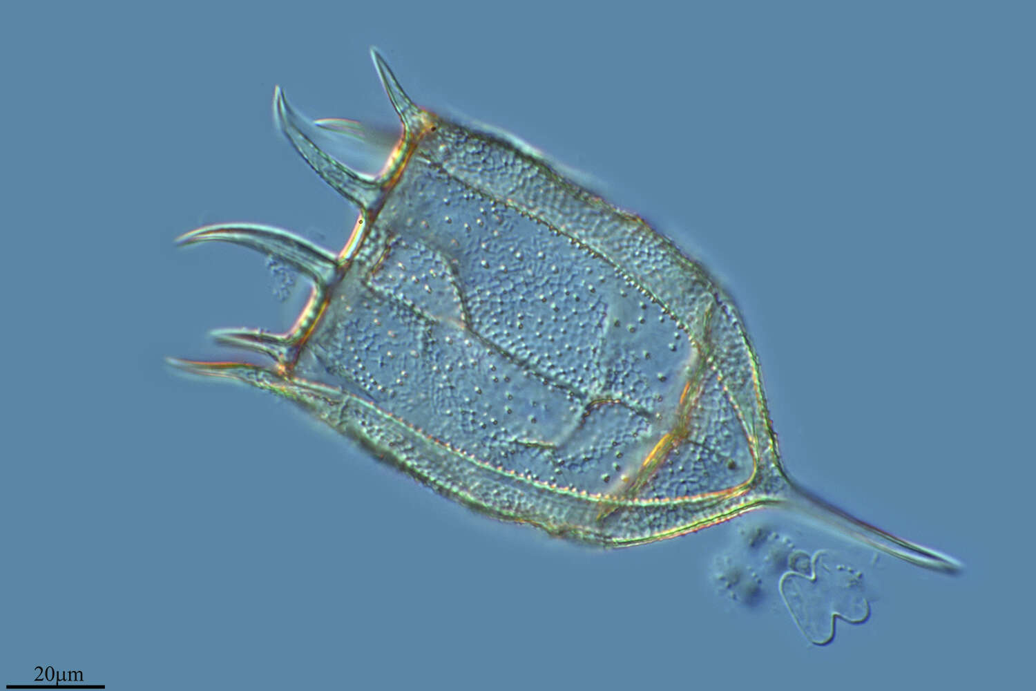
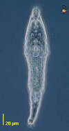
Adineta (add-in-eat-a), a rotifer (metazoa). Rotifers typically have stiffened body wall. The corona is retracted in the anterior part of the body in this specimen - visible as the pair of elliptic structures just above the V-shape of the mouth. Phase contrast, text by Hendrik Seegers.
-
Rattulus elongatus Gosse. Left side.
-
Ribadelago de Franco, Castille and Leon, Spain
-

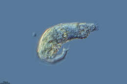
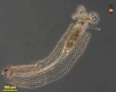
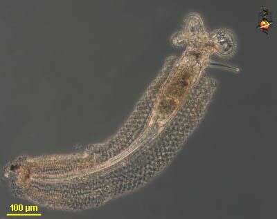
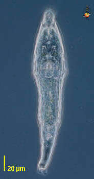
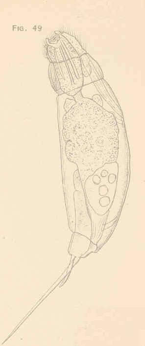
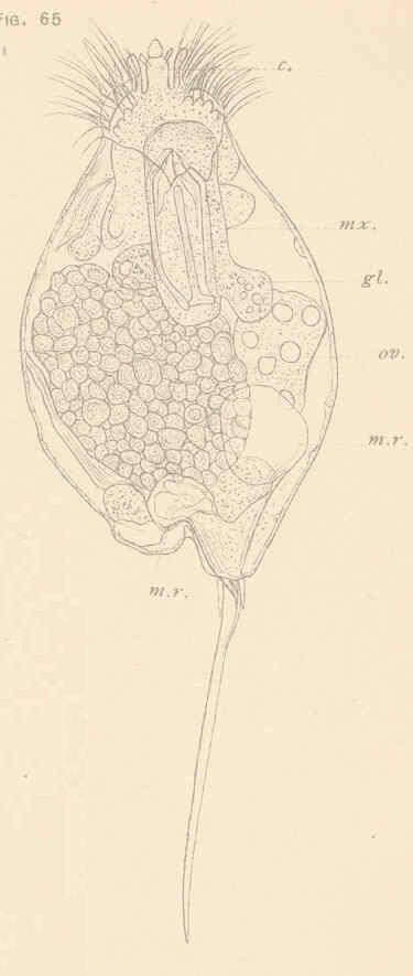
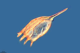
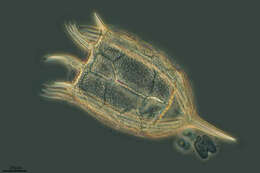
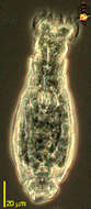
This is a bdelloid rotifer (metazoa), body with a stiffened body wall (lorica) usually in elements that will telescope, posterior end often with two spurs. Very common in aquatic habitats especially those which are prone to dry out - like moss, soils and the contents of pitcher plants. They can resist drying because they can perform cryptobiosis - that is they can dry out completely, and the organisms can come back to life when wetted. Usually eat bacteria or suspended protists, using the cilia of the corona at the front end of the body to sweep particles into the front of the body where an armoured mastax or jawed pharynx helps to grind the food. Rotifers are common members of the microbial communities of many aquatic ecosystems. Although they are multicellular animals, they may be only 100 microns long, and so overlap in size with ciliates. They can be confused with ciliates because they use cilia to capture their food. However, they can be distinguished because many have an exoskeleton and a muscular pharyngeal region just behind the head. Phase contrast.
-
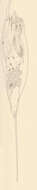
Rattulus gracilis Tessin. Ventral view, trophi extended.
-
Rotifers typically have stiffened body wall. The corona is retracted in the anterior part of the body in this specimen - visible as the pair of elliptic structures just above the V-shape of the mouth. Phase contrast, text by Hendrik Seegers. Phase contrast.
-
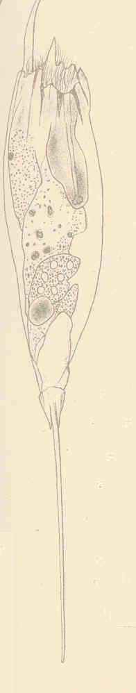
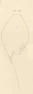
Rattulus latus Jennings. Ventral view of extended specimen.
-
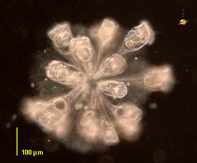
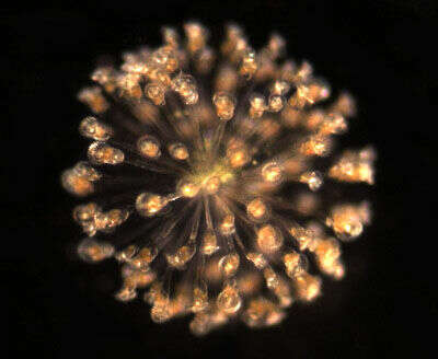
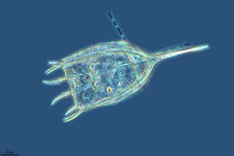
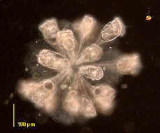
Conochilus (cone-ow-kile-us), this is a colonial rotifer (metazoa), in which the basal parts of the rotifers are embedded in a common gelatinous matrix. The feeding areas, the coronas, project outwards. Rotifers are common members of the microbial communities of many aquatic ecosystems. Although they are multiceullar animals, they may be only be 100 microns long, and so overlap in size with ciliates. They can be confused with ciliates because they use cilia to capture their food. However, they can be distinguished because they have an exoskeleton, usually two posterior toes, and a tough pharyngeal region just behind the head. Dark ground illumination.
-
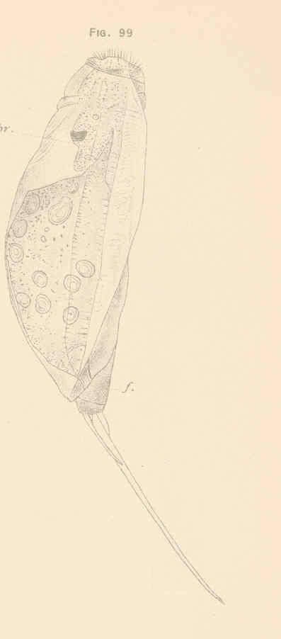
Rattulus latus Jennings. Dorsal view of lorica.
-
Colonial planktonic rotifer forms colonies up to several millimetres in diameter. This is an unconstrained colony.
-
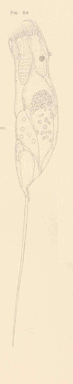
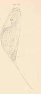
Rattulus longiseta Schrank. Right side view.
-
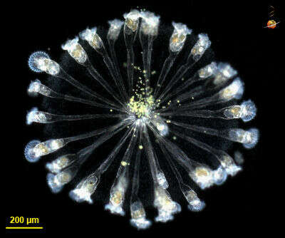
Colonial rotifer, crompressed between slide and coverslip. The organisms are joined at their toes and are embedded in a fine mucus. All colonies at this site had green algae near their cores. The metachronal beat of the cilia is seen in some organisms.
-
Rattulus longiseta Schrank. Ventral view of young specimen, retracted.
-
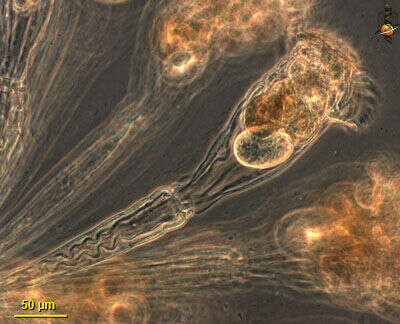
One organism from a colony. Phase contrast optics.
-
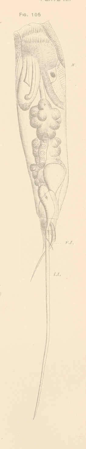
Rattulus lophoessus Gosse. Dorsal view.