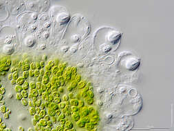
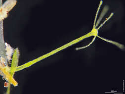
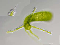
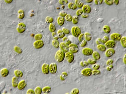
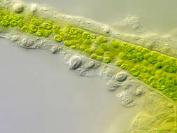
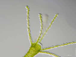
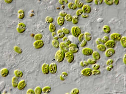
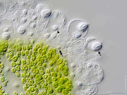
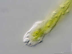
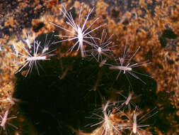
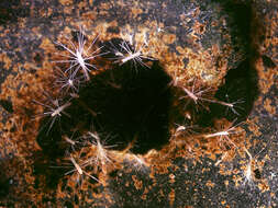
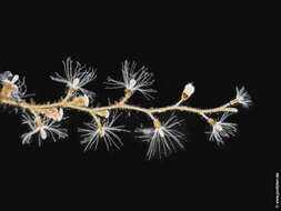
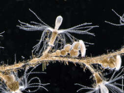
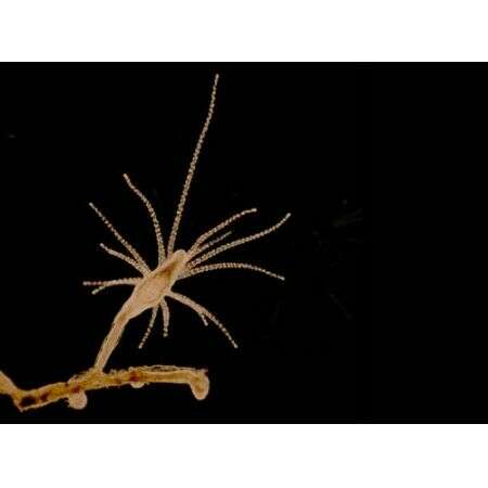
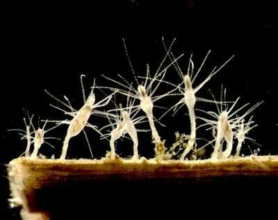
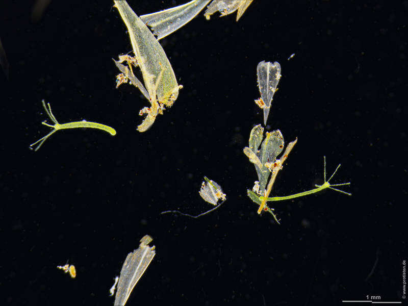
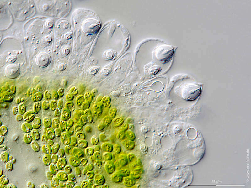
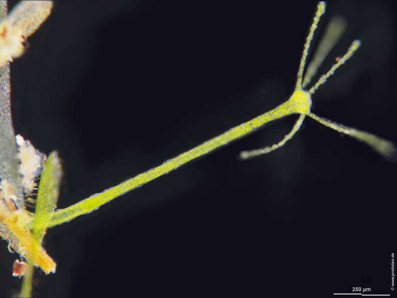
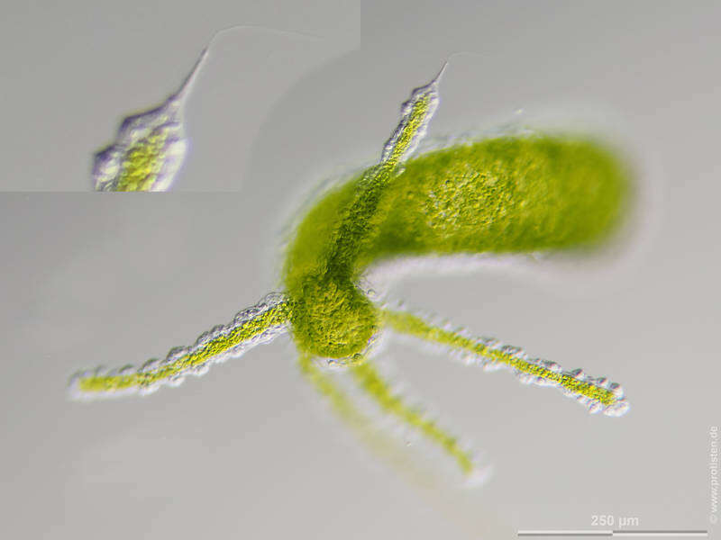
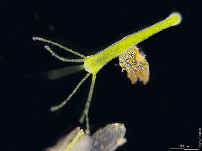
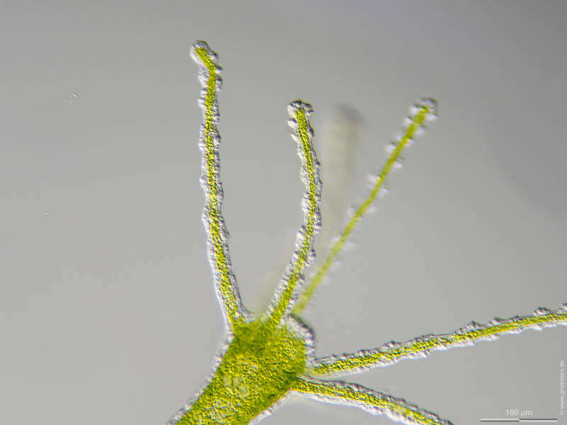
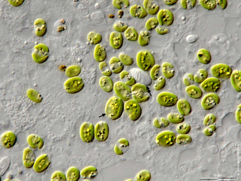
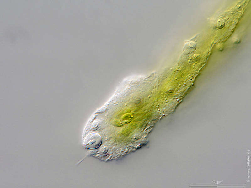
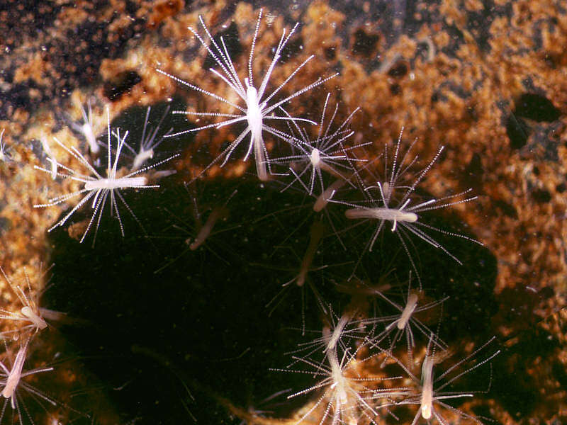
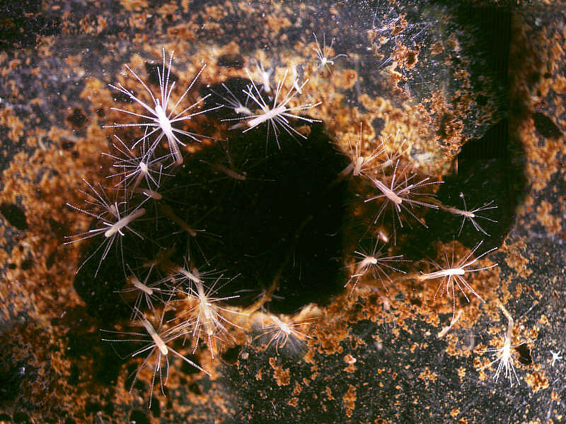
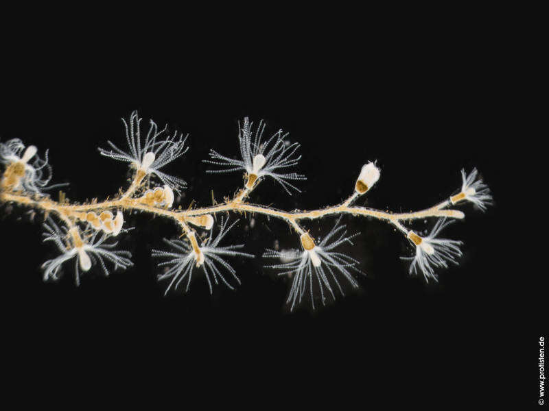
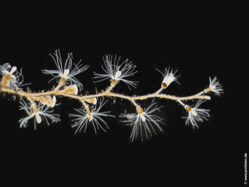
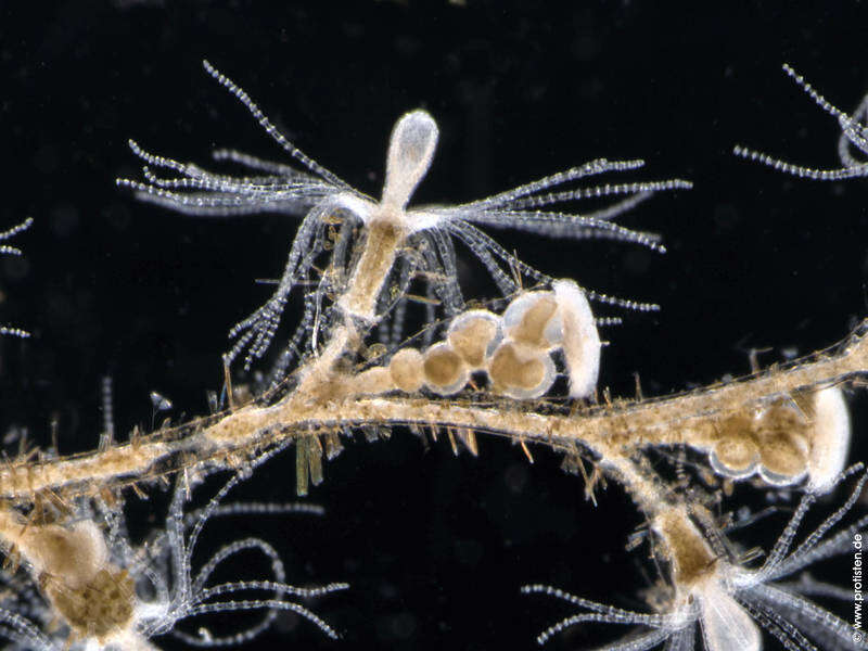
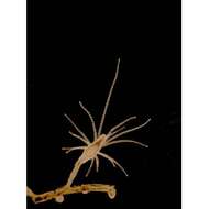
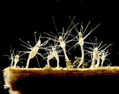
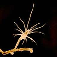
Scale bars indicate 250 µm (1, 2), 25 µm (3-6), 10 µm (7, 8).Four image couples, some with and without marking arrows.First couple:Penetrant nematocysts (arrow), ejected sticky thread of a gultinant ptychocyst (double headed arrow). Second couple:Optical cross section through a tentacle.Third couple:The optical cross section through the body shows all three types of their cnidae: some penetrant nematocysts (arrow) showing shaft and coiled hollow tubule for transportation of the toxin into the prey, some more volvent spirocyst (arrowhead) and one glutinous ptychocysts (double headed arrow).Fourth couple:Nucleus of a zoochlorella (arrow), glutinous ptychocyst for attaching the animal to a surface (double headed arrow).Please click on < or > on the image edges or on the dots at the bottom edge of the images to browse through the slides!Place name: Tropical freshwater aquariumLatitude: 54.3018013 Longitude: 10.07120132Microscope Zeiss Axioplan, camera Olympus OM-D M5 MKII. DOF images.© Wolfgang Bettighofer,images under Creative Commons License V 3.0 (CC BY-NC-SA).For permission to use of (high resolution) images please contact
postmaster@protisten.de.For further information about the image, please click here:
Link to protisten.de page