-
Zongqing Wang, Keliang Wu, Yanli Che
Zookeys
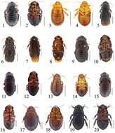
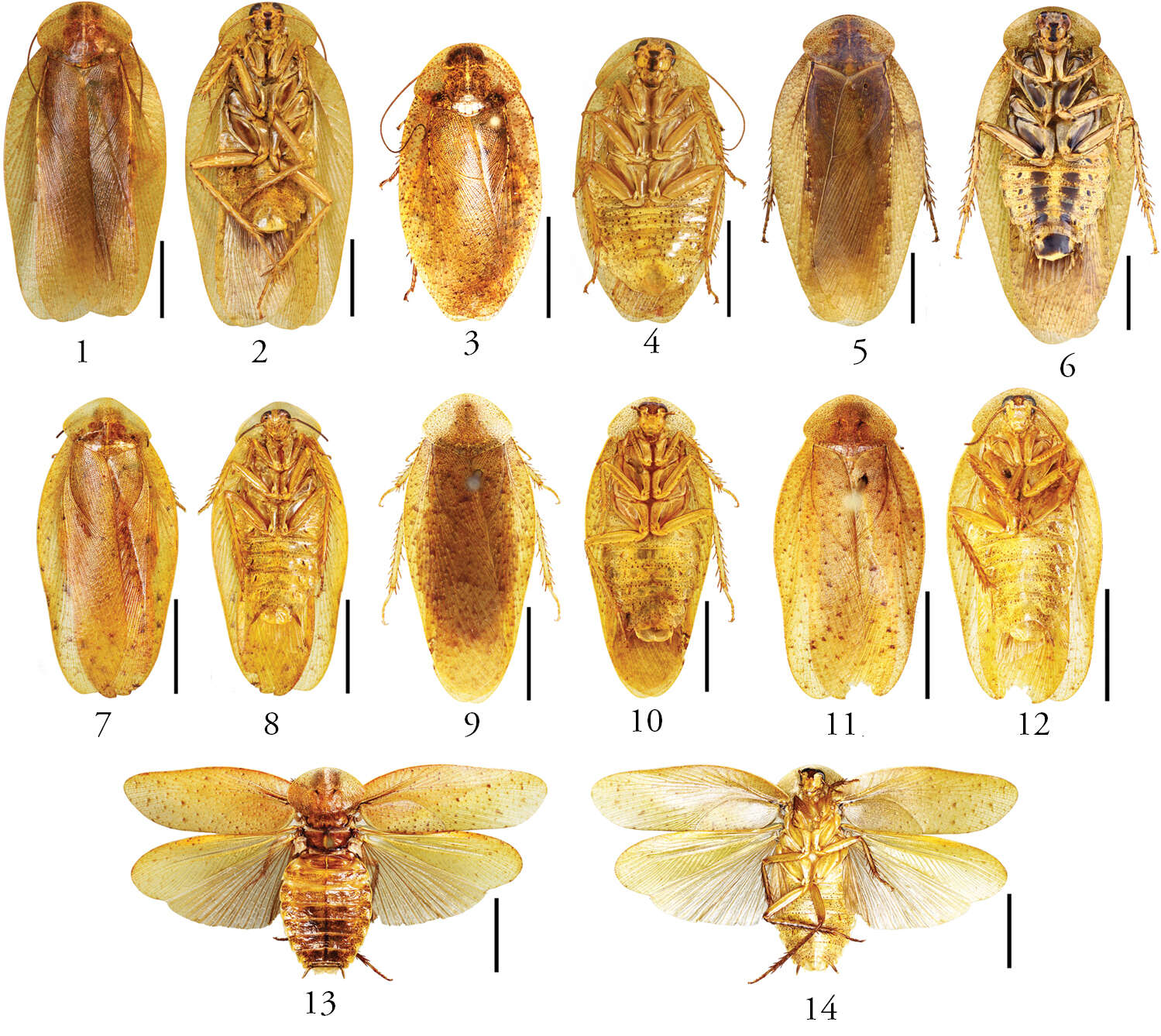
Figures 1–14.1–4 Pseudophoraspis fruhstorferi Shelford, male: 1 dorsal view 2 ventral view; female: 3 dorsal view 4 ventral view; 5–6 Pseudophoraspis tramlapensis Anisyutkin, male: 5 dorsal view 6 ventral view; 7–8 Pseudophoraspis kabakovi Anisyutkin, male: 7 dorsal view 8 ventral view; 9–10 Pseudophoraspis clavellata sp. n., male: 9 holotype, dorsal view 10 holotype, ventral view; 11–12 Pseudophoraspis recurvata sp. n., male: 11 holotype, dorsal view 12 holotype, ventral view; 13–14 Pseudophoraspis incurvata sp. n., male: 13 holotype, dorsal view 14 holotype, ventral view. Scale bars=1cm.
-
Figures 10–13.Genuotermes spinifer: Soldier morphological variations, profile: 10 Fordlândia, PA (MZUSP-8383) 11 Chapada dos Guimarães, MT (MZUSP-6615) 12 Serra do Roncador, MT (MZUSP-7400) 13 UHE Santo Antônio (Módulo de Jirau), RO (MZUSP-16354).
-
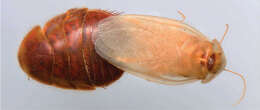
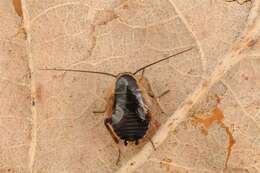
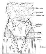
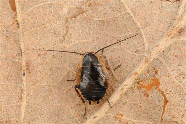
Figure 39.Arenivaga delicata, a dorsal habitus b ventral habitus c pronotum d head.
-
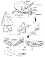
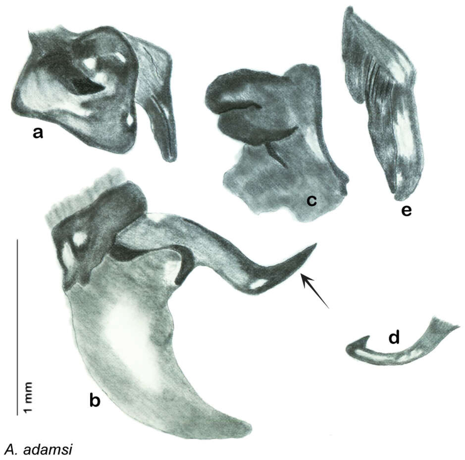
Figure 13.Arenivaga adamsi, genitalia: a right dorsal phallomere b right ventral phallomere c small central sclerite d genital hook e left phallomere. Arrow(s) indicate diagnostic characters (see text).
-
Xiudan Wang, Yan Shi, Zongqing Wang, Yanli Che
Zookeys
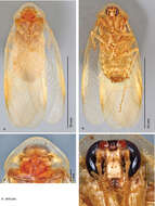
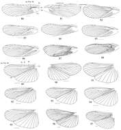
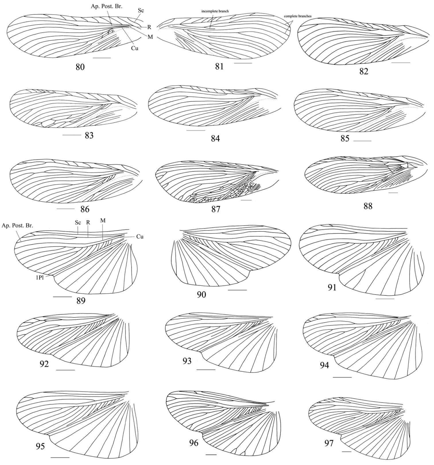
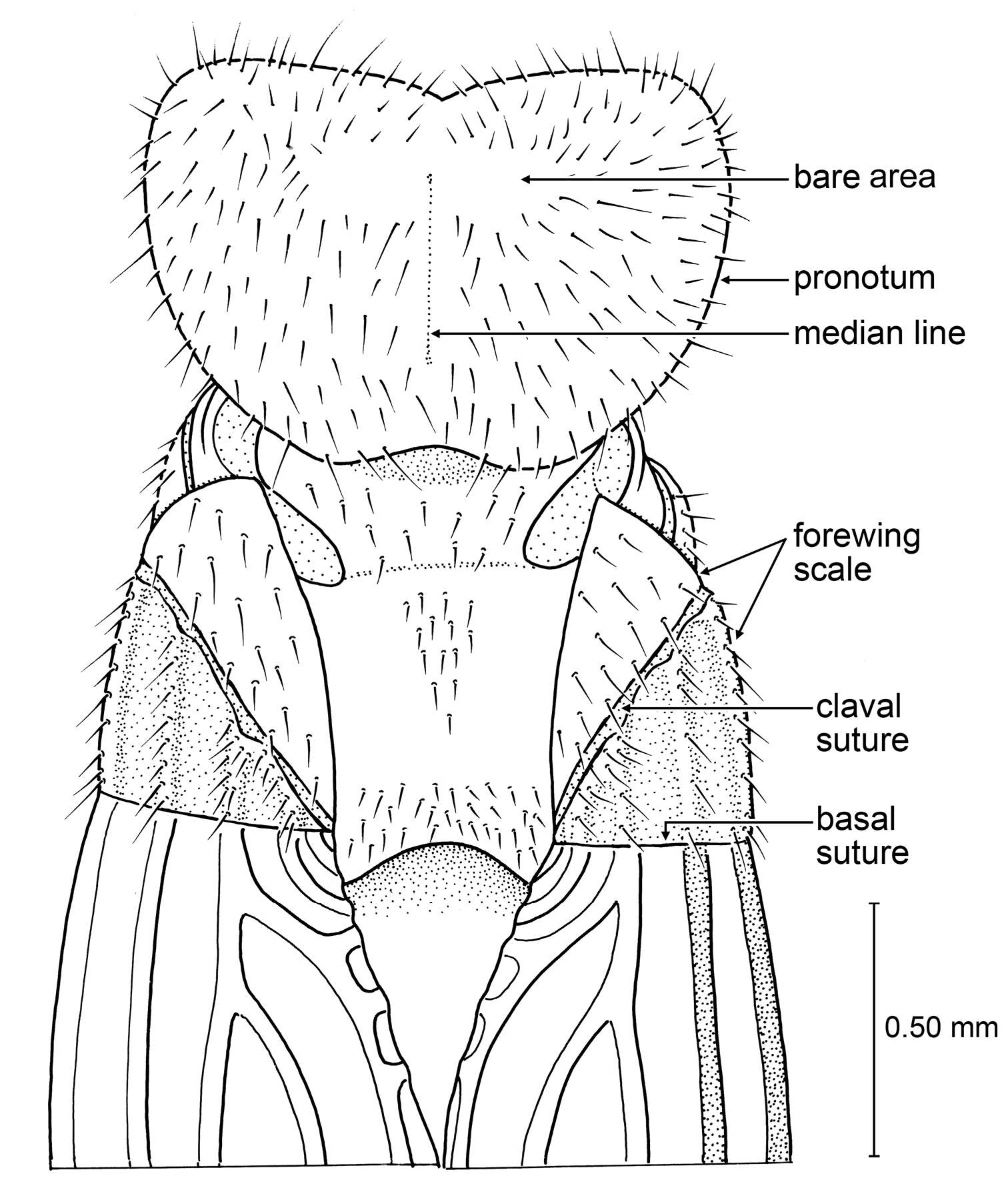
Figures 80–97.80–88 tegmina: 80–81 left and right tegmina of one specimen (Salganea quinquedentata sp. n.), dorsal view 82 Salganea anisodonta sp. n. 83–84 Salganea taiwanensis Roth, 1979 85–86 Salganea incerta (Brunner von Wattenwyl, 1893) 87–88 Salganea raggei Roth, 1979 89–97 wings: 89–90 left and right wings of one specimen (Salganea quinquedentata sp. n.), dorsal view 91 Salganea anisodonta sp. n. 92–93 Salganea taiwanensis Roth, 1979 94–95 Salganea incerta (Brunner von Wattenwyl, 1893) 96–97 Salganea raggei Roth, 1979. Scale bars = 4.0 mm.
-
I kultur
-
Glatved Strand
-
Michael S. Engel, David A. Grimaldi, Paul C. Nascimbene, Hukam Singh
Zookeys
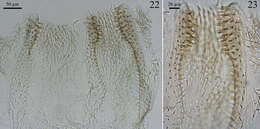
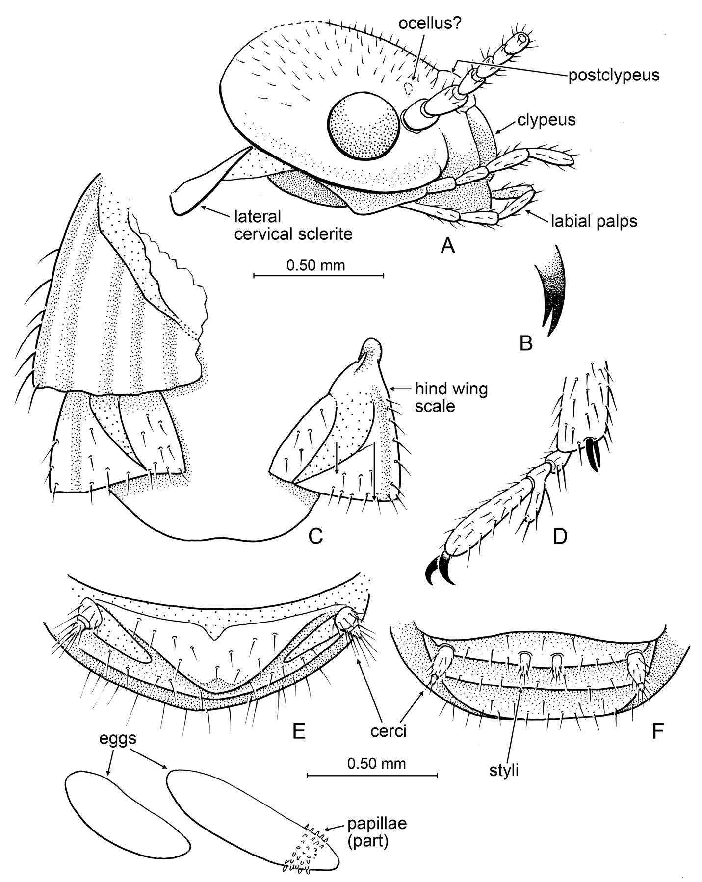
Figure 4.Detail of Prostylotermes kamboja Engel & Grimaldi, gen. et sp. n. (Tad-321C). A Head in lateral aspect B Tip of lacinia C Dorsal view of wing scales (female specimen) D Meso-pretarsus, mesotarsus, and extreme apex of mesotibia (female specimen) E Apex of female abdomen, ventral view, with detail of eggs preserved at abdominal apex F Apex of male abdomen, ventral view. Scale bars are identical and apply to all figures except the detail enlargements of B and D.
-
Zongqing Wang, Keliang Wu, Yanli Che
Zookeys
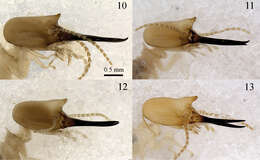
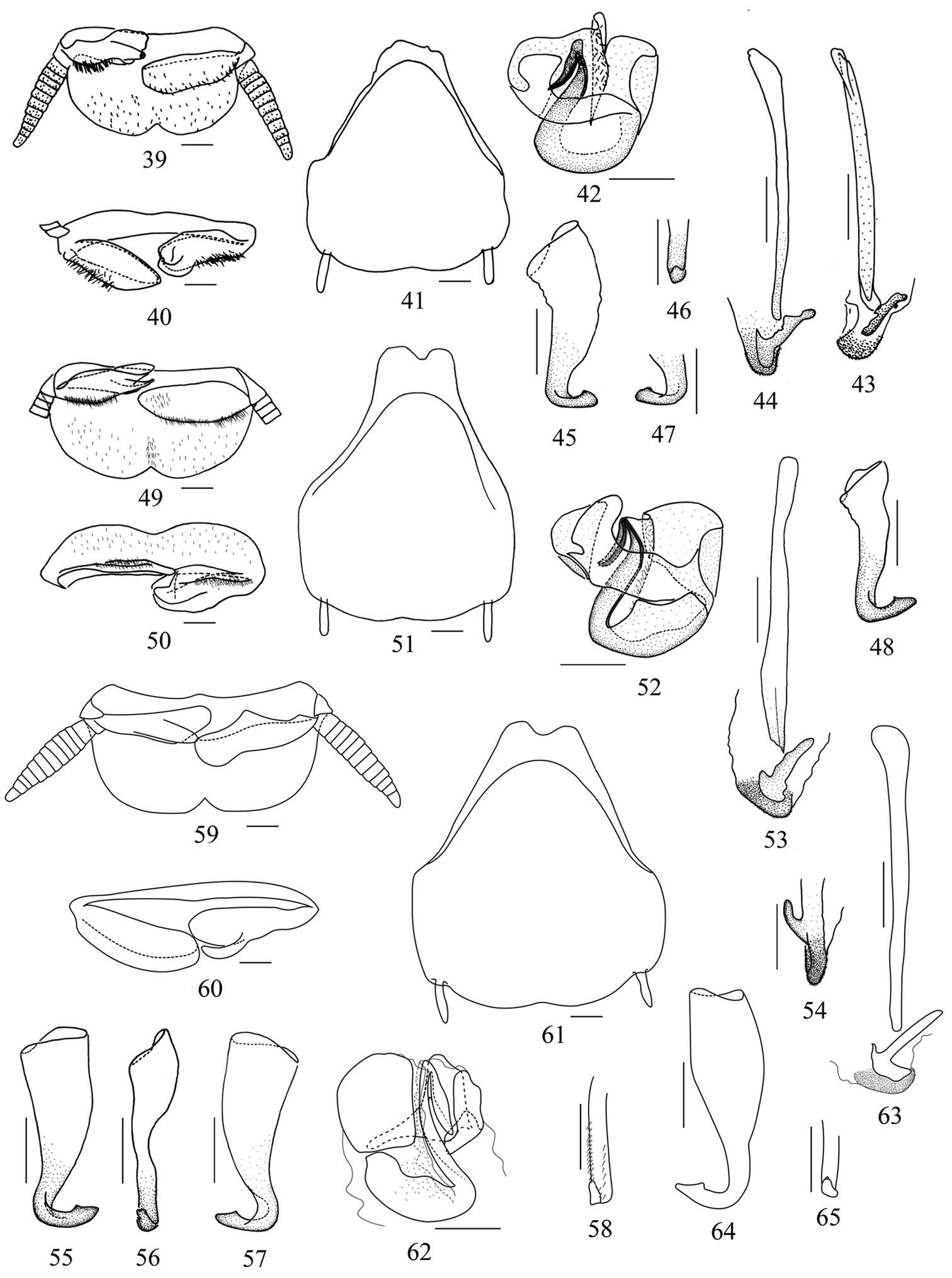
Figures 39–65.39–48 Pseudophoraspis recurvata sp. n. 49–57 Pseudophoraspis incurvata sp. n. 58–65 Pseudophoraspis clavellata sp. n. 39, 49, 59 supra-anal plate, ventral view 40, 50, 60 paraproct, caudal view 41, 51, 61 hypandrium, dorsal view 42, 52, 62 complex L1 43, 44, 53, 63 complex L2, dorsal view (43 specimen from “Hainan” 44 specimen from “Guangxi”) 54 complex L2, lateral view 45–48, 55–57, 64–65 sclerite R2 (45–47 specimen from “Hainan” 48 specimen from “Guangxi”) 58 1st segment of hind tarsus. Scale bars=0.5mm.
-
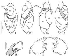
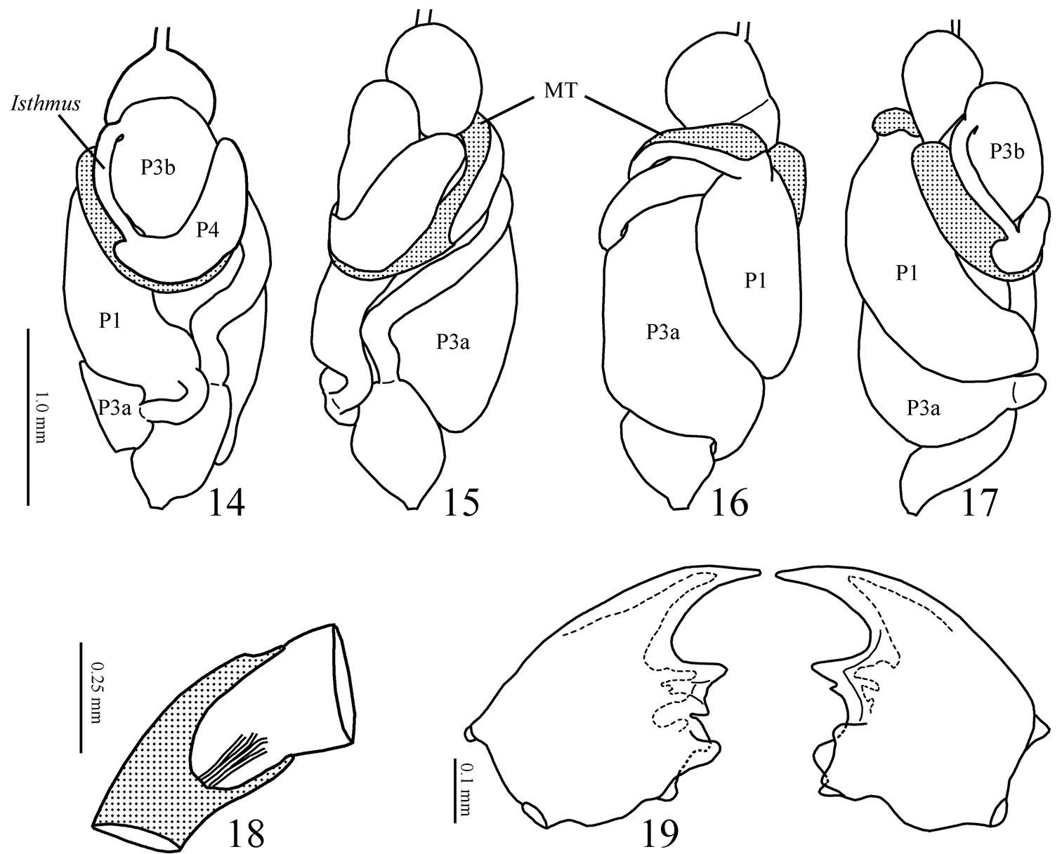
Figures 14–19.14–17 Genuotermes spinifer worker gut in situ. 14 Dorsal 15 Right 16 Ventral 17 Left (MT = mesenteric tongue; P1 = first proctodeal segment (ileum); P3a and b = third proctodeal segment (paunch); P4a = first part of fourth proctodeal segment (colon) 18 Malpighian tubules insertion 19 Worker mandibles, dorsal view.
-
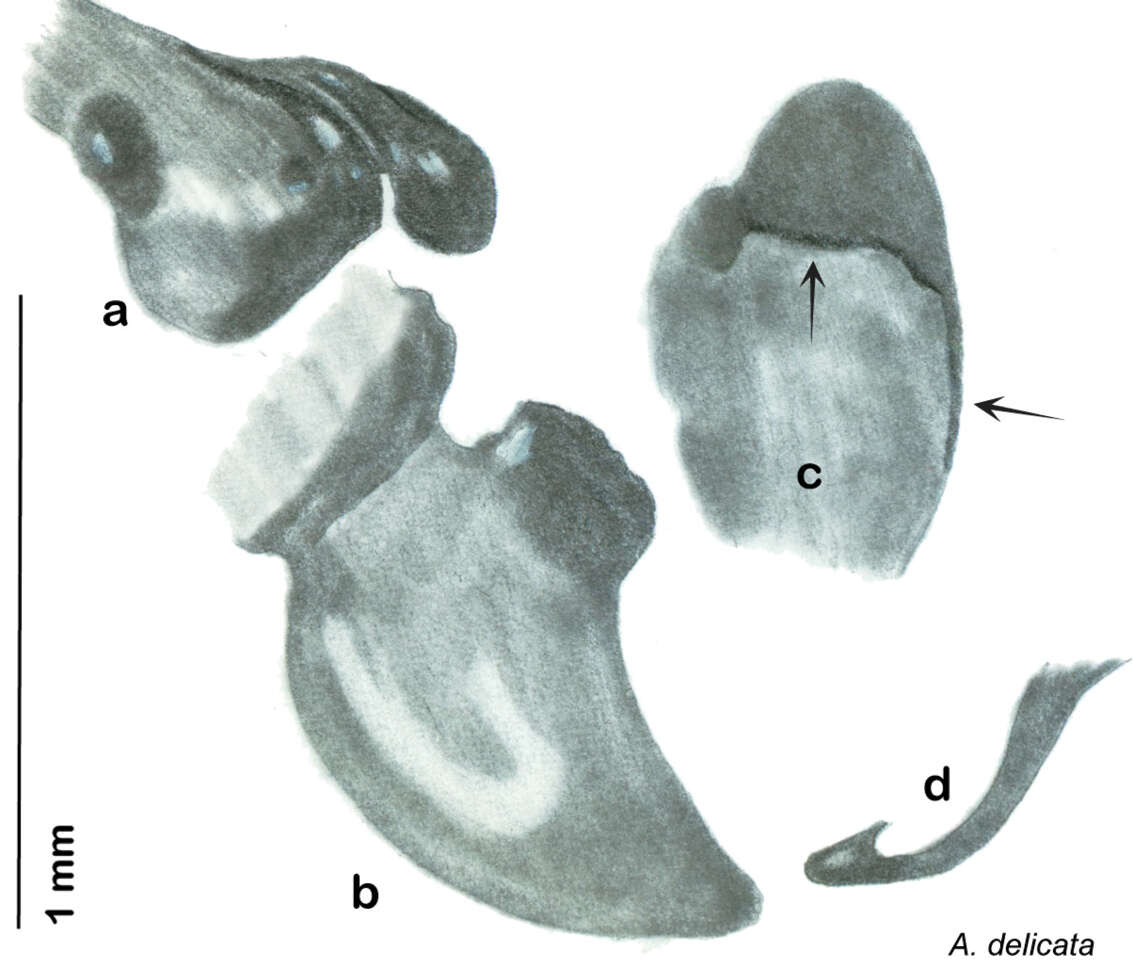
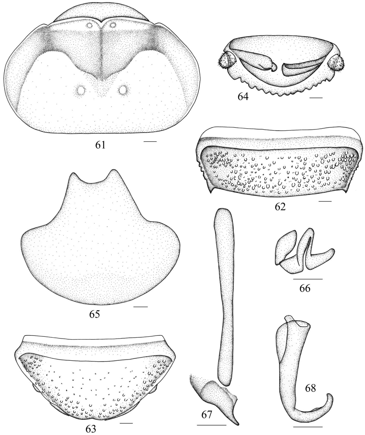
Figure 40.Arenivaga delicata, genitalia: a right dorsal phallomere b right ventral phallomere c small central sclerite d genital hook. Arrow(s) indicate diagnostic characters (see text).
-
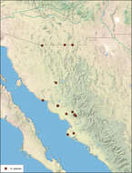
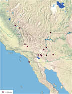
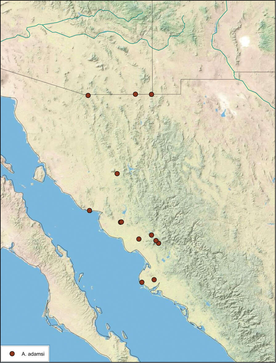
Figure 14.Arenivaga adamsi, distribution.
-
Xiudan Wang, Yan Shi, Zongqing Wang, Yanli Che
Zookeys
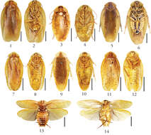
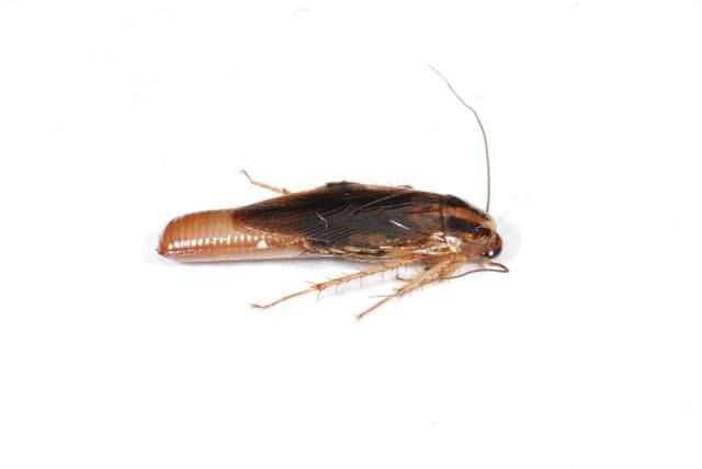
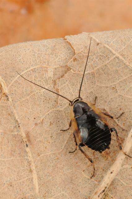
Figures 1–20.1–2 Salganea quinquedentata sp. n., male: 1 holotype, dorsal view 2 same, ventral view 3–4 Salganea quinquedentata sp. n., nymph: 3 paratype, dorsal view 4 same, ventral view 5–6 Salganea anisodonta sp. n., male: 5 holotype, dorsal view 6 same, ventral view 7–8 Salganea taiwanensis Roth, 1979, male: 7 holotype of Panesthia concinna Feng & Woo, 1990, dorsal view 8 same, ventral view 9–10 Salganea guangxiensis (Feng & Woo, 1990), male: 9 holotype of Panesthia guangxiensis Feng & Woo, 1990, dorsal view 10 same, ventral view 11–12 Salganea incerta (Brunner von Wattenwyl, 1893), male: 11 dorsal view 12 ventral view 13–14 Salganea incerta (Brunner von Wattenwyl, 1893), nymph: 13 dorsal view 14 ventral view 15–16 Salganea raggei Roth, 1979, male: 15 dorsal view 16 ventral view 17–18 Salganea raggei Roth, 1979, nymph: 17 dorsal view 18 ventral view 19–20 Salganea flexibilis sp. n., male: 19 holotype, dorsal view 20 same, ventral view. Scale bars = 1.0 cm.
-
Glatved Strand
-
Michael S. Engel, David A. Grimaldi, Paul C. Nascimbene, Hukam Singh
Zookeys
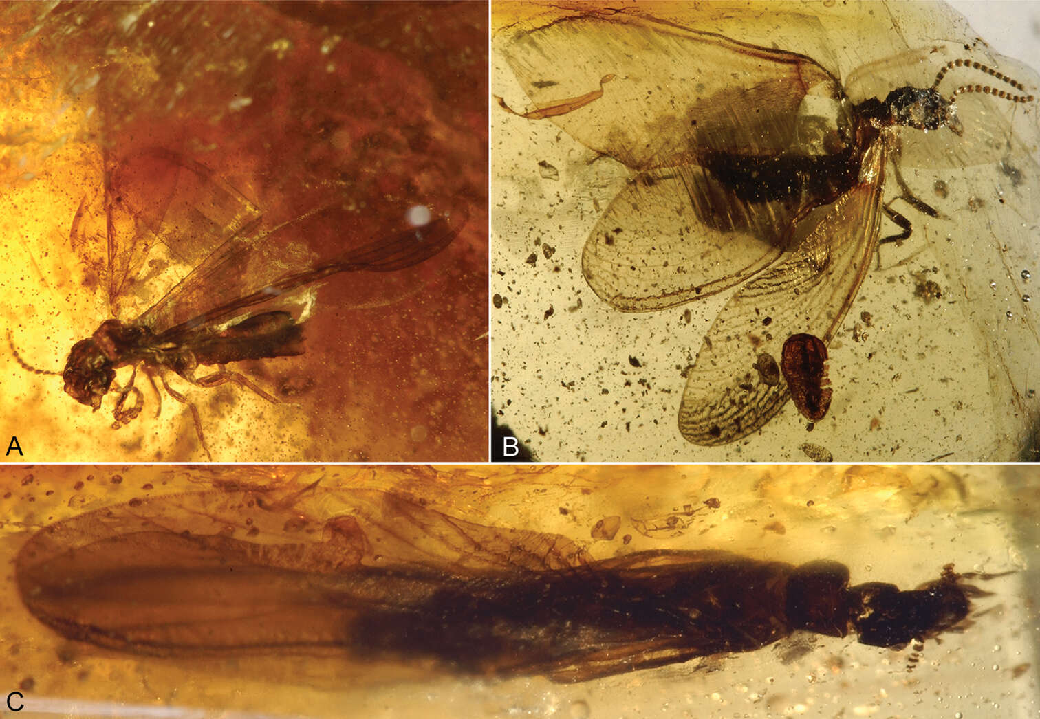
Figure 1.Photomicrographs of Cambay amber (Early Eocene) termites. A Nanotermes isaacae Engel & Grimaldi, gen. et sp. n., holotype (Termitidae: Tad-262) B Parastylotermes krishnai Engel & Grimaldi, sp. n., holotype (Stylotermitidae: Tad-277) C Zophotermes ashoki Engel & Singh, sp. n., holotype (Rhinotermitidae: Tad–42). Not to the same scale.
-
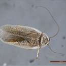
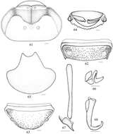
Figures 1–6.1–3 Habitus (dorsal) of the species of genus Muzoa. 1 Muzoa simplex Hebard, 1921, holotype male (ANSP) 2 Muzoa madida Rehn, 1930, holotype male (ANSP). 3 Muzoa curtalata sp. n., holotype male (MUJ). Scale bar 1 cm. 4–6 Heads (ventral) of the species of genus Muzoa 4 Muzoa simplex 5 Muzoa madida 6 Muzoa curtalata sp. n. The arrow is indicating the shape of the vertex. Scale bar 1 mm.
-
Figures 20–21.Genuotermes spinifer worker gizzard: 20 Columnar and pulvillar belts 21 Detail of ornamentation on the columns.
-
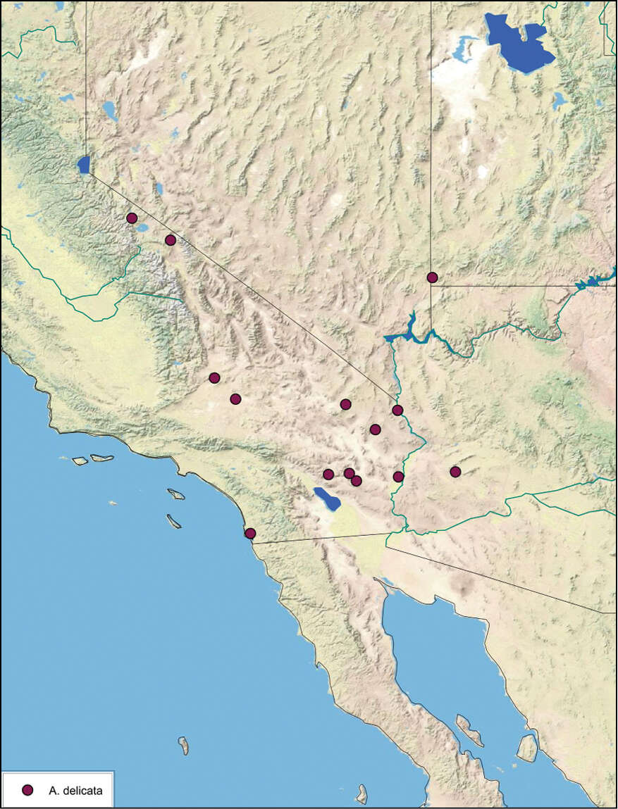
Figure 41.Arenivaga delicata, distribution.
-
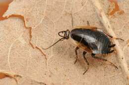
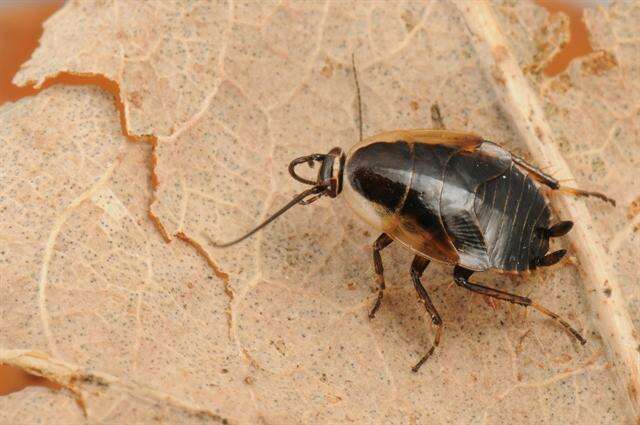
Figure 11.Arenivaga pair in copula.
-
Xiudan Wang, Yan Shi, Zongqing Wang, Yanli Che
Zookeys
Figures 61–68.Salganea raggei Roth, 1979 61 vertex and pronotum 62 abdominal tergum 7, dorsal view 63 abdominal sternite 7, ventral view 64 supra-anal plate and paraprocts, ventral view 65 subgenital plate, dorsal view 66 left phallomere (L1) 67 median phallomere (L2vm and L2d) 68 right phallomere (R2). Scale bars = 1.0 mm (Figs 61–64), 0.5 mm (Figs 65–68).
-
Glatved Strand
-
Michael S. Engel, David A. Grimaldi, Paul C. Nascimbene, Hukam Singh
Zookeys
Figure 5.Detail of Zophotermes ashoki Engel and Singh, sp. n. (Tad-42), dorsal view of thorax and anterior portion of forewings (slightly reconstructed).
-
Figures 7–22.Supra-anal plate (dorsal), subgenital plate (vental), and male genital sclerites of the species of genus Muzoa. Muzoa simplex Hebard, 1921: 7 Supra-anal plate 11 Subgenital plate 14 Median sclerite L2 (dorsal) 18 Hook “hla” of L3 (ventral) 21 Right sclerite R (dorsal). Muzoa madida Rehn, 1930: 8 Supra-anal plate 12 Subgenital plate 15 Median sclerite L2 (dorsal) 19 Hook “hla” of L3 (ventral) 22 Right sclerite R (dorsal). Muzoa curtalata sp. n. (Holotype): 9 Supra-anal plate 10 Supra-anal plate (ventral) with the paraprocts 13 Subgenital plate 16 Median sclerite L2 (dorsal) 17 Hook “hla” of L3 (ventral) 20 Right sclerite R (dorsal) (sclerotized regions R1 [subregions R1c, R1d, R1v, R1t], R2, R3, R4). Scale bar 1 mm.
-
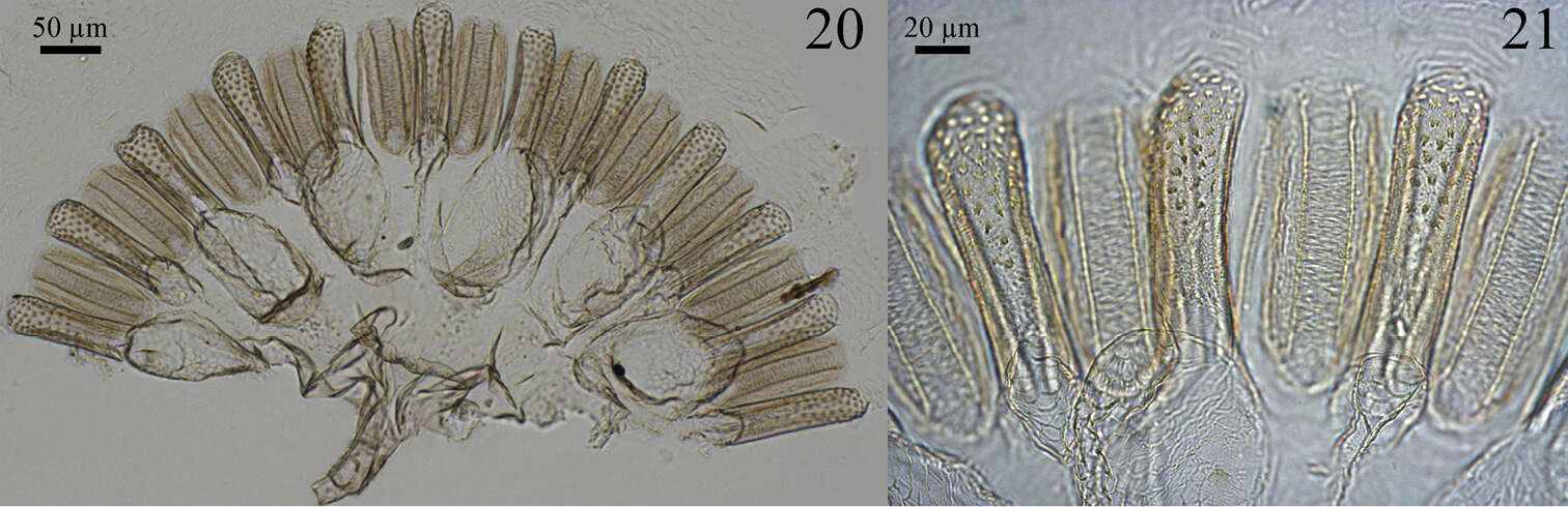
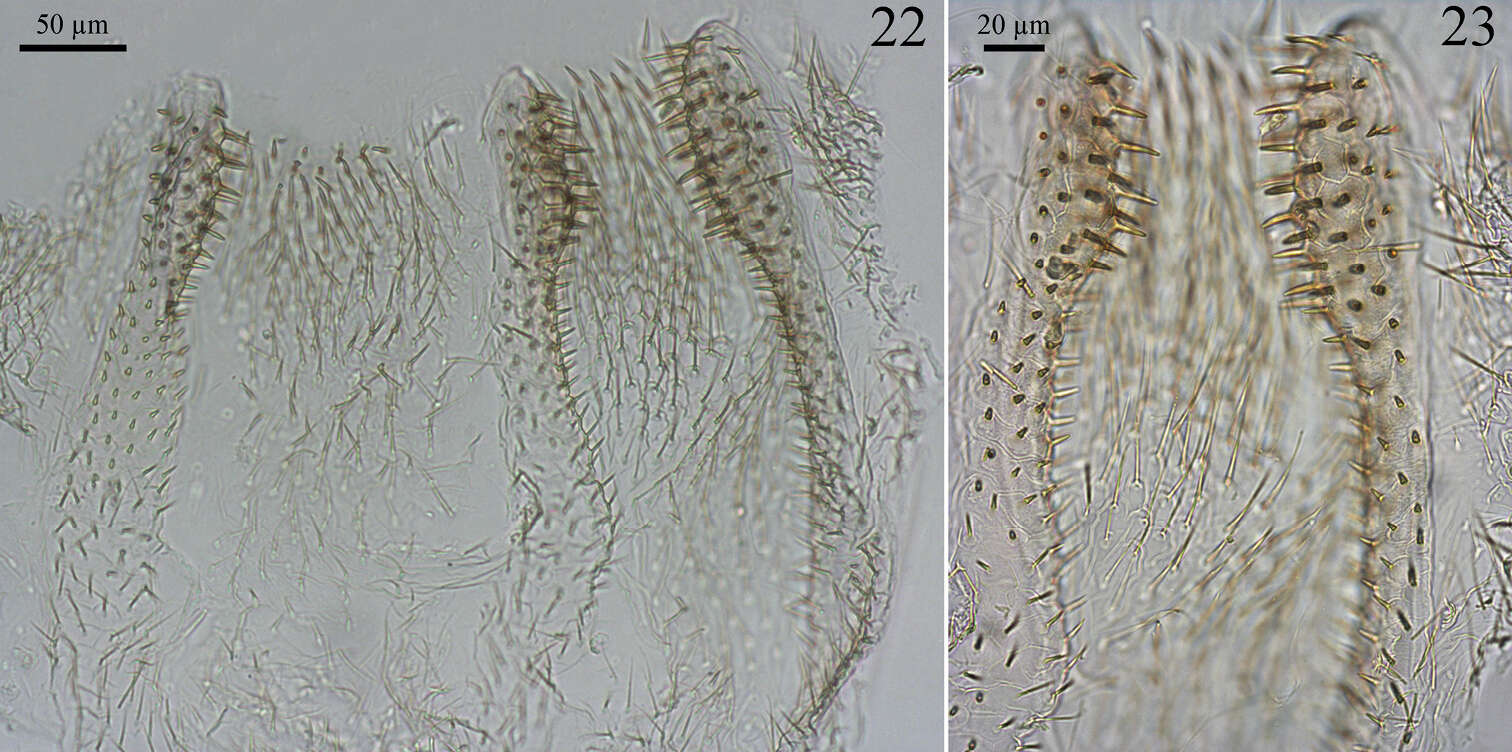
Figures 22–23.Genuotermes spinifer worker enteric valve: 22 Arrangement of the cushions 23 Detail of the cushions.