-
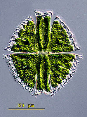
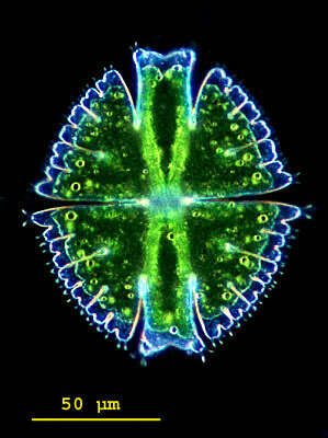
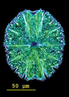
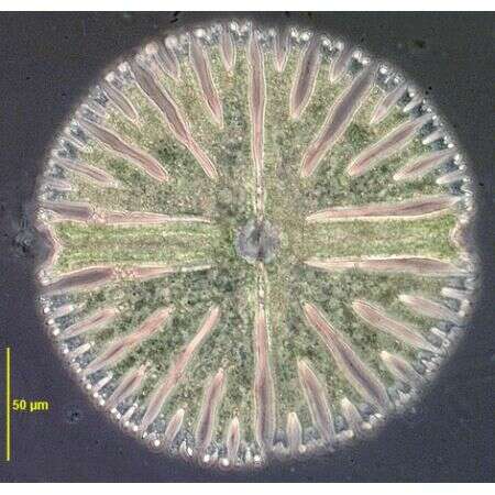
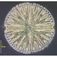
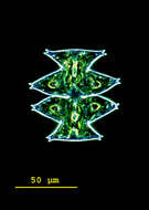
The entire cell wall of the surface is in focus by combining 30 frames with manual stacking technique. Scale bar indicates 50 µm. Sample from sphagnum pond situated in the northern alpine region of Austria near Salzburg. Images were taken using Zeiss Universal with Olympus C7070 CCD camera.
-
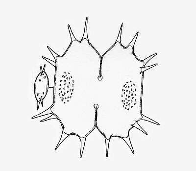
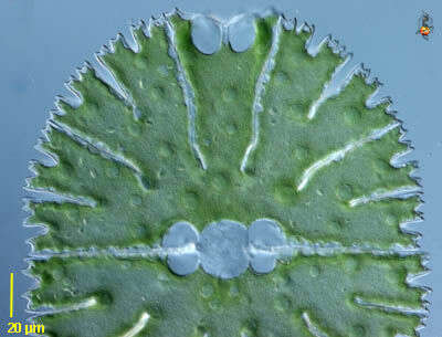
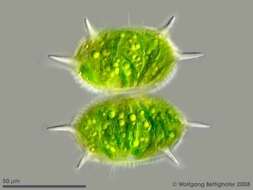
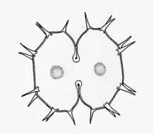
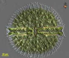
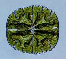
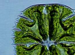
Xanthidium antilopaeum (BREB.) KÃTZ. var. crameri GRÃNBLAD The cells are a little wider than long and octagonal in coarse shape. The central cut is deep and extends strongly outwards. The cell halves are oblong hexagonal with straight or weakly concave sides, the vertices are broadly truncated. At each lateral and apical angle a pair of long pricks originate. Above the center is a flat, often slightly brown colored swelling of the cell wall occupies with small warts. Length without pricks 50 - 60 µm, width without pricks 58 - 63 µm. Occurrence: In littoral zones of mountain lakes in Central Europe rather rarely.
-
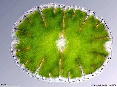
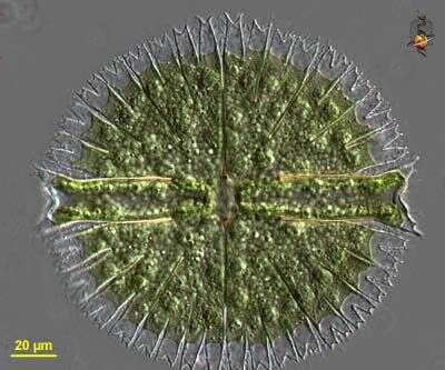
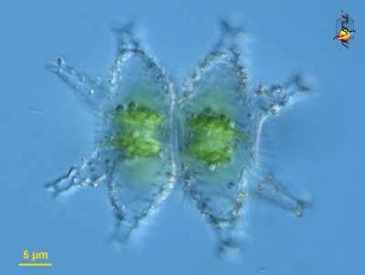
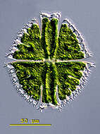
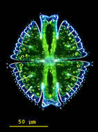
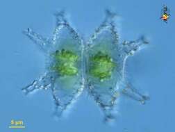
Micrasterias (mike-ras-tear-ee-ass), iconic desmid. The desmids are one type of green algae, often associated with slightly acidic freshwater habitats. As with all desmids, they are formed from two mirror imaged cells. Cellulosic wall, plastid, and pyrenoids are evident. The nucleus is in the centre. Differential interference contrast.
-
Micrasterias(mike-raz-tear-ee-ass) is a genus of unicellular algae in the family Desmidiaceae. The cells are flattened and disc-like. The cells of the genus Micrasterias are organized in two semi-cells that are mirror images of each other. The semicells have a distinctive shape with an intricate lobes and indentation. At the end of the lobes the cell wall may sometimes form notches or short spines. The nucleus is located in the centre between the semicells. Each semicell has a chloroplast with some pyrenoids. Usually found in oligotrophic, acid waters. This specimen of Micrasterias fimbriata was collected in the Salzburger Land, Austria. Differential interference contrast.
-
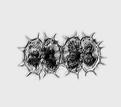
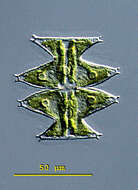
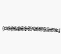
Non-filamentous desmid. The depht of focus picture shows a cell with stelloid chloroplasts. Multi layer image (DOF) using about 65 frames generating depth of focus, stacked manually using Corel Photopaint. Sample from sphagnum pond Dosenmoor near Neumuenster (Schleswig-Holstein, Germany). This image was taken using Zeiss Universal with Olympus C7070 CCD camera.
-
Micrasterias (mike-ras-tear-ee-ass), iconic desmid. The desmids are one type of green algae, often associated with slightly acidic freshwater habitats. As with most desmids, they are formed from two mirror imaged cells. Cellulosic wall, plastid, and pyrenoids are evident. Differential interference contrast.
-
Micrasterias(mike-raz-tear-ee-ass) is a genus of unicellular algae in the family Desmidiaceae. The cells are flattened and disc-like. The cells of the genus Micrasterias are organized in two semi-cells that are mirror images of each other. The semicells have a distinctive shape with an intricate lobes and indentation. At the end of the lobes the cell wall may sometimes form notches or short spines. The nucleus is located in the centre between the semicells. Each semicell has a chloroplast with some pyrenoids. Usually found in oligotrophic, acid waters. This specimen of Micrasterias fimbriata was collected in the Salzburger Land, Austria. Dark ground illumination.
-
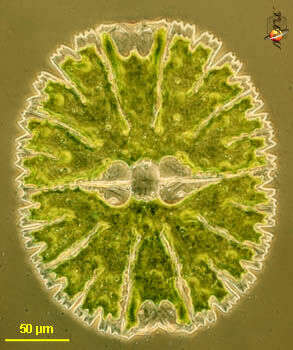
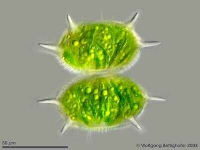
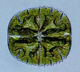
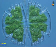
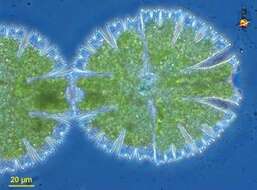
Xanthidium cristatum BREB. In RALFS The cells are little longer than wide, octagonal in coarse shape with straight or weakly concave sides. The central cut is strongly extended outwards. On each side of each cell half origins one prick . The lateral and apical angles likewise have one pair of pricks each. In the center of the cell halves is flat, hemispheric swelling. Length without pricks 50 - 55 µm, width without pricks 40 - 43 µm. Occurrence: In Central Europe sporadic in moderate acidic waters of fens, siltation zones et cetera.
-
Micrasterias (mike-ras-tear-ee-ass), iconic desmid. The desmids are one type of green algae, often associated with slightly acidic freshwater habitats. As with most desmids, they are formed from two mirror imaged cells. Cellulosic wall, plastid, and pyrenoids are evident. The nucleus is in the centre. Differential interference contrast.
-
Micrasterias(mike-raz-tear-ee-ass) is a genus of unicellular algae in the family Desmidiaceae. The cells are flattened and disc-like. The cells of the genus Micrasterias are organized in two semi-cells that are mirror images of each other. The semicells have a distinctive shape with an intricate lobes and indentation. At the end of the lobes the cell wall may sometimes form notches or short spines. The nucleus is located in the centre between the semicells. Each semicell has a chloroplast with some pyrenoids. Usually found in oligotrophic, acid waters. This specimen of Micrasterias fimbriata collected in the Salzburger Land (Austria), photographed in dark field.
-
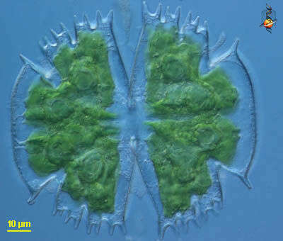
Xanthidium cristatum BREB. In RALFS The cells are little longer than wide, octagonal in coarse shape with straight or weakly concave sides. The central cut is strongly extended outwards. On each side of each cell half origins one prick . The lateral and apical angles likewise have one pair of pricks each. In the center of the cell halves is flat, hemispheric swelling. Length without pricks 50 - 55 µm, width without pricks 40 - 43 µm. Occurrence: In Central Europe sporadic in moderate acidic waters of fens, siltation zones et cetera.
-
Micrasterias (mike-ras-tear-ee-ass), iconic desmid. The desmids are one type of green algae, often associated with slightly acidic freshwater habitats. As with most desmids, they are formed from two mirror imaged cells. Cellulosic wall, plastid, and pyrenoids are evident. The nucleus is in the centre. Phase contrast.
-
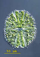
Micrasterias(mike-raz-tear-ee-ass) is a genus of unicellular algae in the family Desmidiaceae. The cells are flattened and disc-like. The cells of the genus Micrasterias are organized in two semi-cells that are mirror images of each other. The semicells have a distinctive shape with an intricate lobes and indentation. At the end of the lobes the cell wall may sometimes form notches or short spines. The nucleus is located in the centre between the semicells. Each semicell has a chloroplast with some pyrenoids. Usually found in oligotrophic, acid waters. This specimen of Micrasterias papillifera collected in the Salzburger Land, Austria. Differential interference contrast.
-
Micrasterias is one of the desmids, flattened green algae in which the organism has a central constriction which gives the organism the appearance of being two cells joined together. Phase contrast micrograph.
-
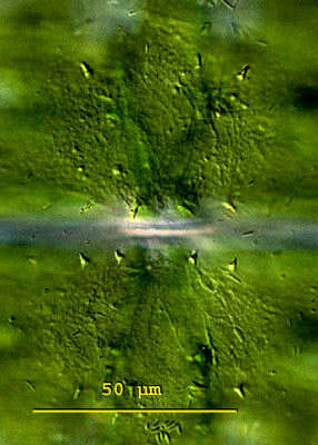
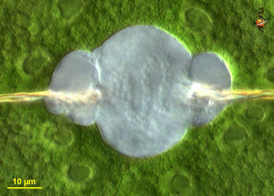
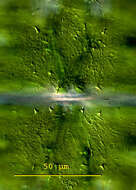
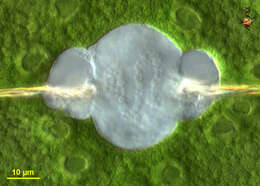
Micrasterias is one of the desmids, flattened green algae in which the organism has a central constriction which gives the organism the appearance of being two cells joined together. This detail showing the central nucleus. Differential interference contrast.
-
Micrasterias(mike-raz-tear-ee-ass) is a genus of unicellular algae in the family Desmidiaceae. The cells are flattened and disc-like. The cells of the genus Micrasterias are organized in two semi-cells that are mirror images of each other. The semicells have a distinctive shape with an intricate lobes and indentation. At the end of the lobes the cell wall may sometimes form notches or short spines. The nucleus is located in the centre between the semicells. Each semicell has a chloroplast with some pyrenoids. Usually found in oligotrophic, acid waters. This specimen of Micrasterias papillifera collected in the Salzburger Land, Austria. Dark ground illumination.
-
Micrasterias is one of the desmids, flattened green algae in which the organism has a central constriction which gives the organism the appearance of being two cells joined together. Differential interference contrast.
-
Differential interference contrast.
-
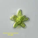
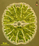
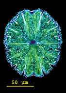
Micrasterias(mike-raz-tear-ee-ass) is a genus of unicellular algae in the family Desmidiaceae. The cells are flattened and disc-like. The cells of the genus Micrasterias are organized in two semi-cells that are mirror images of each other. The semicells have a distinctive shape with an intricate lobes and indentation. At the end of the lobes the cell wall may sometimes form notches or short spines. The nucleus is located in the centre between the semicells. Each semicell has a chloroplast with some pyrenoids. Usually found in oligotrophic, acid waters. This specimen of Micrasterias pinnatifida collected in the Salzburger Land, Austria. Differential interference contrast.
-
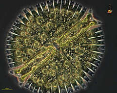
Micrasterias radiosa (RALFS,1848). M. sol (EHRENBERG ex KÅ°TZING,1849) is a junior synonym. Phase contrast.
-
Differential interference contrast.
-
Micrasterias(mike-raz-tear-ee-ass) is a genus of unicellular algae in the family Desmidiaceae. The cells are flattened and disc-like. The cells of the genus Micrasterias are organized in two semi-cells that are mirror images of each other. The semicells have a distinctive shape with an intricate lobes and indentation. At the end of the lobes the cell wall may sometimes form notches or short spines. The nucleus is located in the centre between the semicells. Each semicell has a chloroplast with some pyrenoids. Usually found in oligotrophic, acid waters. This specimen of Micrasterias pinnatifida collected in the Salzburger Land, Austria. Dark ground illumination.
-
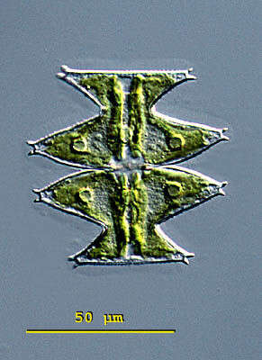
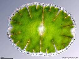
Docidium undulatum BAIL. The cells are 15-25 times longer than wide with truncated ends. The sides are repeatedly flatly undulated. The central cut is little formed. On both sides around the cut runs a ring with tiny, broadly rounded nipples. Dimension: Length 200 â 250 µm, width 12 â 17 µm Ecology: Acidophilic alga, in acidic fens together with Micrasterias jenneri, Micrasterias truncata und Euastrum armatum. Occurrence: Ubiquitous
-
Staurastrum (star-ass-strum) desmid - a green alga with (as is usual for green algae) a cellulose cell wall which has a star-shaped appearance, and bright green chloroplasts. Differential interference contrast.