-
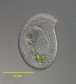
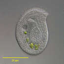
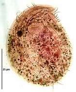
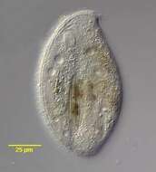
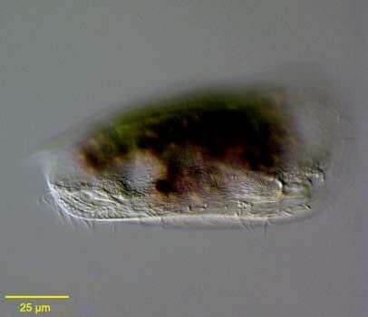
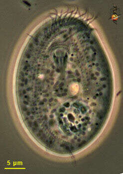
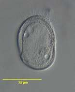
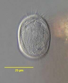
Portrait (ventral surface) of the chilodonellid ciliate Pseudochilodonopsis piscatoris (Blochmann, 1895) Foissner, 1979. The cell is drawn out to the left in a distinct pointed preoral beak. The posterior is broadly rounded. The cell is strongly dorsoventrally compressed. The dorsum is slightly domed and the ventral surface flat. The ciliature is reduced to the ventral surface except for a distinctive dorsal brush which is set back from the anterior edge of the cell and arches across the nearly its entire width. The ventral ciliature consists of right (5) and left (6) kineties separated by a wide bare postoral area. There are two circumoral kineties and a fragmented preoral kinety. The anterior ends of the left somatic kineties abut the transversely oriented fragments of the preoral kinety. These fragments ascend stair-step fashion to the tip of the beak. The cytostome, situated in the anterior 1/4 of the cell, is supported by nematodesmata forming a cyrtos. There are two contractile vacuoles. The nucleus is heteromerous. The genus Pseudochilodonopsis is distinguished from members of the similar genus, Chilodonella, by the fragmented preoral kinety and the long arched dorsal brush. Both features are difficult to appreciate without DIC optics or silver impregnation techniques. P. piscatoris feeds on green algae and diatoms. It is usually found in the surface film of samples collected from a freshwater pond near Boise,Idaho. February 2005. DIC.
-
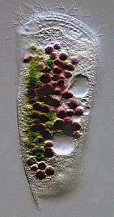
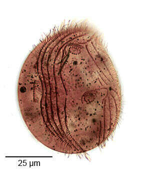
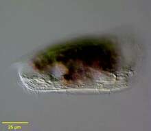
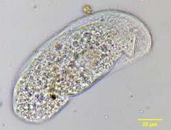
Portrait of the marine Phyllopharyngeid ciliate, Dysteria brasiliensis (Da Cunha, De Faria & Pinto, 1922). This is one of the largest species of this genus (100-130 um). The cell is elongate and dorsoventrally flattened. The dorsum is arched. The anterior end is truncate and curves dorsally. The posterior terminates in a sharp spinous process (seen here) not to be confused with the ventral posterior podite by which the cell attaches to the substrate (not seen in this image)The pellicle is rigid and colorless. The ciliature is reduced to the ventral surface with 3 longitudinal kineties on the right and 7-8 on the left. There are 2 frontoventral kineties. The cytostome is supported by two stout obliquely situated rods with anterior tooth-like projections. The cytoplasm contains food vacuoles brightly colored with green algae and purple sulfur bacteria. There are two contractile vacuoles. There is a central ellipsoid macronucleus. Collected from a commercial saltwater aquarium in Boise, Idaho. March 2004. DIC optics.
-
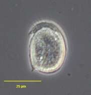
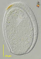
Chlamydodon (clam-ee-doe-don), alga-eating hypostome ciliate. This cell was photographed immediately after dividing. During the division process, the cells do not eat. Food ingested before division is initiated is digested, with the rest that the cytoplasm is very empty and the major organelles can be seen with some clarity: orange zone, ingestion apparatus, macronucleus and railway track. Differential interference contrast.
-
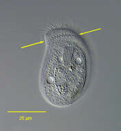
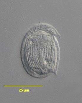
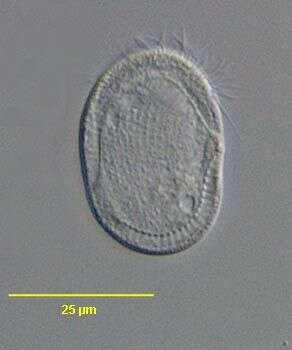
Atopochilodon distichum (Deroux,1976). Collected from a commercial saltwater aquarium in Boise, Idaho. February 2006.Phase contrast.
-

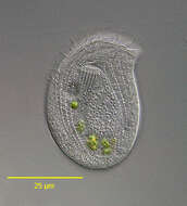
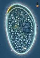
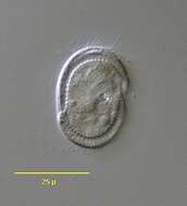
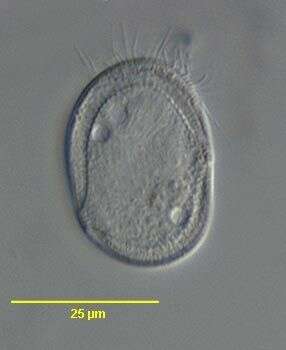
Portrait (dorsal surface) of the chilodonellid ciliate Pseudochilodonopsis piscatoris (Blochmann, 1895) Foissner, 1979. The cell is drawn out to the left in a distinct pointed preoral beak. The posterior is broadly rounded. The cell is strongly dorsoventrally compressed. The dorsum is slightly domed and the ventral surface flat. The ciliature is reduced to the ventral surface except for a distinctive dorsal brush which is set back from the anterior edge of the cell and arches across the nearly its entire width(seen in this image). The ventral ciliature consists of right (5) and left (6) kineties separated by a wide bare postoral area. There are two circumoral kineties and a fragmented preoral kinety. The anterior ends of the left somatic kineties abut the transversely oriented fragments of the preoral kinety. These fragments ascend stair-step fashion to the tip of the beak. The cytostome, situated in the anterior 1/4 of the cell, is supported by nematodesmata forming a cyrtos. There are two contractile vacuoles. The nucleus is heteromerous. The genus Pseudochilodonopsis is distinguished from members of the similar genus, Chilodonella, by the fragmented preoral kinety and the long arched dorsal brush (visible here between arrows). Both features are difficult to appreciate without DIC optics or silver impregnation techniques. P. piscatoris feeds on green algae and diatoms. It is usually found in the surface film of samples Collected from a freshwater pond near Boise, Idaho February 2005. DIC.
-
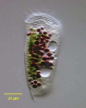
Portrait of the marine Phyllopharyngeid ciliate, Dysteria brasiliensis Da Cunha, De Faria & Pinto, 1922. This is one of the largest species of this genus (100-130 um). The cell is elongate and dorsoventrally flattened. The dorsum is arched. The anterior end is truncate and curves dorsally. The posterior terminates in a sharp spinous process (seen here) not to be confused with the ventral posterior podite by which the cell attaches to the substrate (not seen in this image)The pellicle is rigid and colorless. The ciliature is reduced to the ventral surface with 3 longitudinal kineties on the right and 7-8 on the left. There are 2 frontoventral kineties. The cytostome is supported by two stout obliquely situated rods with anterior tooth-like projections. The cytoplasm contains food vacuoles brightly colored with green algae and purple sulfur bacteria. There are two contractile vacuoles. There is a central ellipsoid macronucleus. Collected from a commercial saltwater aquarium in Boise, Idaho. March 2004. DIC.
-
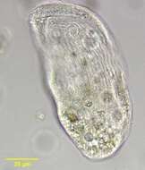
Chlamydodon (clam-ee-doe-don), alga-eating hypostome ciliate, so called because the mouth opens on the ventral surface of the cell. Common in marine habitats. The mouth is used to ingest filamentous algae - and a separate series of images illustrates this process. Phase contrast.
-
Atopochilodon distichum (Deroux,1976). Collected from a commercial saltwater aquarium in Boise, Idaho. February 2006.DIC.
-

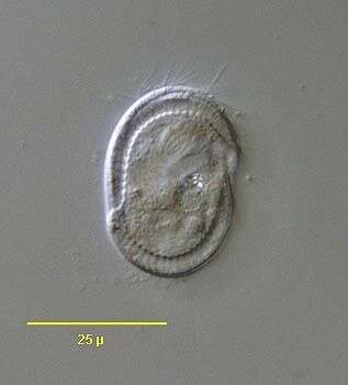
Infraciliature (ventral surface) of the chilodonellid ciliate Pseudochilodonopsis piscatoris (Blochmann, 1895) Foissner, 1979. The cell is drawn out to the left in a distinct pointed preoral beak. The posterior is broadly rounded. The cell is strongly dorsoventrally compressed. The dorsum is slightly domed and the ventral surface flat. The ciliature is reduced to the ventral surface except for a distinctive dorsal brush which is set back from the anterior edge of the cell and arches across the nearly its entire width. The ventral ciliature consists of right (5) and left (6) kineties separated by a wide bare postoral area. There are two circumoral kineties and a fragmented preoral kinety. The anterior ends of the left somatic kineties abut the transversely oriented fragments of the preoral kinety. These fragments ascend stair-step fashion to the tip of the beak. The cytostome, situated in the anterior 1/4 of the cell, is supported by nematodesmata forming a cyrtos. There are two contractile vacuoles. The nucleus is heteromerous. The genus Pseudochilodonopsis is distinguished from members of the similar genus, Chilodonella, by the fragmented preoral kinety and the long arched dorsal brush. Both features are difficult to appreciate without DIC optics or silver impregnation techniques. P. piscatoris feeds on green algae and diatoms. It is usually found in the surface film of samples Collected from a freshwater pond near Boise, Idaho February 2005.Stained by the silver carbonate technic (see Foissner, W.Europ. J. Protistol.27,313-330;1991). Brightfield.
-
Surface detail of the marine Phyllopharyngeid ciliate, Dysteria brasiliensis Da Cunha, De Faria & Pinto, 1922. This is one of the largest species of this genus (100-130 um).The posterior terminates in a sharp spinous process (slightly out of focus here) not to be confused with the ventral posterior podite by which the cell attaches to the substrate. The podite is angled anteriorly in this image (the the viewer's right).Collected from a commercial saltwater aquarium in Boise, Idaho. March 2004. DIC.
-
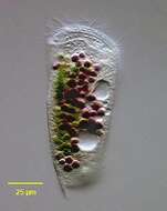
Chlamydodon (clam-ee-doe-don), alga-eating hypostome like many other hypostome ciliates, eats filamentous bacteria - such as filamentous blue green algae. They make contact with the filament, move along up and down until they find an end. They then tip over, pushing the end of the filament into the mouth - a cylindrical structure supported by a palisade of microtubular rods . They then start to suck the filament into the cell. As it hits the posterior margin, the cell is deformed by the stiff filament. The food is stunningly quickly degraded and begins to break and fold so that the cell can pull in a filament very much longer than itself. Yum. Phase contrast.
-
Atopochilodon distichum (Deroux,1976). Collected from a commercial saltwater aquarium in Boise, Idaho. February 2006.DIC.
-
Infraciliature (ventral surface) of the chilodonellid ciliate Pseudochilodonopsis piscatoris (Blochmann, 1895) Foissner, 1979 in early division. Stomatogenesis is of the telokinetal type in which the oral apparatus of the posterior daughter cell (opisthe) derives from fragments of several left sided kineties. As can be seen here, stomatogenesis precedes cytokinesis. Collected from a freshwater pond near Boise, Idaho February 2005.Stained by the silver carbonate technic (see Foissner, W.Europ. J. Protistol.27,313-330;1991). Brightfield.
-
-
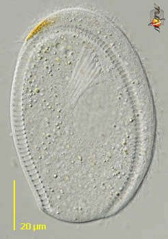
Ventral surface of the marine phyllopharyngiid ciliate, Coeloperix sleighi (Gong and Song,2004).The cell is broadly ovoid in outline and strongly dorsoventrally flattened. The dorsum is slightly convex and the ventral surface flattened. Ciliature is restricted to the ventral surface. The preoral and postoral kineties are separated by a transverse suture and the preoral kineties are transversely oriented. The postoral kineties are continuous , lacking the central bare gap seen in Chlamydodon. There is a peripheral cross-striated band (CSB) similar to that seen in Chlamydodon however the CSB in Coeloperix is interrupted on the right and left sides by two slightly offset gaps. There are 3or 4 short "tentacles" on the posteromedial ventral surface. These are about 5 µm long and are quite difficult to see even with DIC. The anterior ventral cytostome is supported by prominent nematodesmata. There is a central heteromerous macronucleus. There are two contractile vacuoles situated diagonally. They empty through single pores on the ventral surface. Collected from a commercial marine aquarium in Boise, Idaho. pH 7.93. January 2004. DIC.
-
Video showing how this ciiate collected from Cedar Swamp around Woods Hole moves around. Really cute guy.
-
Ventral surface of the marine phyllopharyngiid ciliate, Coeloperix sleighi (Gong and Song,2004).The cell is broadly ovoid in outline and strongly dorsoventrally flattened. The dorsum is slightly convex and the ventral surface flattened. Ciliature is restricted to the ventral surface. The preoral and postoral kineties are separated by a transverse suture and the preoral kineties are transversely oriented. The postoral kineties are continuous , lacking the central bare gap seen in Chlamydodon. There is a peripheral cross-striated band (CSB) similar to that seen in Chlamydodon however the CSB in Coeloperix is interrupted on the right and left sides by two slightly offset gaps. There are 3or 4 short "tentacles" on the posteromedial ventral surface. These are about 5 µm long and are quite difficult to see even with DIC. The anterior ventral cytostome is supported by prominent nematodesmata. There is a central heteromerous macronucleus. There are two contractile vacuoles situated diagonally. They empty through single pores on the ventral surface. Collected from a commercial marine aquarium in Boise, Idaho. pH 7.93. January 2004. DIC.
-
Originally described by Ehrenberg under the name Chlamidodon mnemosyne.
-
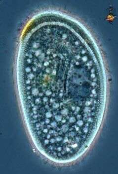
Optical section of the marine phyllopharyngiid ciliate, Coeloperix sleighi (Gong and Song,2004).The cell is broadly ovoid in outline and strongly dorsoventrally flattened. The dorsum is slightly convex and the ventral surface flattened. Ciliature is restricted to the ventral surface. The preoral and postoral kineties are separated by a transverse suture and the preoral kineties are transversely oriented. The postoral kineties are continuous , lacking the central bare gap seen in Chlamydodon. There is a peripheral cross-striated band (CSB) similar to that seen in Chlamydodon however the CSB in Coeloperix is interrupted on the right and left sides by two slightly offset gaps. There are 3or 4 short "tentacles" on the posteromedial ventral surface. These are about 5 µm long and are quite difficult to see even with DIC. The anterior ventral cytostome is supported by prominent nematodesmata. There is a central heteromerous macronucleus. There are two contractile vacuoles situated diagonally. They empty through single pores on the ventral surface. Collected from a commercial marine aquarium in Boise, Idaho. pH 7.93. January 2004. DIC.
-
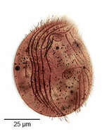
Trithigmostoma, a large hypostome ciliate. The cell is elongate, the right margin curving to meet the relatively straight left margin. The cell is flat ventrally with a slightly domed dorsum. There is a dorsal row of bristles which angles posteriorly toward the left margin (seen clearly here). Protrusible nemadesmata are seen surrounding the cytostome. Multiple small contractile vacuoles are scattered throughout the cytoplasm. From freshwater pond near Boise, Idaho. Brightfield illumination.
-
Optical section of the marine phyllopharyngiid ciliate, Coeloperix sleighi (Gong and Song,2004).The cell is broadly ovoid in outline and strongly dorsoventrally flattened. The dorsum is slightly convex and the ventral surface flattened. Ciliature is restricted to the ventral surface. The preoral and postoral kineties are separated by a transverse suture and the preoral kineties are transversely oriented. The postoral kineties are continuous , lacking the central bare gap seen in Chlamydodon. There is a peripheral cross-striated band (CSB) similar to that seen in Chlamydodon however the CSB in Coeloperix is interrupted on the right and left sides by two slightly offset gaps. There are 3or 4 short "tentacles" on the posteromedial ventral surface. These are about 5 ?m long and are quite difficult to see even with DIC. The anterior ventral cytostome is supported by prominent nematodesmata. There is a central heteromerous macronucleus. There are two contractile vacuoles situated diagonally. They empty through single pores on the ventral surface. Collected from a commercial marine aquarium in Boise, Idaho. pH 7.93. January 2004. DIC.
-
Trithigmostoma, a large hypostome ciliate. The cell is elongate, the right margin curving to meet the relatively straight left margin. The cell is flat ventrally with a slightly domed dorsum. There is a dorsal row of bristles which angles posteriorly toward the left margin (seen clearly here). Protrusible nemadesmata are seen surrounding the cytostome. Multiple small contractile vacuoles are scattered throughout the cytoplasm. From freshwater pond near Boise, Idaho. Brightfield illumination.
-
Ventral view of the chillodonellid ciliate, Pseudochilodonopsis polyvacuolata (Foissner and Didier, 1981). The cell is ovoid. The anterior end is drawn to the left as a bluntly pointed rostrum. The ventral surface is flat and the central dorsal surface is arched. There is a flattened narrow circumferential margin. Ciliature is restricted to the ventral surface except for a short dorsal brush. The 7 left somatic kineties are separated from 5 right somatic kineties by an unciliated postoral bare area. The lateral-most 5 left somatic kineties terminate at a right angle to short separate preoral kineties arranged in stair-step fashion from the cytostome to the tip of the rostrum. The medial two left somatic kineties are shorter. There are two short circumoral kineties. The cyrtos opens ventrally. The heteromerous macronucleus is approximately central with one adherent ovoid micronucleus. There are 7-10 contractile vacuoles each with a single ventral excretory pore. The similar species, P. fluviatilis is smaller and has only two contractile vacuoles.Collected from a freshwater stream with abundant pennate diatoms near Boise, Idaho;43° 34' 41.92" N 116° 08' 50.49" W. March 2006. DIC.
-
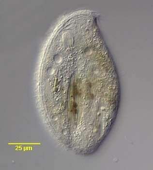
Ventral face, kineties extend to the right and left of the mouth. The mouth is supported by strong microtubular nematodesmata. The granular structure near the rear is the macronucleus. The contractile vacuoles are light structure. the oblong grey organelles are probably mitochondria. Phase contrast microscopy.