-
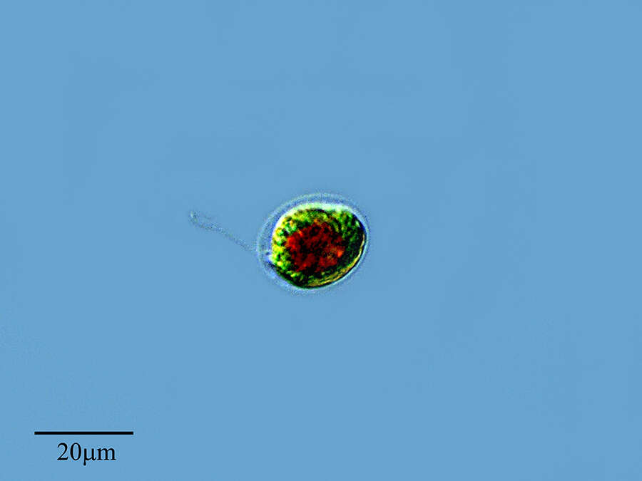
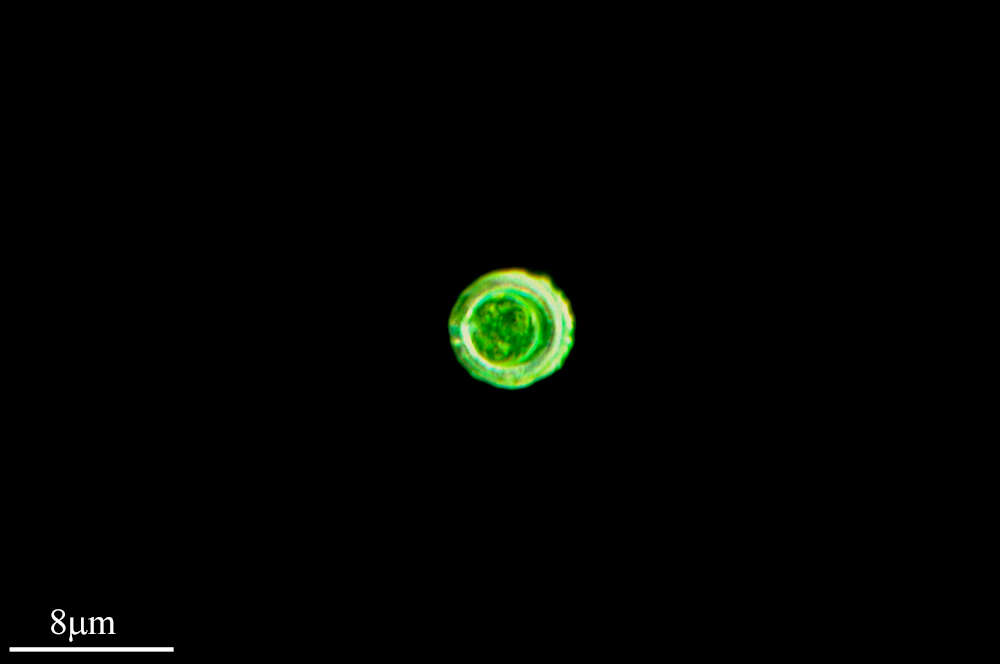
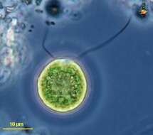
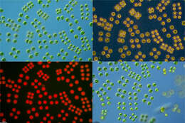
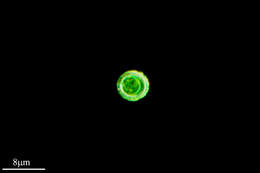
Chlamydomonas (clam-ee-dough-moan-ass) iconic volvocid motile green alga, with two similar flagella inserting into the anterior end of the cell. Photosynthetic pigments include chlorophyll B which gives the cells their bright green colour. Phase contrast micrograph.
-
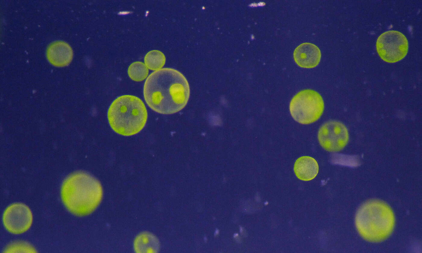
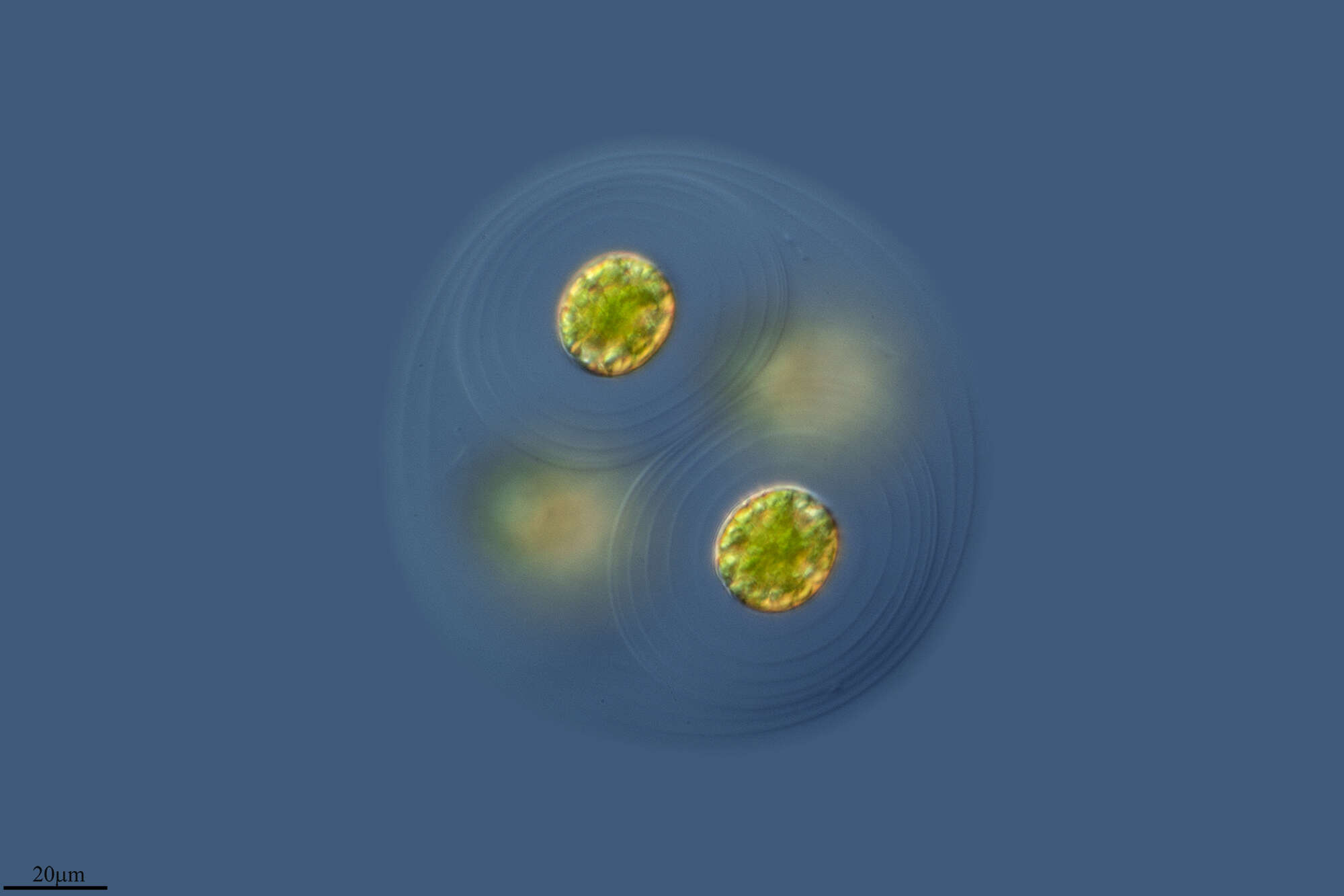
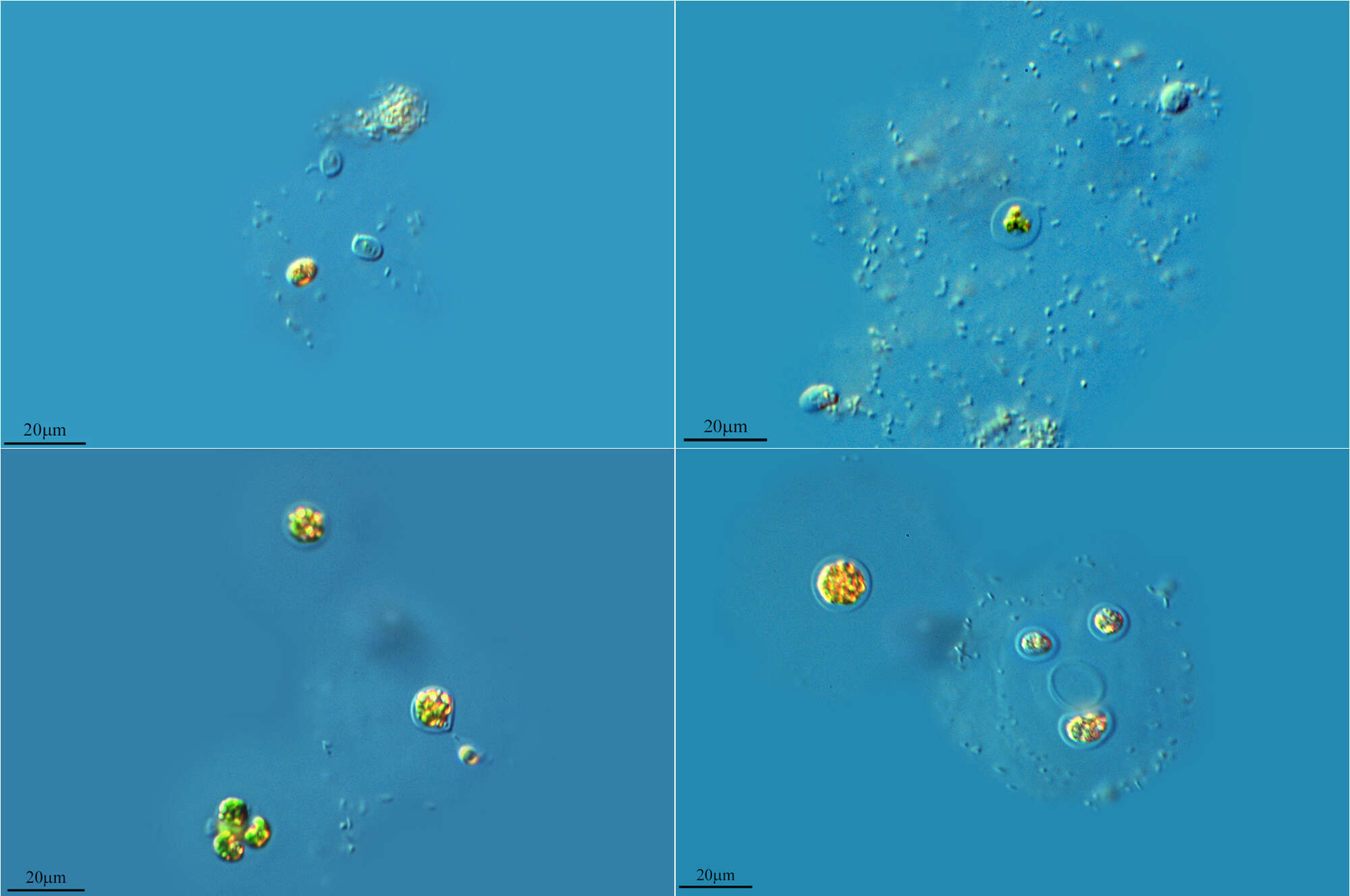
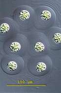
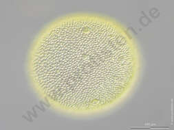
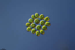
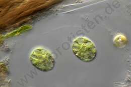
Asterococcus (a-stir-owe-cock-us) superbus. The large round or ellipsoid cells of Asterococcus occur singly or in groups with up to 8 cells embedded in a well-developed mucilage envelope. Groups reach 180 microns in diameter. The gelatinous sheaths are normally colourless and in concentrically arranged layers. The sheaths may be coloured brown by iron hydroxide. The single chloroplast is star-shaped and the pyrenoids are arranged in the center of the cell. The nucleus is located in a gap between the strands of the chlororoplast, together with two contractile vacuoles. No eyespot. Common in freshwater ponds and lakes. This image is of a squashed colony of Asterococcus superbus. The concentrically arranged layers of the gelatinous sheaths are clearly visible. The cells measuring 22 - 26 microns in diameter. Differential interference contrast.
-
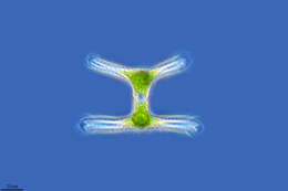
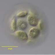
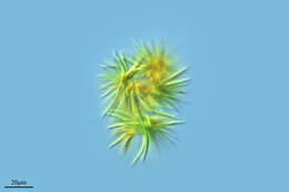
In vivo surface view of the volvocid flagellate, Volvulina steinii Playfair,1915. Collected from a temporary rainwater puddle on a grass lawn in Boise, Idaho 43° 36' 49.03" N 116° 13' 23.77" W elev. 2674 ft. March 2006. DIC.
-
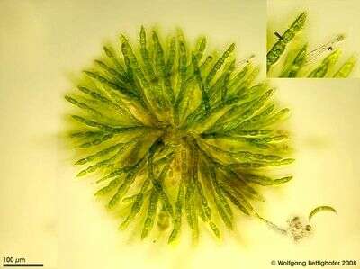
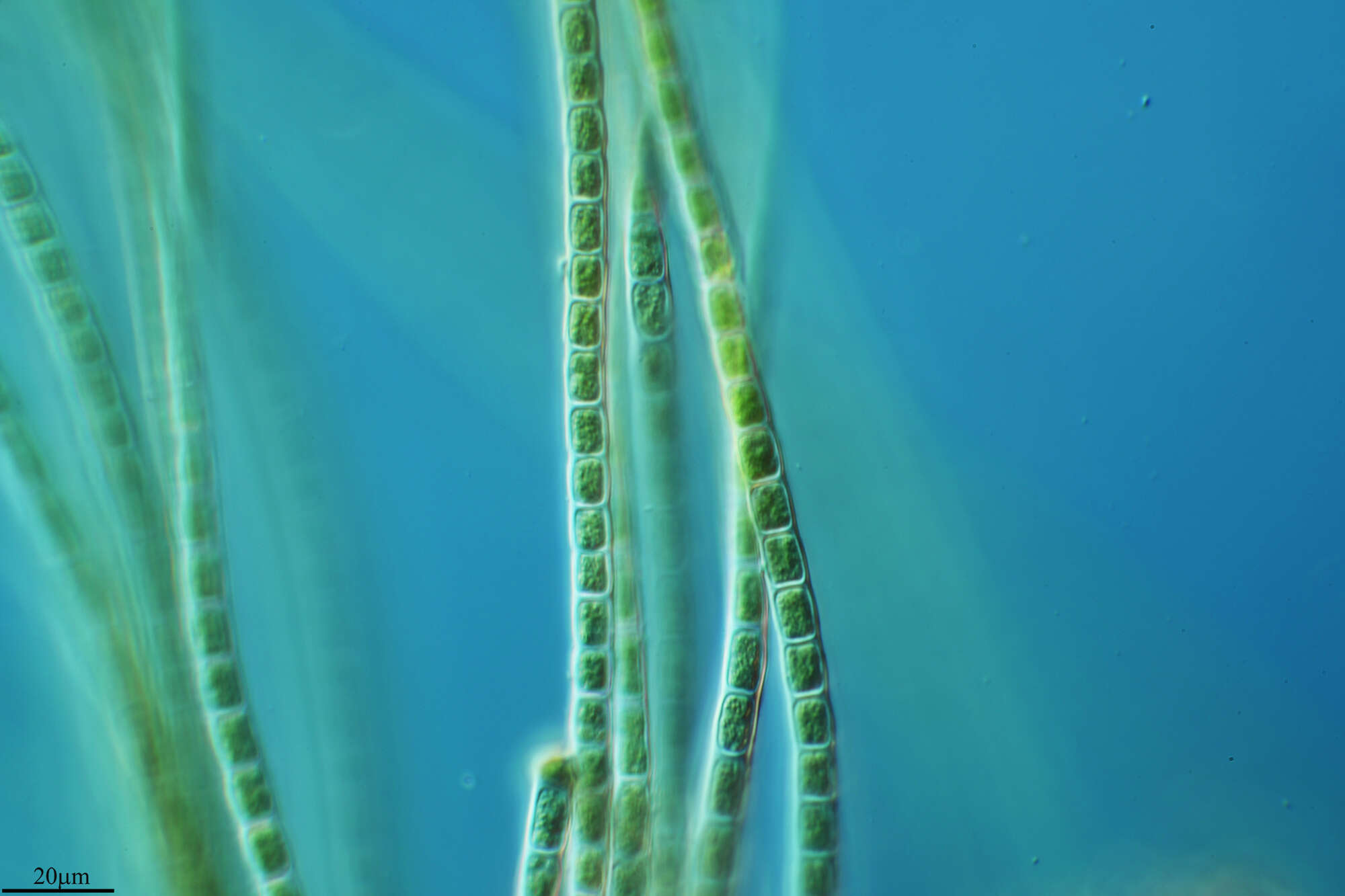
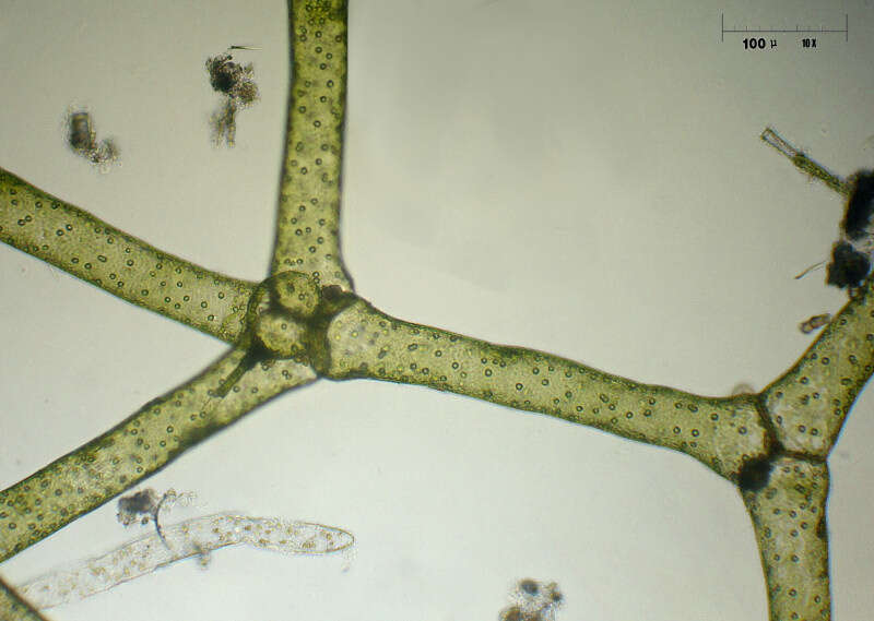
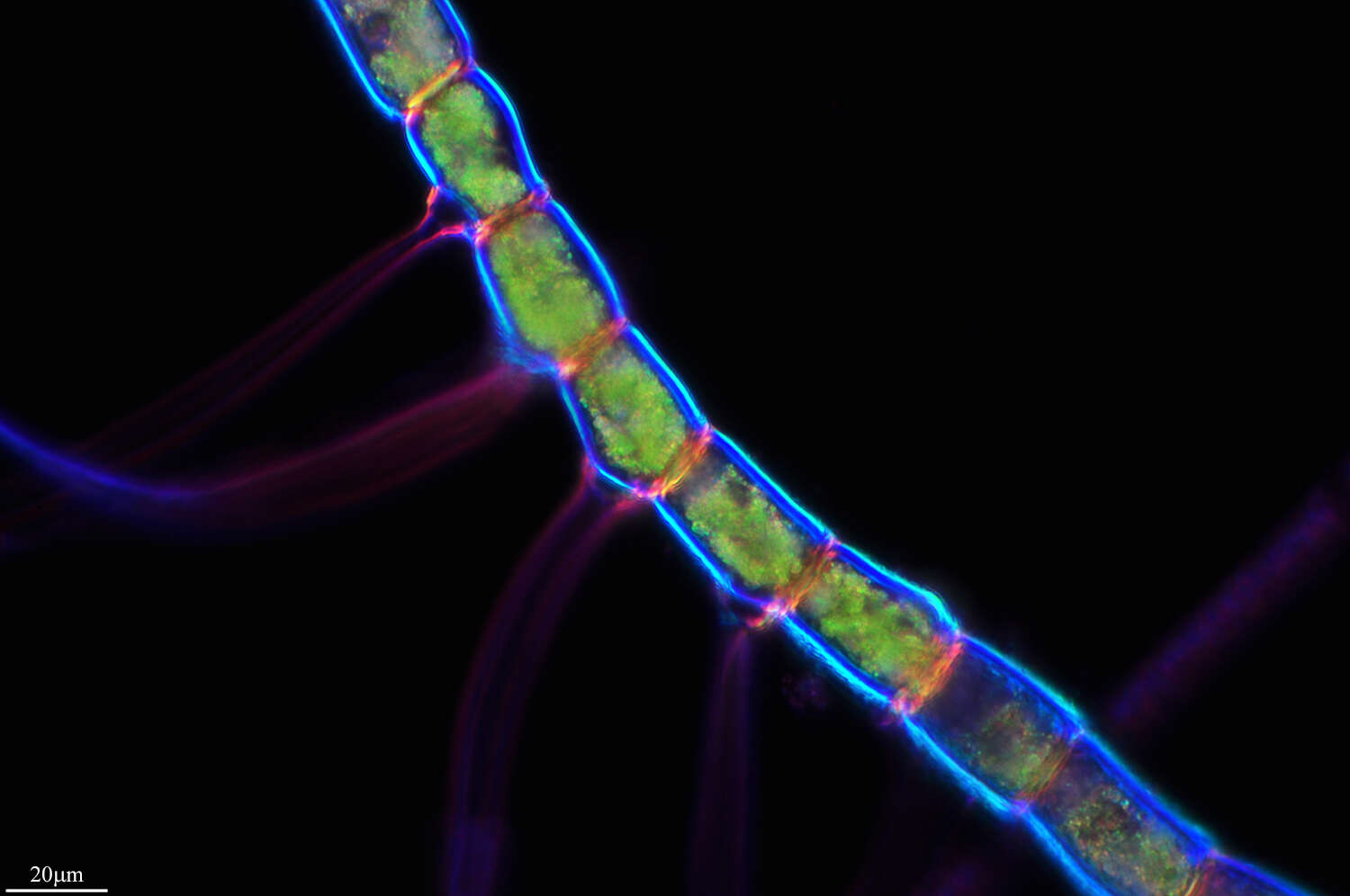
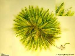
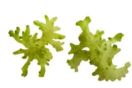
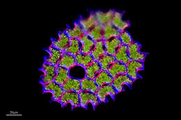
Chaetophora is a branched filamentous alga wholly embedded in mucilage. The species shown forms pads which can grow large enough to be visible to the naked eye as green, jelly globes adhered to stones or plants. The mucilage is a protection against ingestion. On the surface of this jelly globes one can see a lot of colorless cells formed as hairs (chaetae) projecting out of the jelly. This cells contain no chloroplasts and serve for nutrient uptake. The bright field photo doesn´t display the mucilage. This young specimen showed only two projecting cells (see inserted picture). The arrow idicates a pyrenoid within a chloroplast. Collected from Bodden, the brackish waters lying between the isles of Hiddensee and Ruegen (German Baltic Sea). This image was taken using Zeiss Universal with Olympus C7070 CCD camera.
-
Vanserum, Öland, Sverige
-
Villoslada de Cameros, La Rioja, Spain
-
Lumbreras, La Rioja, Spain
-
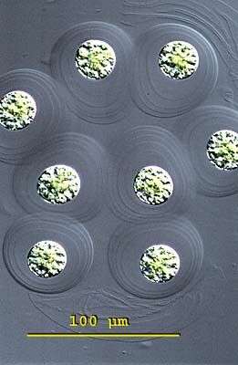
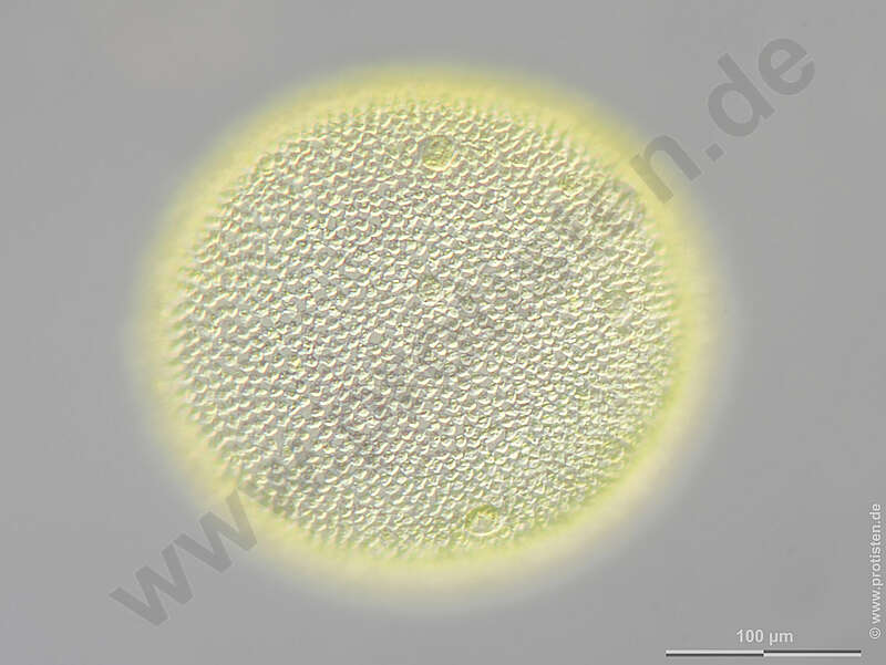
Scale bar indicates 100 m. Sample from the Domnental pond of Kronshagen near Kiel. The image was built up using several photomicrographic frames with manual stacking technique. Images were taken using Zeiss Axioplan with Olympus OM-D-E-M5 MKII.For permission to use of (high-resolution) images please contact postmaster@protisten.de.
-
Lardero, La Rioja, Spain
-
Boone, North Carolina, United States
-
Lumbreras, La Rioja, Spain
-
San Martin De Castaneda, Castille and Leon, Spain
-
Casas de Fadoncino, Castille and Leon, Spain
-
-
Camargo, Cantabria, Spain
-
Neila, Castille and Leon, Spain
-
Lumbreras, La Rioja, Espaa
-
Santa Coloma, La Rioja, Spain
-
Bradford West Gwillimbury, Ontario, Canada
-
Logroo, La Rioja, Espaa
-
Galende, Castille and Leon, Spain
-
Ribadelago de Franco, Castille and Leon, Spain
-
la Parrilla, Madrid, Spain
-
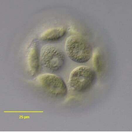
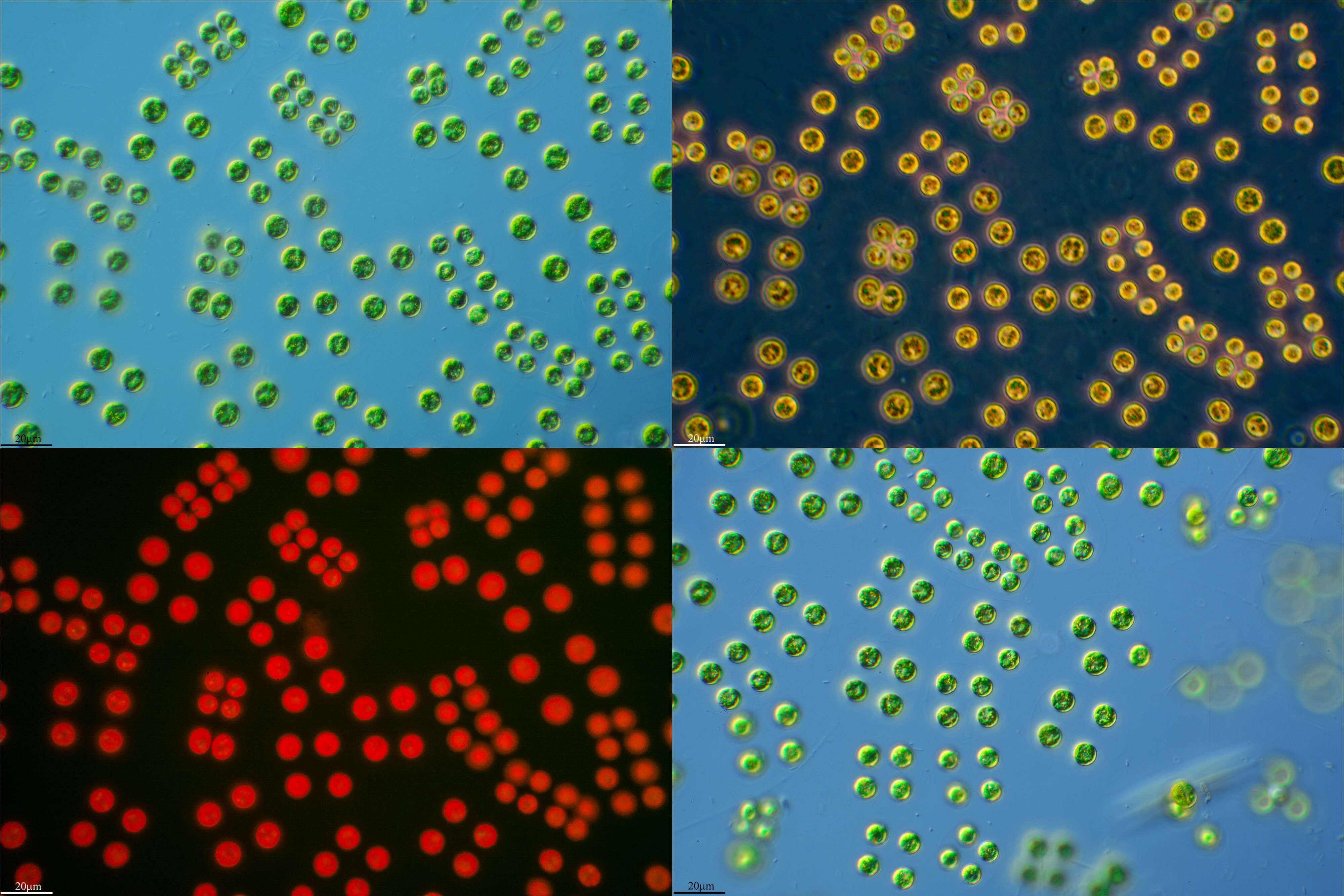
Close-up view. In the center of the cell we see the pyrenoid amidst the stellate chloroplast. The right cell shows, in addition, the nucleus and one of the two contractile vacuoles in maximum extension. The scale bar indicates 10 m. Sample from sphagnum pond Dosenmoor near Neumuenster (Schleswig-Holstein, Germany). Images were taken using Zeiss Axioplan with Canon 600D CCD camera.For permission to use of (high-resolution) images please contact postmaster@protisten.de.