-
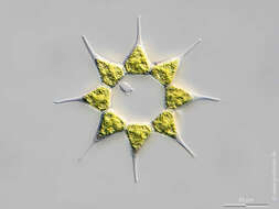
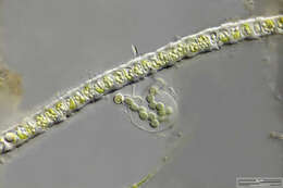
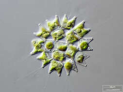
Monactinus simplex Scale bar indicates 25 µm. Sample from the Lake Vollstedter See near Kiel, Germany. Sampling date 8/2018. The image was built up using several photomicrographic frames with manual stacking technique. Images were taken using Zeiss Axioplan with Olympus OM-D M5 MKII. Image under Creative Commons License V 3.0 (CC BY-NC-SA). Place name: Lake Vollstedter See near Kiel (Germany) Latitude: 54.24105528 Longitude: 9.859339 Multiebenen-Abbildung, manuell gestapelt. Der Messbalken markiert eine Länge von 25 µm. Probe aus dem Vollstedter See bei Groß Vollstedt. Datum der Aufsammlung: 8/2018. Mikrotechnik: Zeiss Axioplan, Kamera: Olympus OM-D M5 MKII. Creative Commons License V 3.0 (CC BY-NC-SA). For permission to use of (high-resolution) images please contact postmaster@protisten.de.
-

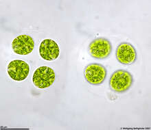
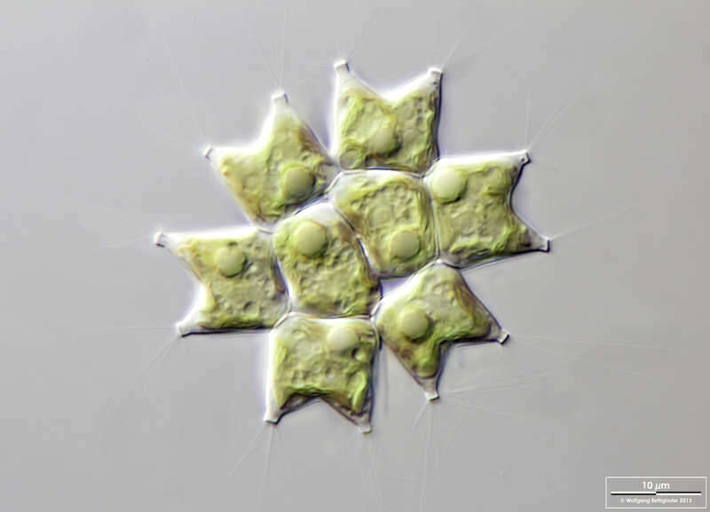
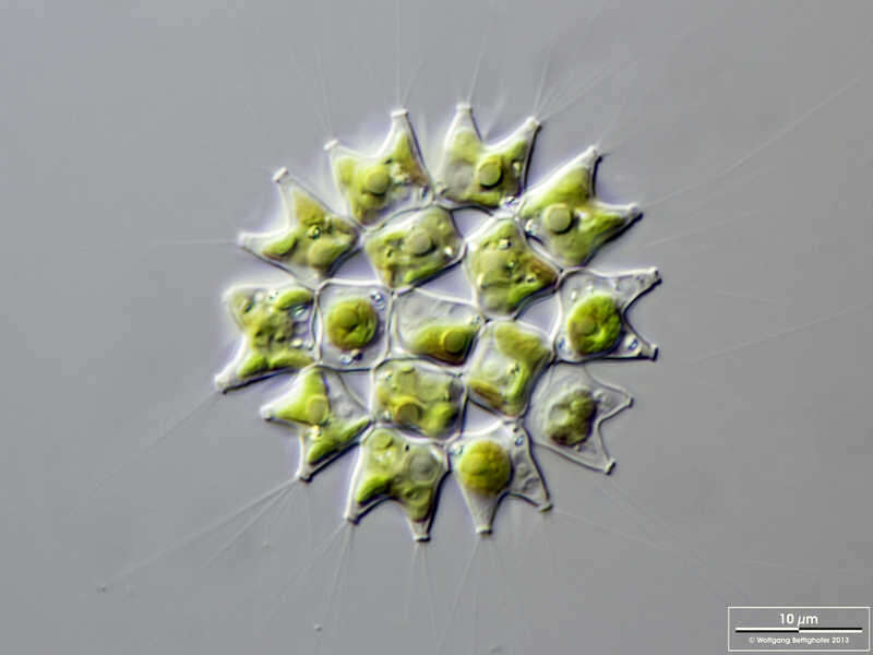
Nephrochlamys rostrata Cells usually solitary, cell grouped after binary fission by the old cell wall of the mother cell. Optical sections using DIC through the cell group:bottom layer showing nucleus of one cell, the old cell wall of the mother cell, and chloroplasts.Please press the MORE button for skipping to the annotated version.Scale bar indicates 10 µm. The specimen was gathered in a pond in the forest of Altenholz-Stift near Kiel. Sampling date 06/2022. The image was built up using several photomicrographic frames with manual stacking technique. Images were taken using Zeiss Axioskop with Olympus OM-D M5 MKII. Image under Creative Commons License V 3.0 (CC BY-NC-SA). Place name: Pond in the forest of Altenholz-Stift (Schleswig-Holstein, Germany) Latitude: 54.384913 Longitude: 10.125691 Zellen üblicherweise einzeln, die Zellgruppierung wurde nach zwei aufeinander folgenden Zellteilungen durch die alte Zellwand der Mutterzelle zusammengehalten. Optische Schnitte mittels DIC durch den Zellverband:untere Schicht mit Kern einer Zelle, der alten Zellwand der Mutterzelle und Chloroplasten.Bitte drücken Sie die Schaltfläche MORE, um zur kommentierten Version zu gelangen.Der Messbalken markiert eine Länge von 10 µm. Die Probe wurde in einem Waldteich bei Altenholz-Stift (nahe Kiel) gesammelt. Datum der Aufsammlung: 06/2022. Mikrotechnik: Zeiss Axioplan, Kamera: Olympus OM-D M5 MKII. Creative Commons License V 3.0 (CC BY-NC-SA). For permission to use of (high-resolution) images please contact postmaster@protisten.de.
-
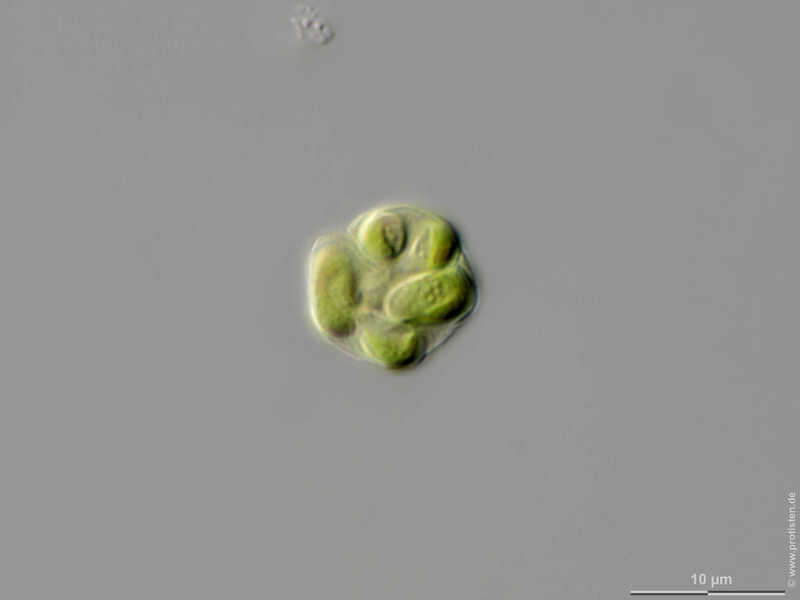
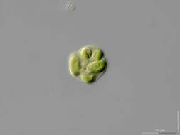
Nephrochlamys rostrata Cells usually solitary, cell grouped after binary fission by the old cell wall of the mother cell. Optical sections using DIC through the cell group:top view. Scale bar indicates 10 µm. The specimen was gathered in a pond in the forest of Altenholz-Stift near Kiel. Sampling date 06/2022. The image was built up using several photomicrographic frames with manual stacking technique. Images were taken using Zeiss Axioskop with Olympus OM-D M5 MKII. Image under Creative Commons License V 3.0 (CC BY-NC-SA). Place name: Pond in the forest of Altenholz-Stift (Schleswig-Holstein, Germany) Latitude: 54.384913 Longitude: 10.125691 Zellen üblicherweise einzeln, die Zellgruppierung wurde nach zwei aufeinander folgenden Zellteilungen durch die alte Zellwand der Mutterzelle zusammengehalten.Der Messbalken markiert eine Länge von 10 µm. Die Probe wurde in einem Waldteich bei Altenholz-Stift (nahe Kiel) gesammelt. Datum der Aufsammlung: 06/2022. Mikrotechnik: Zeiss Axioplan, Kamera: Olympus OM-D M5 MKII. Creative Commons License V 3.0 (CC BY-NC-SA). For permission to use of (high-resolution) images please contact postmaster@protisten.de.
-
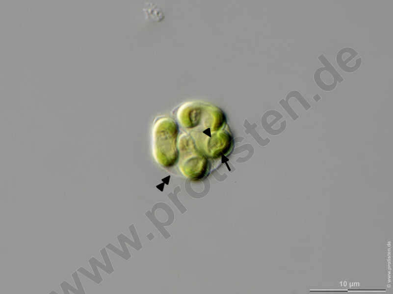
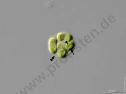
Nephrochlamys rostrata Cells usually solitary, cell grouped after binary fission by the old cell wall of the mother cell. Optical sections using DIC through the cell group:bottom layer showing nucleus of one cell (arrowhead), the old cell wall of the mother cell (double arrowhead), and chloroplasts (arrow). Scale bar indicates 10 µm. The specimen was gathered in a pond in the forest of Altenholz-Stift near Kiel. Sampling date 06/2022. The image was built up using several photomicrographic frames with manual stacking technique. Images were taken using Zeiss Axioskop with Olympus OM-D M5 MKII. Image under Creative Commons License V 3.0 (CC BY-NC-SA). Place name: Pond in the forest of Altenholz-Stift (Schleswig-Holstein, Germany) Latitude: 54.384913 Longitude: 10.125691 Zellen üblicherweise einzeln, die Zellgruppierung wurde nach zwei aufeinander folgenden Zellteilungen durch die alte Zellwand der Mutterzelle zusammengehalten. Optische Schnitte mittels DIC durch den Zellverband:untere Schicht mit Kern einer Zelle (Pfeilspitze), der alten Zellwand der Mutterzelle (Doppelpfeilspitze) und Chloroplasten (Pfeil).Der Messbalken markiert eine Länge von 10 µm. Die Probe wurde in einem Waldteich bei Altenholz-Stift (nahe Kiel) gesammelt. Datum der Aufsammlung: 06/2022. Mikrotechnik: Zeiss Axioplan, Kamera: Olympus OM-D M5 MKII. Creative Commons License V 3.0 (CC BY-NC-SA). For permission to use of (high-resolution) images please contact postmaster@protisten.de.
-

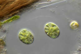
Nephrochlamys rostrata Cells usually solitary, cell grouped after binary fission by the old cell wall of the mother cell. Optical sections using DIC through the cell group:optical transversal cut. Scale bar indicates 10 µm. The specimen was gathered in a pond in the forest of Altenholz-Stift near Kiel. Sampling date 06/2022. The image was built up using several photomicrographic frames with manual stacking technique. Images were taken using Zeiss Axioskop with Olympus OM-D M5 MKII. Image under Creative Commons License V 3.0 (CC BY-NC-SA). Place name: Pond in the forest of Altenholz-Stift (Schleswig-Holstein, Germany) Latitude: 54.384913 Longitude: 10.125691 Zellen üblicherweise einzeln, die Zellgruppierung wurde nach zwei aufeinander folgenden Zellteilungen durch die alte Zellwand der Mutterzelle zusammengehalten. Optische Schnitte mittels DIC durch den Zellverband:optischer Querschnitt.Der Messbalken markiert eine Länge von 10 µm. Die Probe wurde in einem Waldteich bei Altenholz-Stift (nahe Kiel) gesammelt. Datum der Aufsammlung: 06/2022. Mikrotechnik: Zeiss Axioplan, Kamera: Olympus OM-D M5 MKII. Creative Commons License V 3.0 (CC BY-NC-SA). For permission to use of (high-resolution) images please contact postmaster@protisten.de.
-
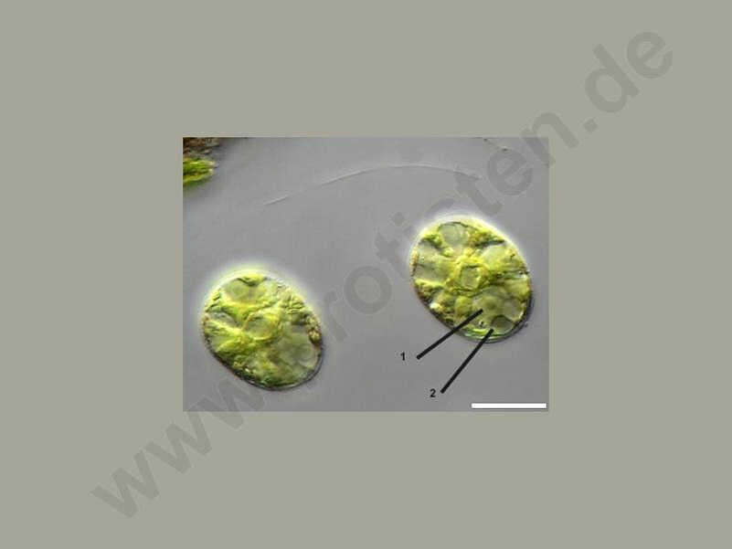
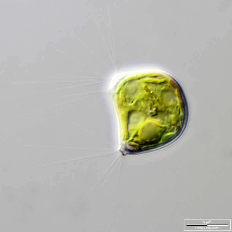
Asterococcus limneticus Close-up view. In the center of the cell we see a pyrenoid amidst the stellate chloroplast. In addition, the right cell shows the nucleus (1) and the two contractile vacuoles (2). The scale bar indicates 10 µm. Sample from sphagnum pond Dosenmoor near Neumuenster (Schleswig-Holstein, Germany). Images were taken using Zeiss Axioplan with Canon 600D CCD camera.Image under Creative Commons License V 3.0 (CC BY-NC-SA). Place name: Bog Dosenmoor near Neumuenster (Schleswig-Holstein, Germany) Latitude: 54.136219 Longitude: 10.026433 Nahaufnahme. In der Mitte der Zelle sehen wir den Pyrenoid inmitten des sternförmigen Chloroplasten. Darüber hinaus zeigt die rechte Zelle den Kern (1) und die beiden kontraktilen Vakuolen (2). Der Messbalken markiert eine Länge von 10 µm. Probe aus dem Dosenmoor in der Nähe von Neumünster. Mikrotechnik: Zeiss Axioplan, Kamera: Canon 600D. Creative Commons License V 3.0 (CC BY-NC-SA). For permission to use of (high-resolution) images please contact postmaster@protisten.de.
-
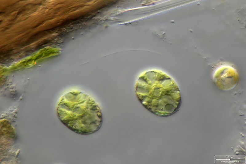
Asterococcus limneticus Close-up view. In the center of the cell we see the pyrenoid amidst the stellate chloroplast. The right cell shows, in addition, the nucleus and one of the two contractile vacuoles in maximum extension. The scale bar indicates 10 µm. Sample from sphagnum pond Dosenmoor near Neumuenster (Schleswig-Holstein, Germany). Images were taken using Zeiss Axioplan with Canon 600D CCD camera.Image under Creative Commons License V 3.0 (CC BY-NC-SA). Place name: Bog Dosenmoor near Neumuenster (Schleswig-Holstein, Germany) Latitude: 54.136219 Longitude: 10.026433 Nahaufnahme. In der Mitte der Zelle sehen wir den Pyrenoid inmitten des sternförmigen Chloroplasten. Die rechte Zelle zeigt zusätzlich den Zellkern und einer der beiden kontraktilen Vakuolen in maximaler Ausdehnung. Der Messbalken markiert eine Länge von 10 µm. Probe aus dem Dosenmoor in der Nähe von Neumünster. Mikrotechnik: Zeiss Axioplan, Kamera: Canon 600D. Creative Commons License V 3.0 (CC BY-NC-SA). For permission to use of (high-resolution) images please contact postmaster@protisten.de.
-
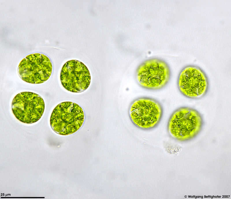
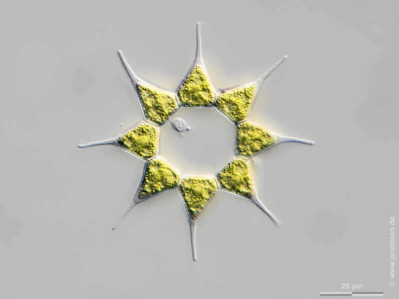
Asterococcus limneticus This alga is characterized by layers of mucilaginous envelopes around each cell and another gelatinous jacket covering the whole colony. The picture also shows contractile vacuoles. Each cell bears two of them.The scale bar indicates 25 µm. Sample from sphagnum pond situated in the northern alpine region of Austria near Salzburg. Images were taken using Zeiss Universal with Olympus C7070 CCD camera.Image under Creative Commons License V 3.0 (CC BY-NC-SA). Place name: Bogs near Salzburg (Austria) Latitude: 48.068516 Longitude: 12.954134 Charakteristisch für diese Algenart ist die Verpackung der Kolonie in mehrere Gallertschichten. Bei jeder Zellteilung werden die Teilungsprodukte in einen Gallertschicht eingepackt. Das Bild zeigt auch kontraktile Vakuolen. Jede Zelle besitzt zwei davon. Der Messbalken markiert eine Länge von 25 µm. Probe aus einem Moor in den nördlichen Kalkalpen von Österreich in der Nähe von Salzburg. Mikrotechnik: Zeiss Universal, Kamera: Olympus C7070. Creative Commons License V 3.0 (CC BY-NC-SA). For permission to use of (high-resolution) images please contact postmaster@protisten.de.
-
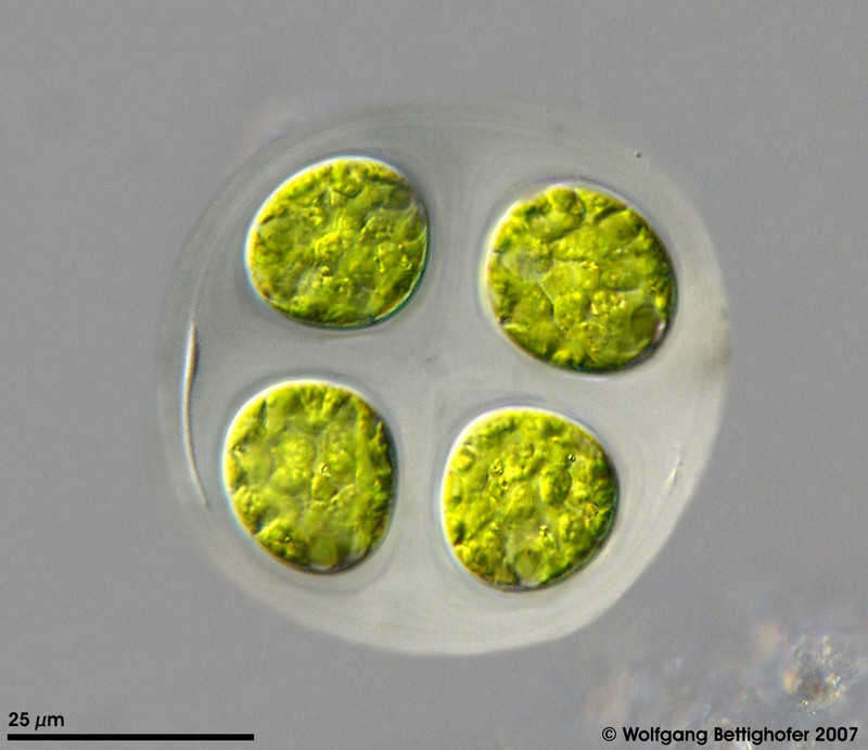
Asterococcus limneticus This alga is characterized by layers of mucilaginous envelopes around each cell and another gelatinous jacket covering the whole colony. The picture also shows contractile vacuoles. Each cell bears two of them. Sample from sphagnum pond situated in the northern alpine region of Austria near Salzburg. Images were taken using Zeiss Universal with Olympus C7070 CCD camera.Image under Creative Commons License V 3.0 (CC BY-NC-SA). Place name: Bogs near Salzburg (Austria) Latitude: 48.068516 Longitude: 12.954134 Charakteristisch für diese Algenart ist die Verpackung der Kolonie in mehrere Gallertschichten. Bei jeder Zellteilung werden die Teilungsprodukte in einen Gallertschicht eingepackt. Das Bild zeigt auch kontraktile Vakuolen. Jede Zelle besitzt zwei davon. Probe aus einem Moor in den nördlichen Kalkalpen von Österreich in der Nähe von Salzburg. Mikrotechnik: Zeiss Universal, Kamera: Olympus C7070. Creative Commons License V 3.0 (CC BY-NC-SA). For permission to use of (high-resolution) images please contact postmaster@protisten.de.
-
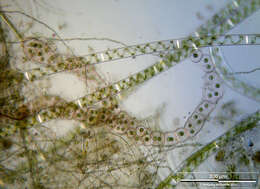
Palmodictyon varium Scale bar indicates 100 µm.Sample from the pond Hegne Moor situated in the vicinity of Lake Constance. The image was built up using several photomicrographic frames with manual stacking technique. Images were taken using Zeiss Universal with Olympus C7070 CCD camera.Image under Creative Commons License V 3.0 (CC BY-NC-SA). Place name: Bog Hegne Moor near Lake Constance (Germany) Latitude: 47.718106 Longitude: 9.093974 Multiebenen-Abbildung, manuell gestapelt. Der Messbalken markiert eine Länge von 100 µm. Probe aus dem Simmelried nahe Konstanz. Mikrotechnik: Zeiss Universal, Kamera: Olympus C7070. Creative Commons License V 3.0 (CC BY-NC-SA). For permission to use of (high-resolution) images please contact postmaster@protisten.de.
-
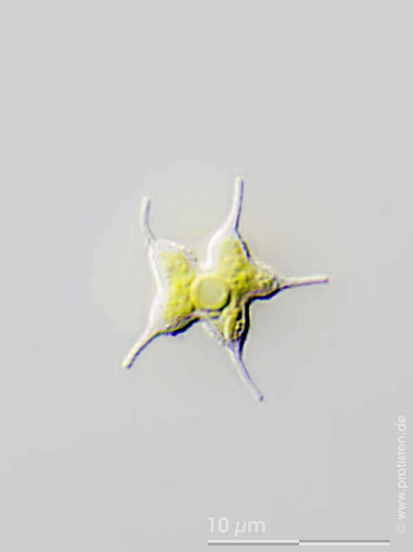
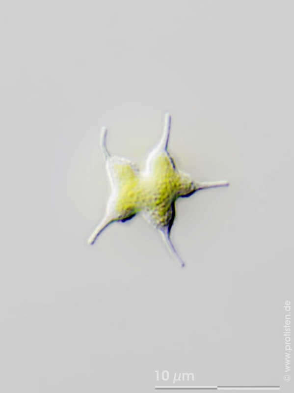
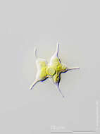
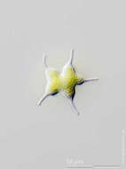
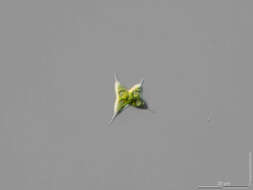
Tetraedron caudatum Scale bar indicates 5 µm.Sample from a pond in Lemkendorf on the isle Fehmarn (Baltic Sea). The image was built up using several photomicrographic frames with manual stacking technique. Images were taken using Zeiss Axioplan with Canon EOS 70D.Image under Creative Commons License V 3.0 (CC BY-NC-SA). Place name: Pond in Lemkendorf, isle Fehmarn (Baltic Sea, Germany) Latitude: 54.472296 Longitude: 11.095662 Multiebenen-Abbildung, manuell gestapelt. Der Messbalken markiert eine Länge von 5 µm. Probe aus einem Teich in Lemkendorf, Fehmarn. Mikrotechnik: Zeiss Axioplan, Kamera: Canon EOS 70D. Creative Commons License V 3.0 (CC BY-NC-SA). For permission to use of (high-resolution) images please contact postmaster@protisten.de.
-
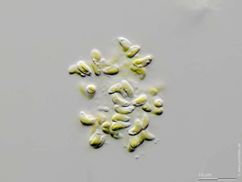
Raphidocelis arcuata Synonym: Kirchneriella arcuata.Scale bar indicates 5 µm. Sample from a pond in Lemkendorf on the isle Fehmarn (Baltic Sea). The image was built up using several photomicrographic frames with manual stacking technique. Images were taken using Zeiss Axioplan with Canon EOS 70D.Image under Creative Commons License V 3.0 (CC BY-NC-SA). Place name: Pond in Lemkendorf, isle Fehmarn (Baltic Sea, Germany) Latitude: 54.472296 Longitude: 11.095662 Synonym: Kirchneriella arcuata.Multiebenen-Abbildung, manuell gestapelt. Der Messbalken markiert eine Länge von 5 µm. Probe aus einem Teich in Lemkendorf, Fehmarn. Mikrotechnik: Zeiss Axioplan, Kamera: Canon EOS 70D. Creative Commons License V 3.0 (CC BY-NC-SA). For permission to use of (high-resolution) images please contact postmaster@protisten.de.
-
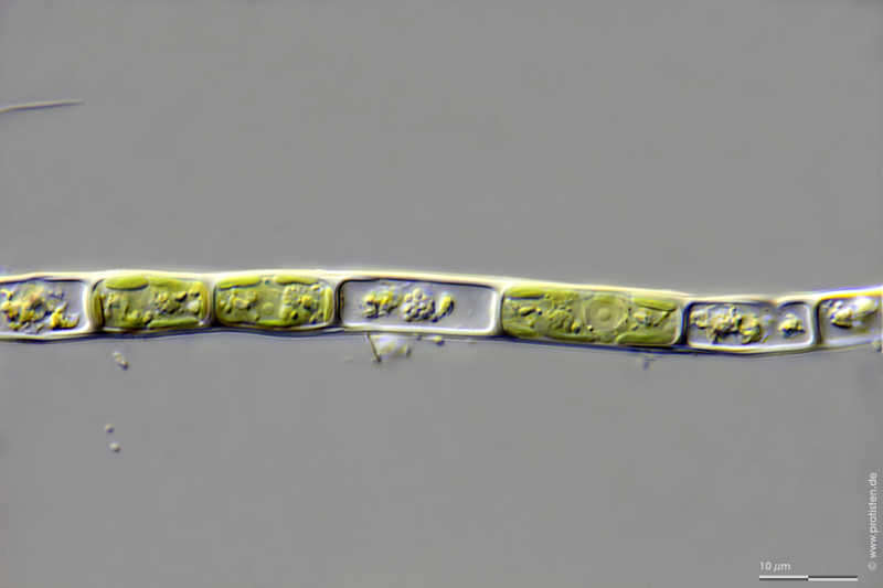
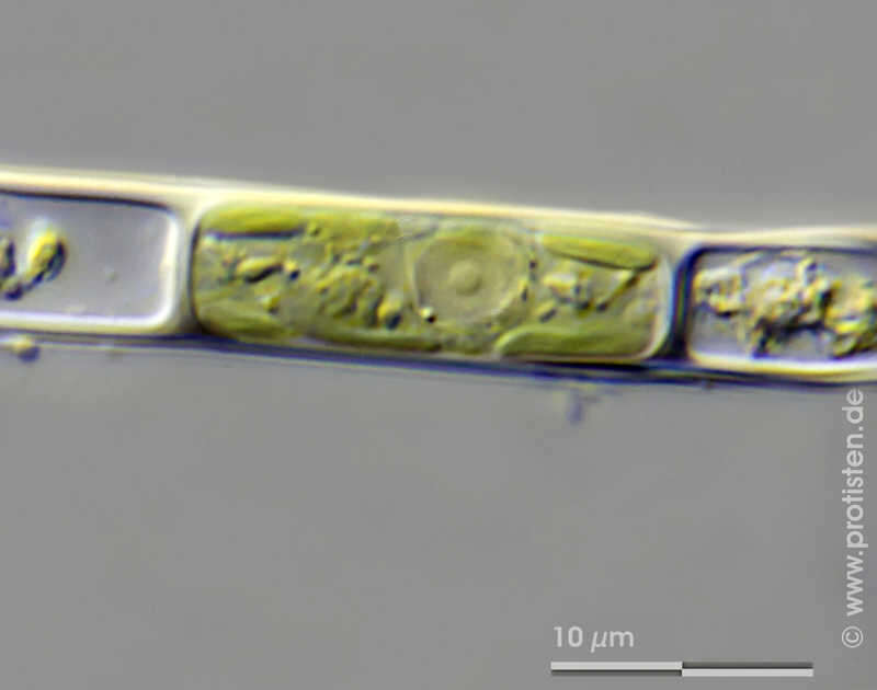
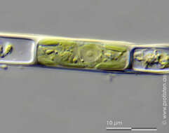
Microspora abbreviata Scale bar indicates 10 µm.Sample from the Pillersee region / Tyrol, pond at Mittermoos. The image was built up using several photomicrographic frames with manual stacking technique. Images were taken using Zeiss Universal with Olympus C7070 CCD camera.Image under Creative Commons License V 3.0 (CC BY-NC-SA). Place name: Wetland Mittermoos near Fieberbrunn (Tyrol, Austria) Latitude: 47.47998695 Longitude: 12.5240922 Multiebenen-Abbildung, manuell gestapelt. Der Messbalken markiert eine Länge von 10 µm. Probe vom Mittermoos im Gebiet Pillersee / Tirol. Mikrotechnik: Zeiss Universal, Kamera: Olympus C7070. Creative Commons License V 3.0 (CC BY-NC-SA). For permission to use of (high-resolution) images please contact postmaster@protisten.de.
-
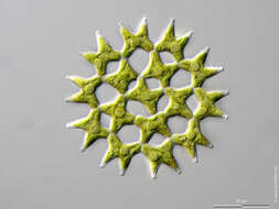
Chlorotetraedron incus Scale bar indicates 25 µm. The specimen was gathered in a pond in the forest of Altenholz-Stift near Kiel. Sampling date 06/2022. The image was built up using several photomicrographic frames with manual stacking technique. Images were taken using Zeiss Axioskop with Olympus OM-D M5 MKII. Image under Creative Commons License V 3.0 (CC BY-NC-SA). Place name: Pond in the forest of Altenholz-Stift (Schleswig-Holstein, Germany) Latitude: 54.384913 Longitude: 10.125691 Der Messbalken markiert eine Länge von 25 µm. Die Probe wurde in einem Waldteich bei Altenholz-Stift (nahe Kiel) gesammelt. Datum der Aufsammlung: 06/2022. Mikrotechnik: Zeiss Axioplan, Kamera: Olympus OM-D M5 MKII. Creative Commons License V 3.0 (CC BY-NC-SA). For permission to use of (high-resolution) images please contact postmaster@protisten.de.
-
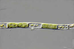
Microspora irregularis Scale bar indicates 25 µm.Sample from the Pillersee region / Tyrol, pond at Mittermoos. The image was built up using several photomicrographic frames with manual stacking technique. Images were taken using Zeiss Universal with Olympus C7070 CCD camera.Image under Creative Commons License V 3.0 (CC BY-NC-SA). Place name: Wetland Mittermoos near Fieberbrunn (Tyrol, Austria) Latitude: 47.47998695 Longitude: 12.5240922 Multiebenen-Abbildung, manuell gestapelt. Der Messbalken markiert eine Länge von 25 µm. Probe vom Mittermoos im Gebiet Pillersee / Tirol. Mikrotechnik: Zeiss Universal, Kamera: Olympus C7070. Creative Commons License V 3.0 (CC BY-NC-SA). For permission to use of (high-resolution) images please contact postmaster@protisten.de.
-
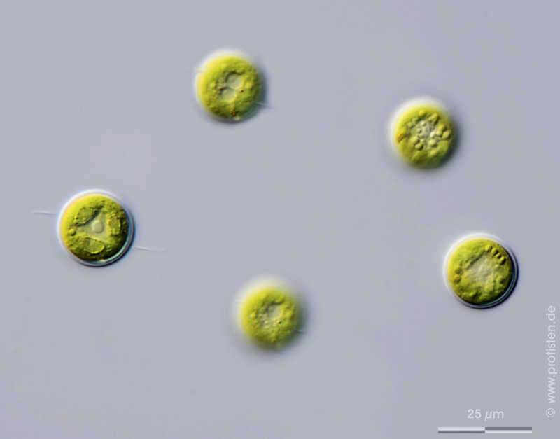
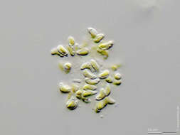
Chlamydomonas sphagnicola The scale bar indicates 25 µm. Sample from sphagnum pond Dosenmoor near Neumuenster (Schleswig-Holstein, Germany). Images were taken using Zeiss Axioplan with Olympus OM-D M5 MKII.Image under Creative Commons License V 3.0 (CC BY-NC-SA). Place name: Bog Dosenmoor near Neumuenster (Schleswig-Holstein, Germany) Latitude: 54.136219 Longitude: 10.026433 Der Messbalken markiert eine Länge von 25 µm. Probe aus dem Dosenmoor in der Nähe von Neumünster. Mikrotechnik: Zeiss Axioplan, Kamera: OM-D M5 MKII. Creative Commons License V 3.0 (CC BY-NC-SA). For permission to use of (high-resolution) images please contact postmaster@protisten.de.
-
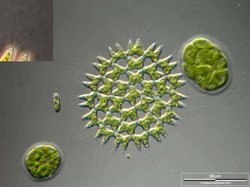
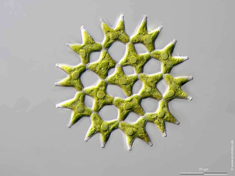
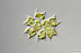
Pediastrum duplex Scale bar indicates 50 µm. The inset at upper left shows fine cellulose spines which support flotation. The specimen was gathered in the wetlands of Nationalpark Unteres Odertal (100 km north east of Berlin). The image was built up using several photomicrographic frames with manual stacking technique. Images were taken using Zeiss Universal with Olympus C7070 CCD camera. Image under Creative Commons License V 3.0 (CC BY-NC-SA). Place name: Creek in Oder valley 100 km north east of Berlin (Germany) Latitude: 53.135032 Longitude: 14.348738 Multiebenen-Abbildung, manuell gestapelt. Der Messbalken markiert eine Länge von 50 µm. Der Ausschnitt oben rechts zeigt feine Schwebefortsätze aus Zellulose.Die Probe wurde in den Feuchtgebieten des Nationalpark Unteres Odertal (100 km nordöstlich von Berlin) gesammelt. Mikrotechnik: Zeiss Universal, Kamera: Olympus C7070. Creative Commons License V 3.0 (CC BY-NC-SA). For permission to use of (high-resolution) images please contact postmaster@protisten.de.
-
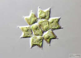
Pediastrum duplex Scale bar indicates 25 µm. The specimen was gathered in a pond in the forest of Altenholz-Stift near Kiel (Schleswig-Holstein, Germany). Sampling date 1/2018. The image was built up using several photomicrographic frames with manual stacking technique. Images were taken using Zeiss Axioskop with Olympus OM-D M5 MKII. Image under Creative Commons License V 3.0 (CC BY-NC-SA). Place name: Pond in the forest of Altenholz-Stift (Schleswig-Holstein, Germany) Latitude: 54.384913 Longitude: 10.125691 Der Messbalken markiert eine Länge von 25 µm. Die Probe wurde in einem Waldteich bei Altenholz-Stift (nahe Kiel) im Januar 2018 gesammelt. Mikrotechnik: Zeiss Axioplan, Kamera: Olympus OM-D M5 MKII. Creative Commons License V 3.0 (CC BY-NC-SA). For permission to use of (high-resolution) images please contact postmaster@protisten.de.
-

Pediastrum duplex The fine cellulose spins which supports floating are visible. Scale bar indicates 5 µm. Specimen from the Algal Culture Collection at the University of Cologne (CCAC), strain number M1253/1. Link: https://www.ccac.uni-koeln.de/. Sample material courtesy of AG Boenigk, University Duisburg-Essen. The image was built up using several photomicrographic frames with manual stacking technique. Images were taken using Zeiss Axioplan with DSLR Canon 600 D. Image under Creative Commons License V 3.0 (CC BY-NC-SA). Place name: Protist culture CCAC University Duisburg-Essen (Germany) Latitude: 51.463807 Longitude: 7.005321 Die feinen Schwebefortsätze aus Zellulose sind sichtbar. Multiebenen-Abbildung, manuell gestapelt. Der Messbalken markiert 5 µm. Exemplar aus der Algenkultursammlung der Universität zu Köln (CCAC), Stammnummer M1253/1. Link: https://www.ccac.uni-koeln.de/. Das Material wurde freundlicherweise von der AG Boenigk, Universität Duisburg-Essen, zur Verfügung gestellt. Mikrotechnik: Zeiss Axioplan, Kamera: Canon 600D. Creative Commons License V 3.0 (CC BY-NC-SA). For permission to use of (high-resolution) images please contact postmaster@protisten.de.
-
Pediastrum duplex The fine cellulose spins which supports floating are visible. Scale bar indicates 10 µm. Specimen from the Algal Culture Collection at the University of Cologne (CCAC), strain number M1253/1. Link: https://www.ccac.uni-koeln.de/. Sample material courtesy of AG Boenigk, University Duisburg-Essen. The image was built up using several photomicrographic frames with manual stacking technique. Images were taken using Zeiss Axioplan with DSLR Canon 600 D. Image under Creative Commons License V 3.0 (CC BY-NC-SA). Place name: Protist culture CCAC University Duisburg-Essen (Germany) Latitude: 51.463807 Longitude: 7.005321 Die feinen Schwebefortsätze aus Zellulose sind sichtbar. Der Messbalken markiert 10 µm. Multiebenen-Abbildung, manuell gestapelt. Exemplar aus der Algenkultursammlung der Universität zu Köln (CCAC), Stammnummer M1253/1. Link: https://www.ccac.uni-koeln.de/. Das Material wurde freundlicherweise von der AG Boenigk, Universität Duisburg-Essen, zur Verfügung gestellt. Mikrotechnik: Zeiss Axioplan, Kamera: Canon 600D. Creative Commons License V 3.0 (CC BY-NC-SA). For permission to use of (high-resolution) images please contact postmaster@protisten.de.
-
Pediastrum duplex The fine cellulose spins which supports floating are visible. Scale bar indicates 5 µm. Specimen from the Algal Culture Collection at the University of Cologne (CCAC), strain number M1253/1. Link: https://www.ccac.uni-koeln.de/. Sample material courtesy of AG Boenigk, University Duisburg-Essen. The image was built up using several photomicrographic frames with manual stacking technique. Images were taken using Zeiss Axioplan with DSLR Canon 600 D. Image under Creative Commons License V 3.0 (CC BY-NC-SA). Place name: Protist culture CCAC University Duisburg-Essen (Germany) Latitude: 51.463807 Longitude: 7.005321 Die feinen Schwebefortsätze aus Zellulose sind gut sichtbar. Multiebenen-Abbildung, manuell gestapelt. Der Messbalken markiert 5 µm. Exemplar aus der Algenkultursammlung der Universität zu Köln (CCAC), Stammnummer M1253/1. Link: https://www.ccac.uni-koeln.de/. Das Material wurde freundlicherweise von der AG Boenigk, Universität Duisburg-Essen, zur Verfügung gestellt. Mikrotechnik: Zeiss Axioplan, Kamera: Canon 600D. Creative Commons License V 3.0 (CC BY-NC-SA). For permission to use of (high-resolution) images please contact postmaster@protisten.de.
-
Pediastrum duplex The fine cellulose spins which supports floating are visible. Scale bar indicates 10 µm. Specimen from the Algal Culture Collection at the University of Cologne (CCAC), strain number M1253/1. Link: https://www.ccac.uni-koeln.de/. Sample material courtesy of AG Boenigk, University Duisburg-Essen. The image was built up using several photomicrographic frames with manual stacking technique. Images were taken using Zeiss Axioplan with DSLR Canon 600 D. Image under Creative Commons License V 3.0 (CC BY-NC-SA). Place name: Protist culture CCAC University Duisburg-Essen (Germany) Latitude: 51.463807 Longitude: 7.005321 Die feinen Schwebefortsätze aus Zellulose sind gut sichtbar. Multiebenen-Abbildung, manuell gestapelt. Messbalken markiert 10 µm. Exemplar aus der Algenkultursammlung der Universität zu Köln (CCAC), Stammnummer M1253/1. Link: https://www.ccac.uni-koeln.de/. Das Material wurde freundlicherweise von der AG Boenigk, Universität Duisburg-Essen, zur Verfügung gestellt.Mikrotechnik: Zeiss Axioplan, Kamera: Canon 600D. Creative Commons License V 3.0 (CC BY-NC-SA). For permission to use of (high-resolution) images please contact postmaster@protisten.de.