-

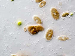
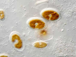
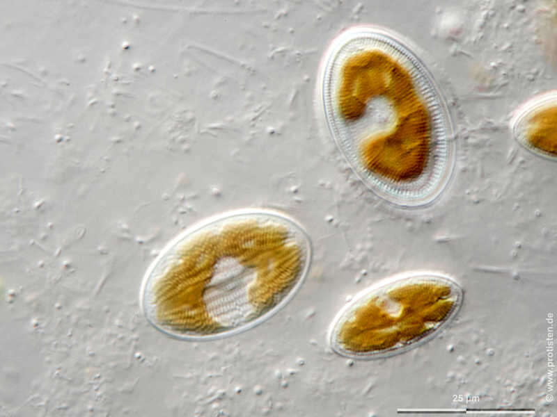
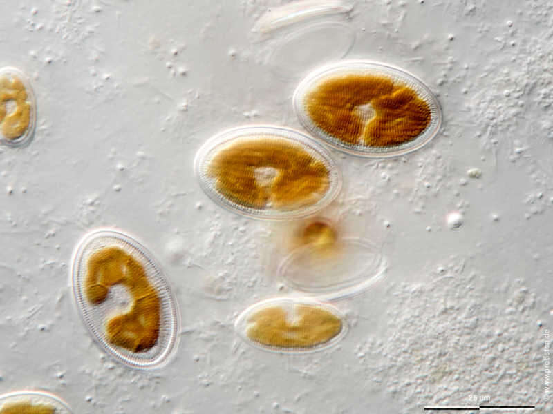
Cocconeis placentula var. euglypta Sexual reproduction of diatoms generating auxospores. Tomographical cross-sections through an auxospore from top to bottom. Chl = chloroplast, N = nucleus, UV = upper valve, LV = lower valve. Scale bar indicates 25 µm. Sample from a tropical freshwater aquarium. Sampling date 3/2021. The image was built up using several photomicrographic frames with manual stacking technique. Images were taken using Zeiss Axioplan with Olympus OM-D M5 MKII. Image under Creative Commons License V 3.0 (CC BY-NC-SA). Place name: Tropical freshwater aquarium Latitude: 54.3018013 Longitude: 10.07120132 Auxosporenbildung, sexuelle Vermehrungsweise von Diatomeen. Schnittbilder durch die Auxospore von oben nach unten. Chl = Chloroplast, N = Kern, UV = obere Halbschale, LV = untere Halbschale. Multiebenen-Abbildung, manuell gestapelt. Der Messbalken markiert eine Länge von 25 µm. Probe aus einem Süßwasseraquarium. Datum der Aufsammlung: 3/2021. Mikrotechnik: Zeiss Axioplan, Kamera: Olympus OM-D M5 MKII. Creative Commons License V 3.0 (CC BY-NC-SA). For permission to use of (high-resolution) images please contact postmaster@protisten.de.
-
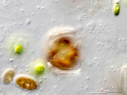
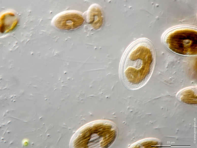
Cocconeis placentula var. euglypta Sexual reproduction of diatoms generating auxospores. Developing state. Tomographical cross-sections. UV = upper valve, LV = lower valve.Scale bar indicates 25 µm. Sample from a tropical freshwater aquarium. Sampling date 3/2021. The image was built up using several photomicrographic frames with manual stacking technique. Images were taken using Zeiss Axioplan with Olympus OM-D M5 MKII. Image under Creative Commons License V 3.0 (CC BY-NC-SA). Place name: Tropical freshwater aquarium Latitude: 54.3018013 Longitude: 10.07120132 Frühes Stadium der Auxosporenbildung, der sexuellen Vermehrungsweise von Diatomeen. Zwei Schnittbilder. UV = obere Halbschale, LV = untere Halbschale. Multiebenen-Abbildung, manuell gestapelt. Der Messbalken markiert eine Länge von 25 µm. Probe aus einem Süßwasseraquarium. Datum der Aufsammlung: 3/2021. Mikrotechnik: Zeiss Axioplan, Kamera: Olympus OM-D M5 MKII. Creative Commons License V 3.0 (CC BY-NC-SA). For permission to use of (high-resolution) images please contact postmaster@protisten.de.
-
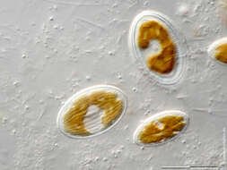
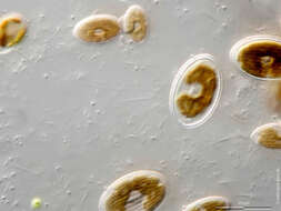
Cocconeis placentula var. euglypta Upper valve without raphe.Scale bar indicates 25 µm. Sample from a tropical freshwater aquarium. Sampling date 3/2021. The image was built up using several photomicrographic frames with manual stacking technique. Images were taken using Zeiss Axioplan with Olympus OM-D M5 MKII. Image under Creative Commons License V 3.0 (CC BY-NC-SA). Place name: Tropical freshwater aquarium Latitude: 54.3018013 Longitude: 10.07120132 Die obere Halbschale ist raphenlos. Multiebenen-Abbildung, manuell gestapelt. Der Messbalken markiert eine Länge von 25 µm. Probe aus einem Süßwasseraquarium. Datum der Aufsammlung: 3/2021. Mikrotechnik: Zeiss Axioplan, Kamera: Olympus OM-D M5 MKII. Creative Commons License V 3.0 (CC BY-NC-SA). For permission to use of (high-resolution) images please contact postmaster@protisten.de.
-
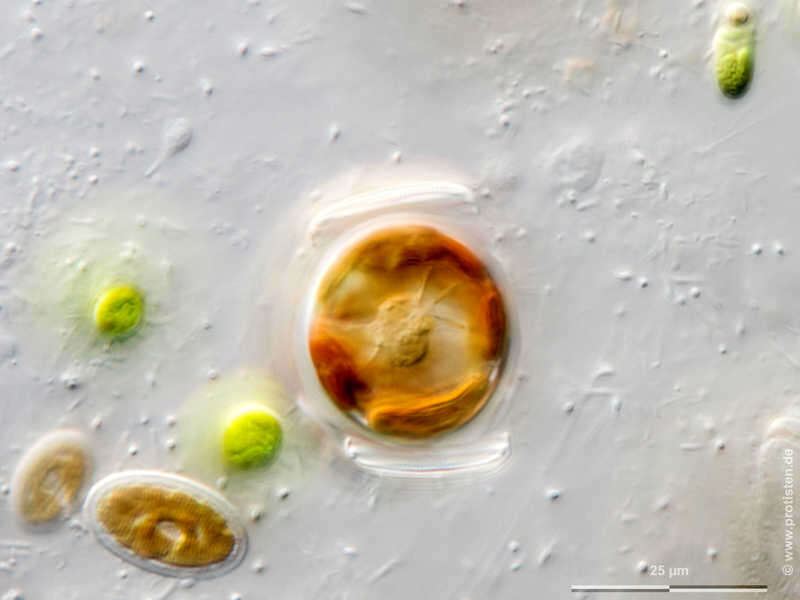
Cocconeis placentula var. euglypta Sexual reproduction of diatoms generating auxospores. Tomographical cross-sections through an auxospore from top to bottom. Chl = chloroplast, N = nucleus, UV = upper valve, LV = lower valve. Scale bar indicates 25 µm. Sample from a tropical freshwater aquarium. Sampling date 3/2021. The image was built up using several photomicrographic frames with manual stacking technique. Images were taken using Zeiss Axioplan with Olympus OM-D M5 MKII. Image under Creative Commons License V 3.0 (CC BY-NC-SA). Place name: Tropical freshwater aquarium Latitude: 54.3018013 Longitude: 10.07120132 Auxosporenbildung, sexuelle Vermehrungsweise von Diatomeen. Schnittbilder durch die Auxospore von oben nach unten. Chl = Chloroplast, N = Kern, UV = obere Halbschale, LV = untere Halbschale. Multiebenen-Abbildung, manuell gestapelt. Der Messbalken markiert eine Länge von 25 µm. Probe aus einem Süßwasseraquarium. Datum der Aufsammlung: 3/2021. Mikrotechnik: Zeiss Axioplan, Kamera: Olympus OM-D M5 MKII. Creative Commons License V 3.0 (CC BY-NC-SA). For permission to use of (high-resolution) images please contact postmaster@protisten.de.
-
Cocconeis placentula var. euglypta Lower valve with raphe which enables slow movement.Scale bar indicates 25 µm. Sample from a tropical freshwater aquarium. Sampling date 3/2021. The image was built up using several photomicrographic frames with manual stacking technique. Images were taken using Zeiss Axioplan with Olympus OM-D M5 MKII. Image under Creative Commons License V 3.0 (CC BY-NC-SA). Place name: Tropical freshwater aquarium Latitude: 54.3018013 Longitude: 10.07120132 Die untere Halbschale mit Raphe ermöglicht langesame Fortbewegung. Multiebenen-Abbildung, manuell gestapelt. Der Messbalken markiert eine Länge von 25 µm. Probe aus einem Süßwasseraquarium. Datum der Aufsammlung: 3/2021. Mikrotechnik: Zeiss Axioplan, Kamera: Olympus OM-D M5 MKII. Creative Commons License V 3.0 (CC BY-NC-SA). For permission to use of (high-resolution) images please contact postmaster@protisten.de.
-
Cocconeis placentula var. euglypta Sexual reproduction of diatoms generating auxospores. Developing state. Tomographical cross-sections. UV = upper valve, LV = lower valve.Scale bar indicates 25 µm. Sample from a tropical freshwater aquarium. Sampling date 3/2021. The image was built up using several photomicrographic frames with manual stacking technique. Images were taken using Zeiss Axioplan with Olympus OM-D M5 MKII. Image under Creative Commons License V 3.0 (CC BY-NC-SA). Place name: Tropical freshwater aquarium Latitude: 54.3018013 Longitude: 10.07120132 Frühes Stadium der Auxosporenbildung, der sexuellen Vermehrungsweise von Diatomeen. Zwei Schnittbilder. UV = obere Halbschale, LV = untere Halbschale. Multiebenen-Abbildung, manuell gestapelt. Der Messbalken markiert eine Länge von 25 µm. Probe aus einem Süßwasseraquarium. Datum der Aufsammlung: 3/2021. Mikrotechnik: Zeiss Axioplan, Kamera: Olympus OM-D M5 MKII. Creative Commons License V 3.0 (CC BY-NC-SA). For permission to use of (high-resolution) images please contact postmaster@protisten.de.
-

Cocconeis placentula var. euglypta Sexual reproduction of diatoms generating auxospores. Tomographical cross-sections through an auxospore from top to bottom. Chl = chloroplast, N = nucleus, UV = upper valve, LV = lower valve. Scale bar indicates 25 µm. Sample from a tropical freshwater aquarium. Sampling date 3/2021. The image was built up using several photomicrographic frames with manual stacking technique. Images were taken using Zeiss Axioplan with Olympus OM-D M5 MKII. Image under Creative Commons License V 3.0 (CC BY-NC-SA). Place name: Tropical freshwater aquarium Latitude: 54.3018013 Longitude: 10.07120132 Auxosporenbildung, sexuelle Vermehrungsweise von Diatomeen. Schnittbilder durch die Auxospore von oben nach unten. Chl = Chloroplast, N = Kern, UV = obere Halbschale, LV = untere Halbschale. Multiebenen-Abbildung, manuell gestapelt. Der Messbalken markiert eine Länge von 25 µm. Probe aus einem Süßwasseraquarium. Datum der Aufsammlung: 3/2021. Mikrotechnik: Zeiss Axioplan, Kamera: Olympus OM-D M5 MKII. Creative Commons License V 3.0 (CC BY-NC-SA). For permission to use of (high-resolution) images please contact postmaster@protisten.de.
-
Cocconeis placentula var. euglypta Lower valve with raphe which enables slow movement.Scale bar indicates 25 µm. Sample from a tropical freshwater aquarium. Sampling date 3/2021. The image was built up using several photomicrographic frames with manual stacking technique. Images were taken using Zeiss Axioplan with Olympus OM-D M5 MKII. Image under Creative Commons License V 3.0 (CC BY-NC-SA). Place name: Tropical freshwater aquarium Latitude: 54.3018013 Longitude: 10.07120132 Die untere Halbschale mit Raphe ermöglicht langesame Fortbewegung. Multiebenen-Abbildung, manuell gestapelt. Der Messbalken markiert eine Länge von 25 µm. Probe aus einem Süßwasseraquarium. Datum der Aufsammlung: 3/2021. Mikrotechnik: Zeiss Axioplan, Kamera: Olympus OM-D M5 MKII. Creative Commons License V 3.0 (CC BY-NC-SA). For permission to use of (high-resolution) images please contact postmaster@protisten.de.
-

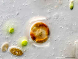
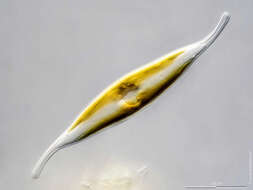
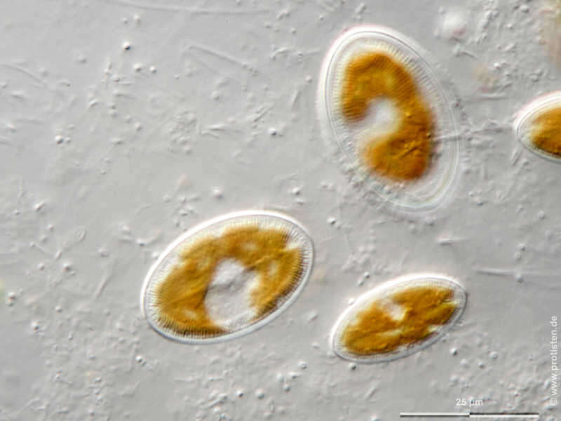
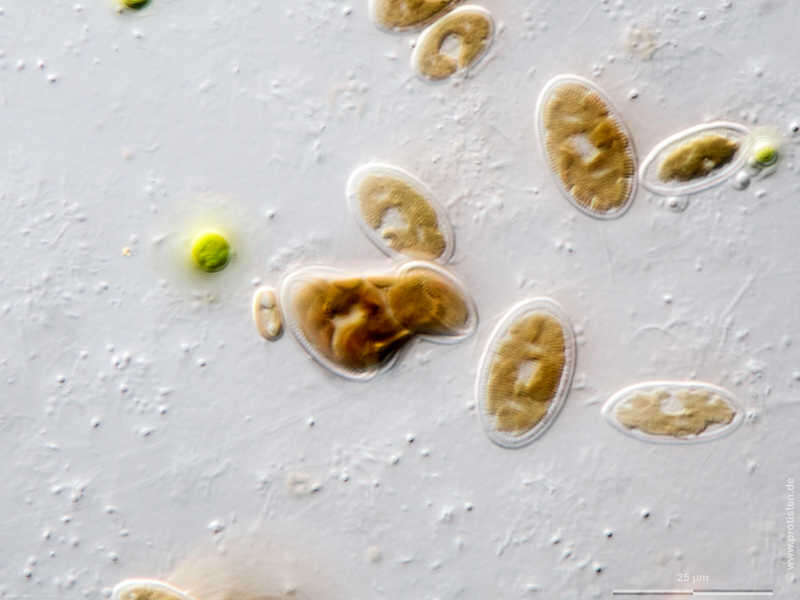
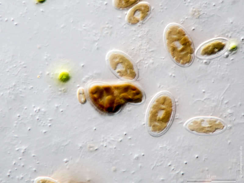
Cocconeis placentula var. euglypta Sexual reproduction of diatoms generating auxospores, first phase (arrow). UV = upper valve, LV = lower valve.Scale bar indicates 25 µm. Sample from a tropical freshwater aquarium. Sampling date 3/2021. The image was built up using several photomicrographic frames with manual stacking technique. Images were taken using Zeiss Axioplan with Olympus OM-D M5 MKII. Image under Creative Commons License V 3.0 (CC BY-NC-SA). Place name: Tropical freshwater aquarium Latitude: 54.3018013 Longitude: 10.07120132 Erste Phase der Auxosporenbildung, der sexuellen Vermehrungsweise von Diatomeen (siehe Pfeil). UV = obere Halbschale, LV = untere Halbschale. Multiebenen-Abbildung, manuell gestapelt. Der Messbalken markiert eine Länge von 25 µm. Probe aus einem Süßwasseraquarium. Datum der Aufsammlung: 3/2021. Mikrotechnik: Zeiss Axioplan, Kamera: Olympus OM-D M5 MKII. Creative Commons License V 3.0 (CC BY-NC-SA). For permission to use of (high-resolution) images please contact postmaster@protisten.de.
-
Cocconeis placentula var. euglypta Upper valve without raphe.Scale bar indicates 25 µm. Sample from a tropical freshwater aquarium. Sampling date 3/2021. The image was built up using several photomicrographic frames with manual stacking technique. Images were taken using Zeiss Axioplan with Olympus OM-D M5 MKII. Image under Creative Commons License V 3.0 (CC BY-NC-SA). Place name: Tropical freshwater aquarium Latitude: 54.3018013 Longitude: 10.07120132 Die obere Halbschale ist raphenlos. Multiebenen-Abbildung, manuell gestapelt. Der Messbalken markiert eine Länge von 25 µm. Probe aus einem Süßwasseraquarium. Datum der Aufsammlung: 3/2021. Mikrotechnik: Zeiss Axioplan, Kamera: Olympus OM-D M5 MKII. Creative Commons License V 3.0 (CC BY-NC-SA). For permission to use of (high-resolution) images please contact postmaster@protisten.de.
-
Cocconeis placentula var. euglypta Sexual reproduction of diatoms generating auxospores, first phase (arrow). UV = upper valve, LV = lower valve.Scale bar indicates 25 µm. Sample from a tropical freshwater aquarium. Sampling date 3/2021. The image was built up using several photomicrographic frames with manual stacking technique. Images were taken using Zeiss Axioplan with Olympus OM-D M5 MKII. Image under Creative Commons License V 3.0 (CC BY-NC-SA). Place name: Tropical freshwater aquarium Latitude: 54.3018013 Longitude: 10.07120132 Erste Phase der Auxosporenbildung, der sexuellen Vermehrungsweise von Diatomeen (siehe Pfeil). UV = obere Halbschale, LV = untere Halbschale. Multiebenen-Abbildung, manuell gestapelt. Der Messbalken markiert eine Länge von 25 µm. Probe aus einem Süßwasseraquarium. Datum der Aufsammlung: 3/2021. Mikrotechnik: Zeiss Axioplan, Kamera: Olympus OM-D M5 MKII. Creative Commons License V 3.0 (CC BY-NC-SA). For permission to use of (high-resolution) images please contact postmaster@protisten.de.
-
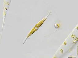
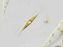
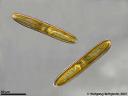
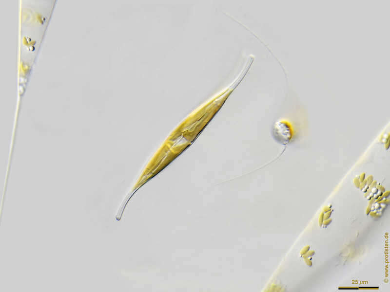
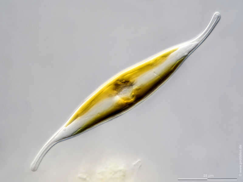
Gyrosigma fasciola Scale bar indicates 25 µm. The specimen was gathered in the Kieler Förde (German Baltic Sea). Sampling date 4/2018. The image was built up using several photomicrographic frames with manual stacking technique. Images were taken using Zeiss Axioplan with Olympus OM-D M5 MKII. Image under Creative Commons License V 3.0 (CC BY-NC-SA). Place name: Baltic Sea, Kieler Förde, Kiel Fjord (Germany) Latitude: 54.3894126 Longitude: 10.1749055 Multiebenen-Abbildung, manuell gestapelt. Der Messbalken markiert eine Länge von 25 µm. Probe aus der Kieler Förde. Datum der Aufsammlung: 4/2018. Mikrotechnik: Zeiss Axioplan, Kamera: Olympus OM-D M5 MKII. Creative Commons License V 3.0 (CC BY-NC-SA). For permission to use of (high-resolution) images please contact postmaster@protisten.de.
-
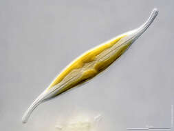
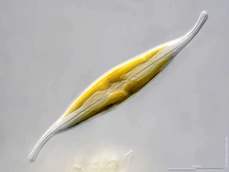
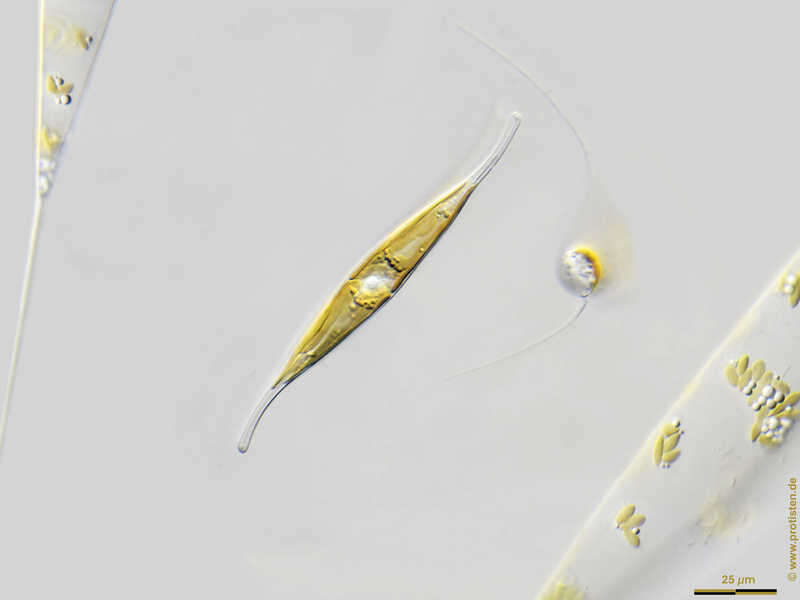
Gyrosigma fasciola Scale bar indicates 25 µm. Collected from Bodden, the brackish waters lying between the isles of Hiddensee and Ruegen (German Baltic Sea). Sampling date 9/2022. The image was built up using several photomicrographic frames with manual stacking technique. Images were taken using Zeiss Standard with Olympus OM-D M5 MKII. Image under Creative Commons License V 3.0 (CC BY-NC-ND). Place name: Hiddensee Bodden (Germany) Latitude: 54.582633 Longitude: 13.115051 Multiebenen-Abbildung, manuell gestapelt. Der Messbalken markiert eine Länge von 25 µm. Probe aus dem Hiddenseer Bodden. Datum der Aufsammlung: 9/2022. Mikrotechnik: Zeiss Standard, Kamera: Olympus OM-D M5 MKII. Creative Commons License V 3.0 (CC BY-NC-ND). For permission to use of (high-resolution) images please contact postmaster@protisten.de.
-
Gyrosigma fasciola Scale bar indicates 25 µm. The specimen was gathered in the Kieler Förde (German Baltic Sea). Sampling date 4/2018. The image was built up using several photomicrographic frames with manual stacking technique. Images were taken using Zeiss Axioplan with Olympus OM-D M5 MKII. Image under Creative Commons License V 3.0 (CC BY-NC-SA). Place name: Baltic Sea, Kieler Förde, Kiel Fjord (Germany) Latitude: 54.3894126 Longitude: 10.1749055 Multiebenen-Abbildung, manuell gestapelt. Der Messbalken markiert eine Länge von 25 µm. Probe aus der Kieler Förde. Datum der Aufsammlung: 4/2018. Mikrotechnik: Zeiss Axioplan, Kamera: Olympus OM-D M5 MKII. Creative Commons License V 3.0 (CC BY-NC-SA). For permission to use of (high-resolution) images please contact postmaster@protisten.de.
-
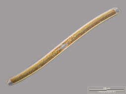
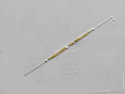
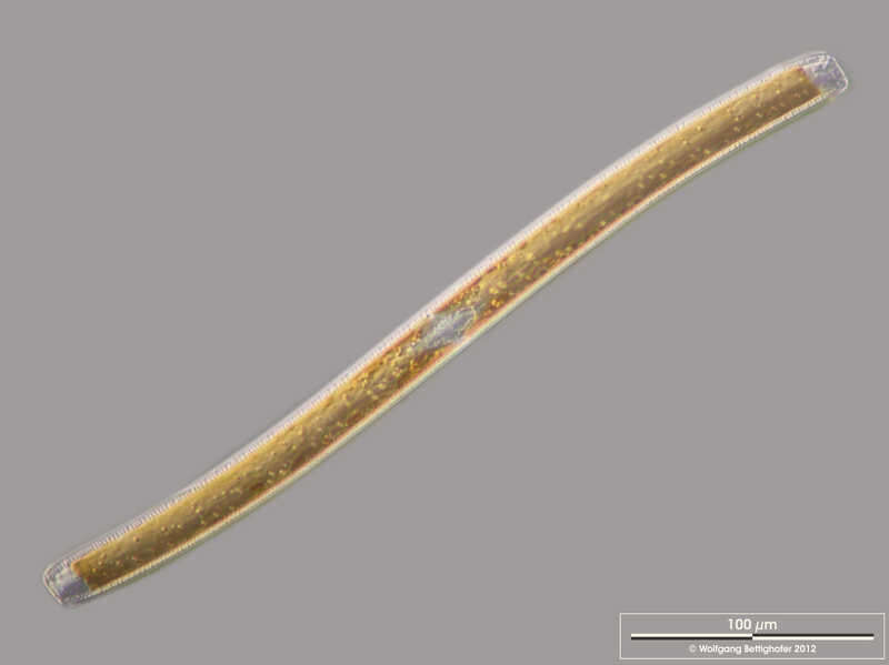
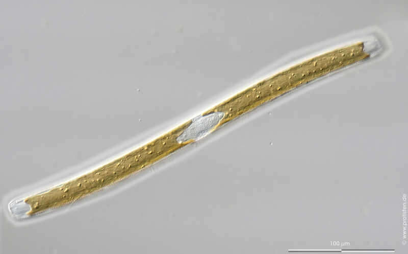
Nitzschia sigmoidea Cingular view. Scale bar indicates 100 µm. Sample from a wetland at the Pillersee (Tyrol, Austria). The image was built up using several photomicrographic frames with manual stacking technique. Images were taken using Zeiss Universal with Olympus C7070 CCD camera.Image under Creative Commons License V 3.0 (CC BY-NC-SA). Place name: Pond Suploch, Hiddensee (Germany) Latitude: 54.538638 Longitude: 13.097802 Gürtelbandansicht. Multiebenen-Abbildung, manuell gestapelt. Der Messbalken markiert eine Länge von 100 µm. Probe aus dem Pillersee in Tirol. Mikrotechnik: Zeiss Universal, Kamera: Olympus C7070. Creative Commons License V 3.0 (CC BY-NC-SA). For permission to use of (high-resolution) images please contact postmaster@protisten.de.
-
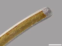
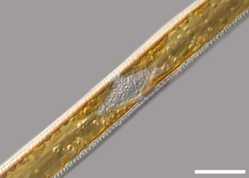
Nitzschia sigmoidea Detail: Center of the cell in cingular view with nucleus, cloroplasts with oil droplets (energy store) and the canal raphes. Scale bar indicates 25 µm. Sample from a wetland at the Pillersee (Tyrol, Austria). The image was built up using several photomicrographic frames with manual stacking technique. Images were taken using Zeiss Universal with Olympus C7070 CCD camera.Image under Creative Commons License V 3.0 (CC BY-NC-SA). Place name: Pond Suploch, Hiddensee (Germany) Latitude: 54.538638 Longitude: 13.097802 Detail: Zellmitte in Gürtelbandansicht mit Zellkern, den Chloroplasten mit Öltröpfchen (Speicherstoff) und den beiden Kanalraphen. Multiebenen-Abbildung, manuell gestapelt. Der Messbalken markiert eine Länge von 25 µm. Probe aus dem Pillersee in Tirol. Mikrotechnik: Zeiss Universal, Kamera: Olympus C7070. Creative Commons License V 3.0 (CC BY-NC-SA). For permission to use of (high-resolution) images please contact postmaster@protisten.de.
-
Nitzschia sigmoidea Cingular view. Scale bar indicates 100 µm. Sample from the Domänental pond near Kronshagen (Kiel, Germany). The image was built up using several photomicrographic frames with manual stacking technique. Images were taken using Zeiss Axioplan with Olympus OM-D M5 MKII.Image under Creative Commons License V 3.0 (CC BY-NC-SA). Place name: Pond Domänental near Kronshagen (Kiel, Germany) Latitude: 54.33211 Longitude: 10.060821 Gürtelbandansicht. Multiebenen-Abbildung, manuell gestapelt. Der Messbalken markiert eine Länge von 100 µm. Probe aus dem Domänentalteich bei Kronshagen. Mikrotechnik: Zeiss Axioplan, Kamera: Olympus OM-D M 5 MKII. Creative Commons License V 3.0 (CC BY-NC-SA). For permission to use of (high-resolution) images please contact postmaster@protisten.de.
-
Nitzschia sigmoidea Detail: Apex in cingular view, displaying cingulum (central strae and the two canal raphes on the edges. Scale bar indicates 25 µm. Sample from a wetland at the Pillersee (Tyrol, Austria). The image was built up using several photomicrographic frames with manual stacking technique. Images were taken using Zeiss Universal with Olympus C7070 CCD camera.Image under Creative Commons License V 3.0 (CC BY-NC-SA). Place name: Pond Suploch, Hiddensee (Germany) Latitude: 54.538638 Longitude: 13.097802 Detail: Zellspitze in Gürtelbandansicht. Das Gürtelband ist zentral als feine Striche zu erkennen, an den Außenkanten sind die Kanalraphen zu sehen. Multiebenen-Abbildung, manuell gestapelt. Der Messbalken markiert eine Länge von 25 µm. Probe aus dem Pillersee in Tirol. Mikrotechnik: Zeiss Universal, Kamera: Olympus C7070. Creative Commons License V 3.0 (CC BY-NC-SA). For permission to use of (high-resolution) images please contact postmaster@protisten.de.
-
Pinnularia viridiformis Specimen were selected from a sample of bottom sediments of a rain storage reservoir in Kiel (Schleswig-Holstein, Germany). The scale bar indicates 50 µm. Image was taken using Zeiss Universal with Olympus C7070 CCD camera.Image under Creative Commons License V 3.0 (CC BY-NC-SA). Place name: Pond Demühlen, rain storage reservoir in Kiel-Russee (Schleswig-Holstein, Germany) Latitude: 54.304095 Longitude: 10.086073 Der Messbalken markiert eine Länge von 50 µm. Sedimentprobe aus einem Regenrückhaltebecken in Kiel. Mikrotechnik: Zeiss Universal, Kamera: Olympus C7070.Creative Commons License V 3.0 (CC BY-NC-SA). For permission to use of (high-resolution) images please contact postmaster@protisten.de.
-
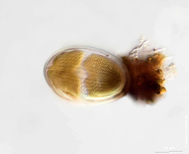
Cocconeis pediculus Scale bar indicates 10 µm. Sample from a pond called Fuhlensee in Schilksee (Kiel, Germany). Sampling date 7/2018. The image was built up using several photomicrographic frames with manual stacking technique. Images were taken using Zeiss Axioplan with Olympus OM-D M5 MKII. Image under Creative Commons License V 3.0 (CC BY-NC-SA) Place name: Lake Fuhlensee near Schilksee (Kiel, Germany) Latitude: 54.43136338 Longitude: 10.16243935 Multiebenen-Abbildung, manuell gestapelt. Der Messbalken markiert eine Länge von 10 µm. Probe aus dem Feuchtbiotop Fuhlensee bei Schilksee/Kiel. Datum der Aufsammlung: 7/2018. Mikrotechnik: Zeiss Axioplan, Kamera: Olympus OM-D M5 MKII. Creative Commons License V 3.0 (CC BY-NC-SA). For permission to use of (high-resolution) images please contact postmaster@protisten.de.
-
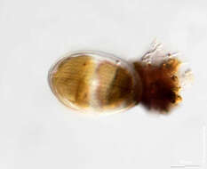
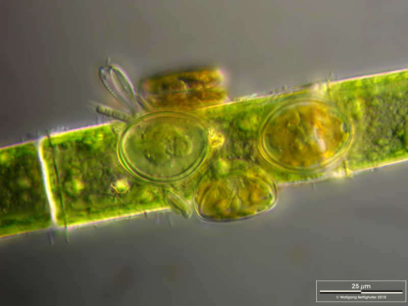
Cocconeis pediculus Cocconeis pediculus living on the green alga Oedogonium.There is also a filamentous coloniy of cyanobacteria Homeothrix spec. Scale bar indicates 25 µm. Sample from Lake Constance near Bodman. Images were taken using Zeiss Universal with Olympus C7070 CCD camera.Image under Creative Commons License V 3.0 (CC BY-NC-SA). Place name: Lake Constance vicinity of Bodman (Germany) Latitude: 47.796494 Longitude: 9.047656 Cocconeis pediculus auf der Grünalge Oedogonium spec. Des Weiteren sind fädige Kolonien der Blaualge Homeothrix spec. zu sehen. Der Messbalken markiert eine Länge von 25 µm. Probe aus dem Bodensee bei Bodman. Mikrotechnik: Zeiss Universal, Kamera: Olympus C7070.Creative Commons License V 3.0 (CC BY-NC-SA). For permission to use of (high-resolution) images please contact postmaster@protisten.de.
-
Cocconeis pediculus Scale bar indicates 10 µm. Sample from a pond called Fuhlensee in Schilksee (Kiel, Germany). Sampling date 7/2018. The image was built up using several photomicrographic frames with manual stacking technique. Images were taken using Zeiss Axioplan with Olympus OM-D M5 MKII. Image under Creative Commons License V 3.0 (CC BY-NC-SA) Place name: Lake Fuhlensee near Schilksee (Kiel, Germany) Latitude: 54.43136338 Longitude: 10.16243935 Multiebenen-Abbildung, manuell gestapelt. Der Messbalken markiert eine Länge von 10 µm. Probe aus dem Feuchtbiotop Fuhlensee bei Schilksee/Kiel. Datum der Aufsammlung: 7/2018. Mikrotechnik: Zeiss Axioplan, Kamera: Olympus OM-D M5 MKII. Creative Commons License V 3.0 (CC BY-NC-SA). For permission to use of (high-resolution) images please contact postmaster@protisten.de.
-
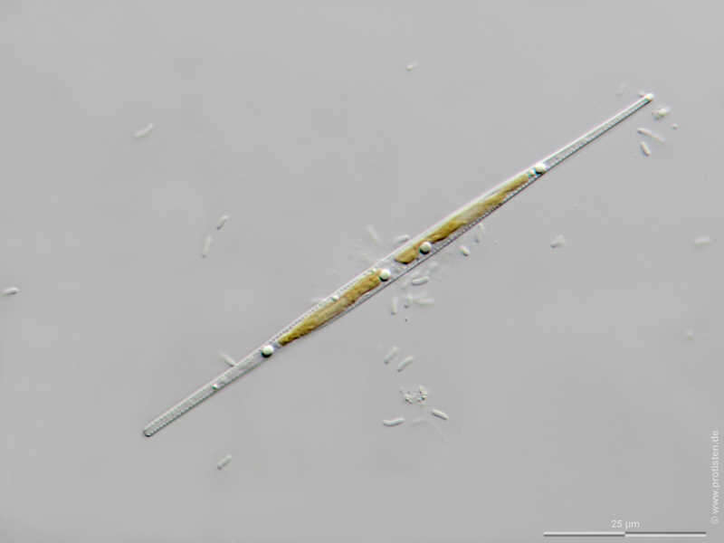
Nitzschia gracilis Scale bar indicates 25 µm. Sample from the Lake Vollstedter See near Kiel, Germany. Sampling date 7/2018. The image was built up using several photomicrographic frames with manual stacking technique. Images were taken using Zeiss Axioplan with Olympus OM-D M5 MKII. Image under Creative Commons License V 3.0 (CC BY-NC-SA). Place name: Lake Vollstedter See near Kiel (Germany) Latitude: 54.24105528 Longitude: 9.859339 Multiebenen-Abbildung, manuell gestapelt. Der Messbalken markiert eine Länge von 25 µm. Probe aus dem Vollstedter See bei Groß Vollstedt. Datum der Aufsammlung: 7/2018. Mikrotechnik: Zeiss Axioplan, Kamera: Olympus OM-D M5 MKII. Creative Commons License V 3.0 (CC BY-NC-SA). For permission to use of (high-resolution) images please contact postmaster@protisten.de.