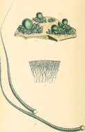
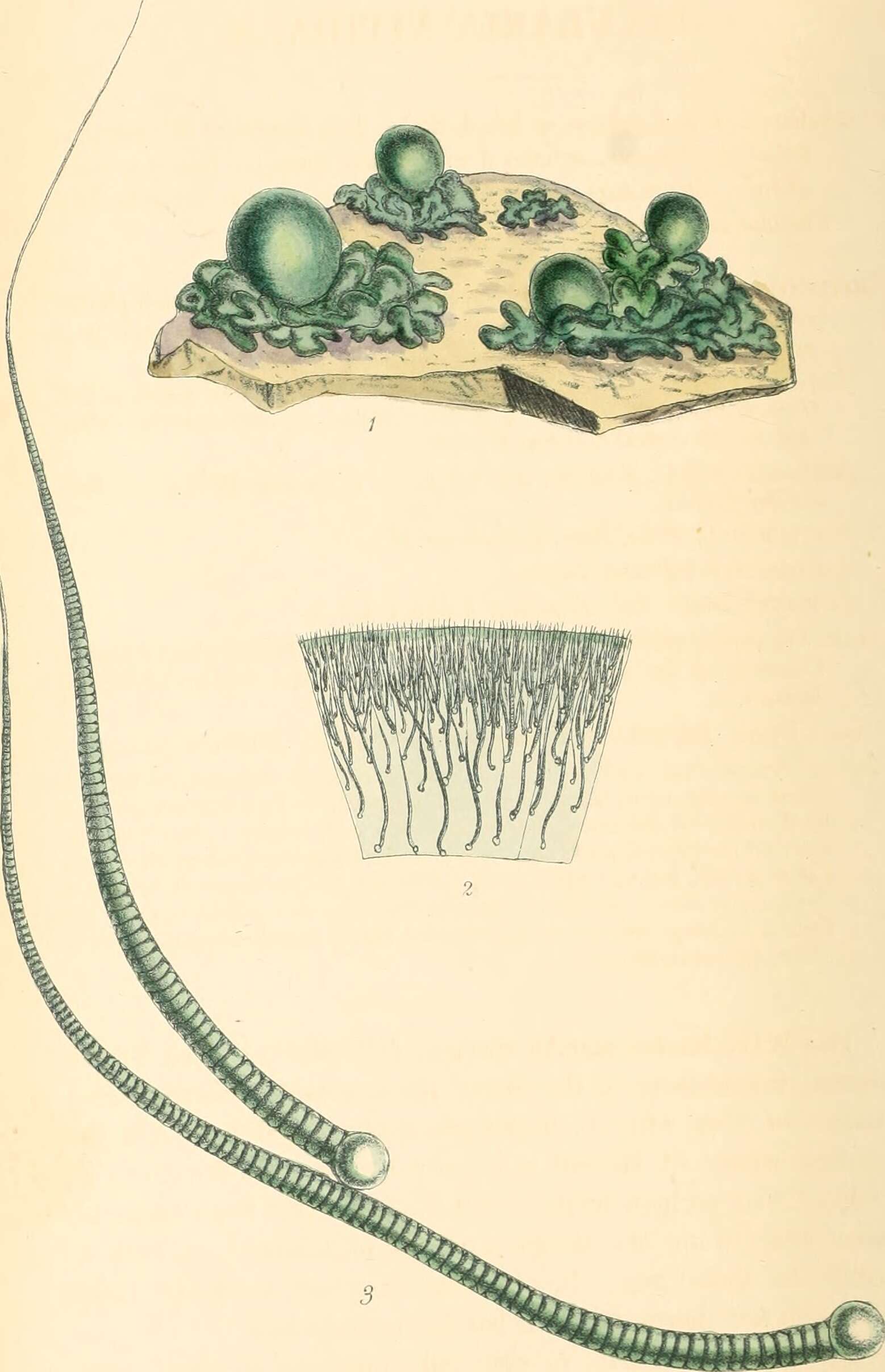
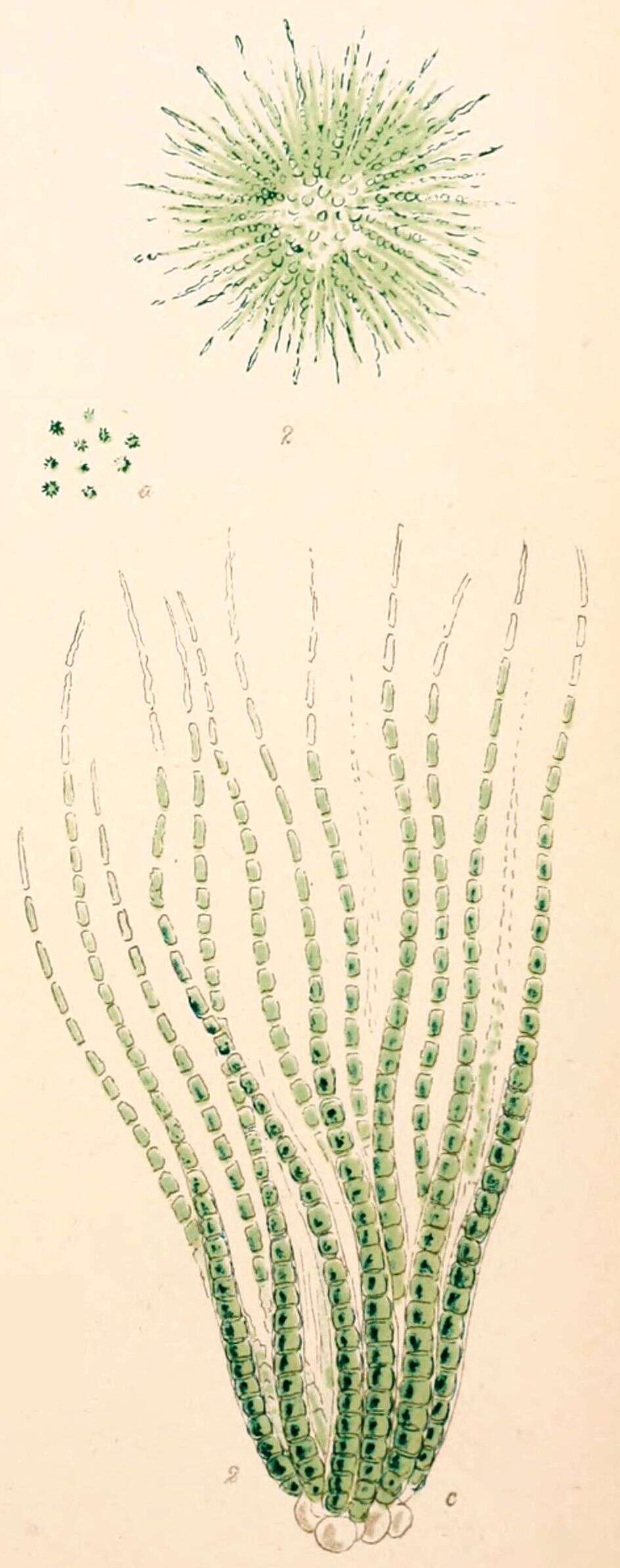
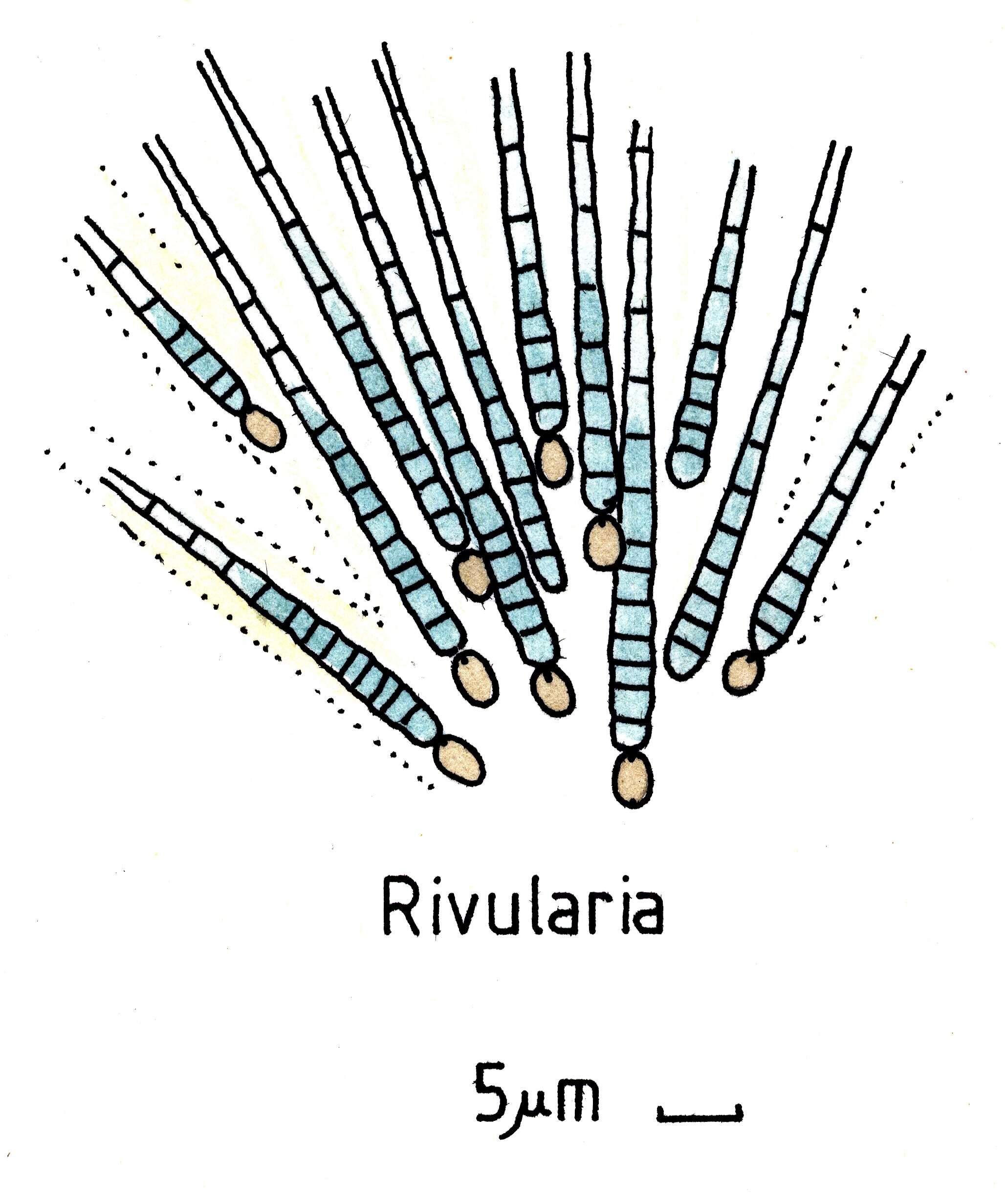
Description: English: Rivularia nitida illustration. Date: 1846. Source: Image from page 257 of "Phycologia britannica, or, A History of British sea-weeds, containing coloured figures, generic and specific characters, synonymes, and descriptions of all the species of algae inhabiting the shores of the British Islands" (1846) Identifier: phycologiabri04harv Title: Phycologia britannica, or, A History of British sea-weeds, containing coloured figures, generic and specific characters, synonymes, and descriptions of all the species of algae inhabiting the shores of the British Islands Year: 1846 (1840s) Authors: Harvey, William H. (William Henry), 1811-1866 Subjects: Marine algae Publisher: London, Reeve and Benham Contributing Library: The LuEsther T Mertz Library, the New York Botanical Garden Digitizing Sponsor: Metropolitan New York Library Council - METRO. Author: Harvey, William H. (William Henry), 1811-1866.
Mlewski EC, Pisapia C, Gomez F, Lecourt L, Soto Rueda E, Benzerara K, Ménez B, Borensztajn S, Jamme F, Réfrégiers M and Gérard E
Wikimedia Commons
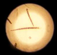
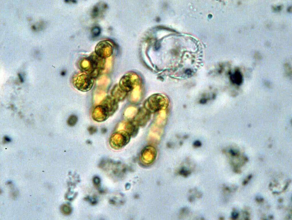
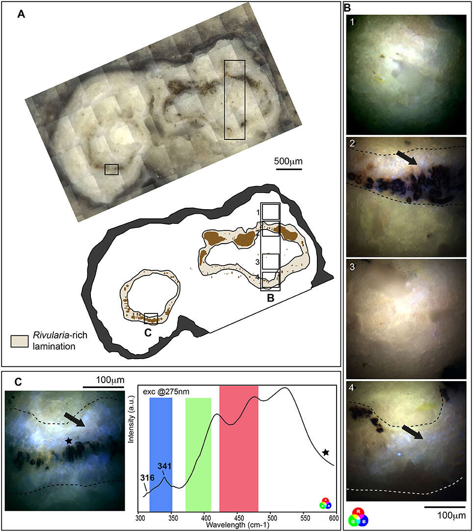
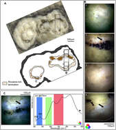
Summary.mw-parser-output table.commons-file-information-table,.mw-parser-output.fileinfotpl-type-information{border:1px solid #a2a9b1;background-color:#f8f9fa;padding:5px;font-size:95%;border-spacing:2px;box-sizing:border-box;margin:0;width:100%}.mw-parser-output table.commons-file-information-table>tbody>tr,.mw-parser-output.fileinfotpl-type-information>tbody>tr{vertical-align:top}.mw-parser-output table.commons-file-information-table>tbody>tr>td,.mw-parser-output table.commons-file-information-table>tbody>tr>th,.mw-parser-output.fileinfotpl-type-information>tbody>tr>td,.mw-parser-output.fileinfotpl-type-information>tbody>tr>th{padding:4px}.mw-parser-output.fileinfo-paramfield{background:#ccf;text-align:right;padding-right:0.4em;width:15%;font-weight:bold}.mw-parser-output.commons-file-information-table+table.commons-file-information-table,.mw-parser-output.commons-file-information-table+div.commons-file-information-table>table{border-top:0;padding-top:0;margin-top:-8px}@media only screen and (max-width:719px){.mw-parser-output table.commons-file-information-table,.mw-parser-output.commons-file-information-table.fileinfotpl-type-information{border-spacing:0;padding:0;word-break:break-word;width:100%!important}.mw-parser-output.commons-file-information-table>tbody,.mw-parser-output.fileinfotpl-type-information>tbody{display:block}.mw-parser-output.commons-file-information-table>tbody>tr>td,.mw-parser-output.commons-file-information-table>tbody>tr>th,.mw-parser-output.fileinfotpl-type-information>tbody>tr>td,.mw-parser-output.fileinfotpl-type-information>tbody>tr>th{padding:0.2em 0.4em;text-align:left;text-align:start}.mw-parser-output.commons-file-information-table>tbody>tr,.mw-parser-output.fileinfotpl-type-information>tbody>tr{display:flex;flex-direction:column}.mw-parser-output.commons-file-information-table+table.commons-file-information-table,.mw-parser-output.commons-file-information-table+div.commons-file-information-table>table{margin-top:-1px}.mw-parser-output.fileinfo-paramfield{box-sizing:border-box;flex:1 0 100%;width:100%}} Description: English: Fluorescence emission signal after synchrotron-based deep UV excitation at 275 nm of a cross section of an oncoid associated with a black pustular mat. (A) Optical microscopy observation and schematic representation of the cross section highlighting Rivularia-rich laminations with encrusted Rivularia-like cells. The localization of areas of interest analyzed by S-DUV fluorescence imaging is given. (B) Composite RGB images of 4 areas of interest were reconstructed using the fluorescence signal collected with filters between 327 and 353 nm (Blue), 370 and 410 nm (Green) and 420 and 480 nm (Red). They were surimposed on the optical image. An intense fluorescence signal was observed using the blue filter between 327 and 353 nm (black arrows) in Rivularia-rich laminations (underlined by dot lines in B,C) while the rest of the matrix is marked by a mixed fluorescence collected using the green and red filters. (C) The fluorescence emission signal associated with Rivularia-rich laminations was recorded and showed two bands at 316 and 341 cm−1. The fluorescence ranges covered by the three filters are indicated with respective colors and the precise localization where the spectrum was collected is given by a black star on the associated full-field RGB fluorescence image. Date: 24 March 2018, 23:33:37. Source: Mlewski EC, Pisapia C, Gomez F, Lecourt L, Soto Rueda E, Benzerara K, Ménez B, Borensztajn S, Jamme F, Réfrégiers M and Gérard E (2018) Characterization of Pustular Mats and Related Rivularia-Rich Laminations in Oncoids From the Laguna Negra Lake (Argentina). Front. Microbiol. 9:996. doi: 10.3389/fmicb.2018.00996. Author: Mlewski EC, Pisapia C, Gomez F, Lecourt L, Soto Rueda E, Benzerara K, Ménez B, Borensztajn S, Jamme F, Réfrégiers M and Gérard E.
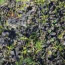
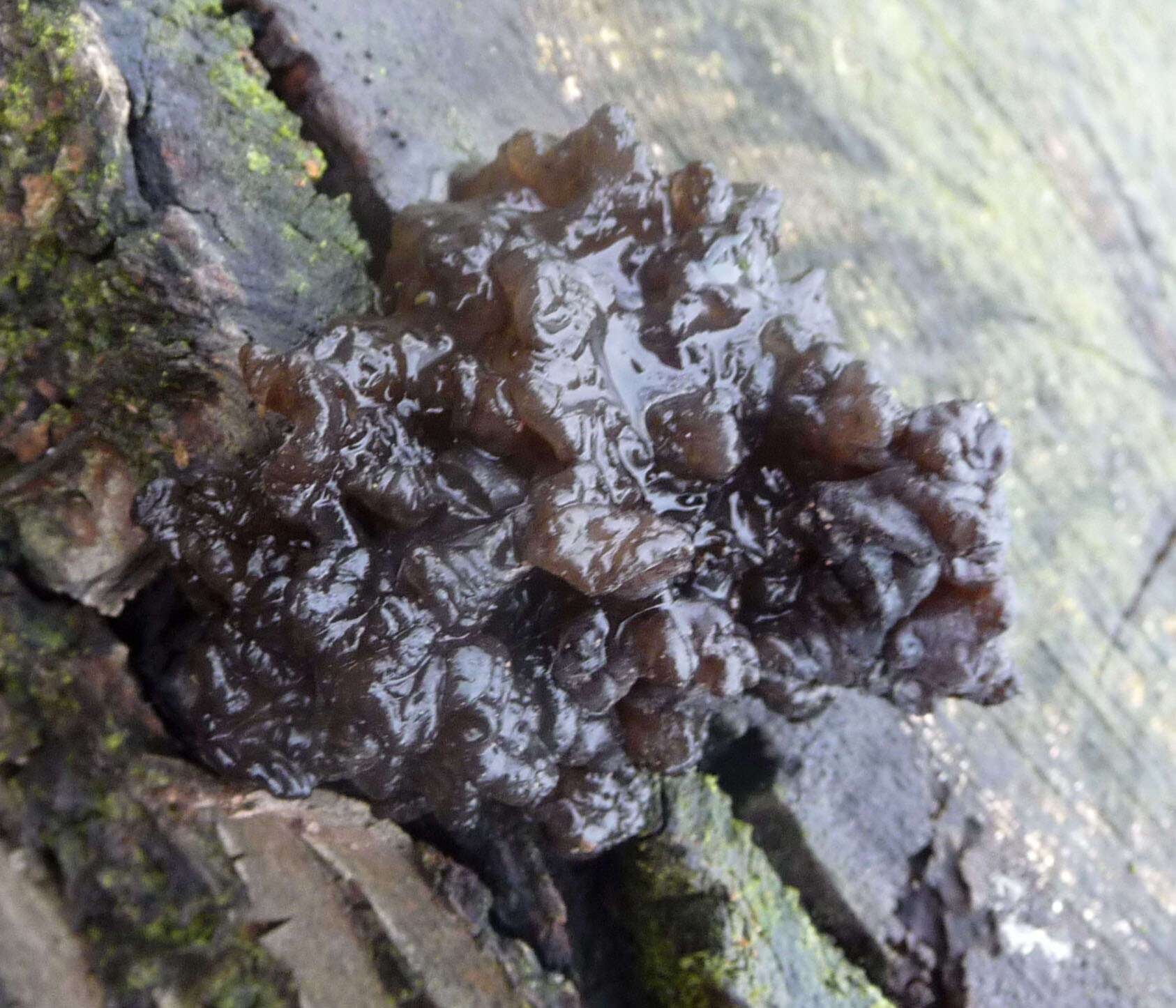
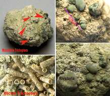
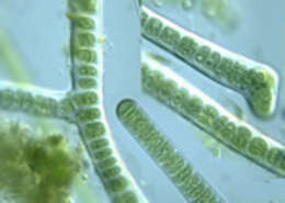
Description: English: recent oncoid, location: river "Obere-Alz", Truchtlaching, Bavaria, Germany Deutsch: rezenter Onkoid, Fundort: "Obere-Alz", Truchtlaching, Bayern. Date: 3 August 2012. Source: Own work. Author: Hans Lauterbach
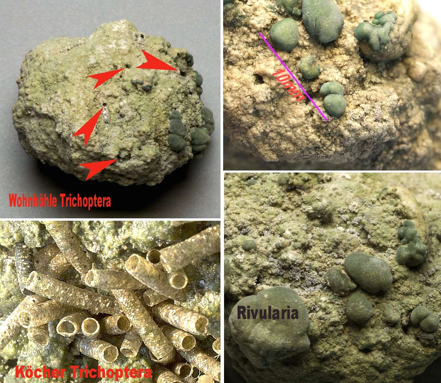
Furchenstein. Größe: 4,5 x 3,5 x 3,0cm, rezenter Onkoid, Darstellung der Oberfläche, Besiedelung mit unterschiedlichen Kalkalgen, unter anderem die auffälligen Rivularia, mit Köchern von Köcherfliegenlarven, markiert die Wohnhöhlen von Trichoptera-Larven Licensing[
edit] : This file is licensed under the
Creative Commons Attribution-Share Alike 3.0 Unported license.:. https://creativecommons.org/licenses/by-sa/3.0 CC BY-SA 3.0 Creative Commons Attribution-Share Alike 3.0 truetrue.


_BHL11346758.jpg)
_(20231600079).jpg)
.jpg)
.jpg)