-
Galende, Castile and Len, Spain
-
Castille and Leon, Spain
-
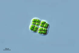
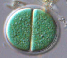
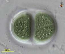
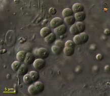
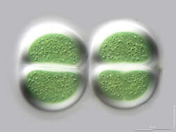
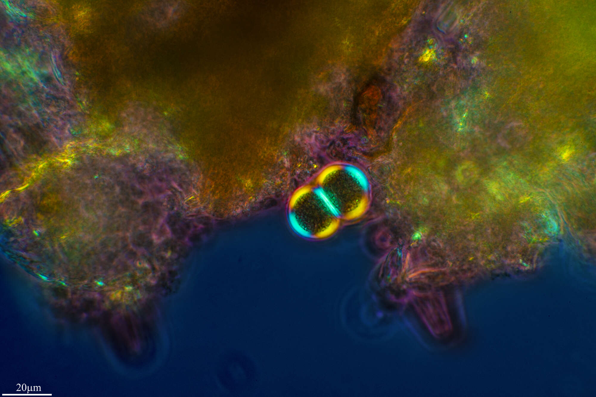
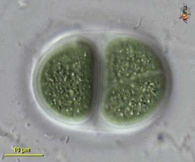
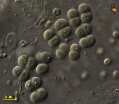
Chroococcus turgidus, (Cyanobacteria, Chroococales), from Lake Kinneret pelagic waters, April 2006, showing 2 daughter cells after division by simple binary fission â as characteristic for most Chroococcales species. This species is common in the plankton of Lake Kinneret throughout the year. Usually there are 2 â 8 cells in a colony. Clearly delimited colorless mucilaginous envelopes surround the individual cells following their contours, and the entire colony. Cell diameter: 8 â 11 µm.
-
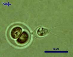
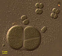
Chroococcus turgidus (Cyanobacteria, Chroococcales) is common in Lake Kinneret in recent years. In this unusual photograph a choanoflagellate is attached to its outer shell.
-
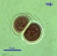
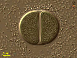
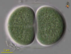
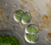
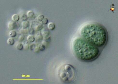
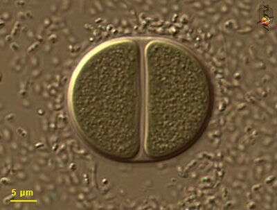
Chroococcus (crow-o-cock-us), large cyanobacterium, typically two (but sometimes one) cells enclosed within a mucus sheath. Photosynthetic pigment distributed through cytoplasm, which may have a granular texture, but does not have subcompartments (organelles). Differential interference contrast.
-
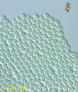
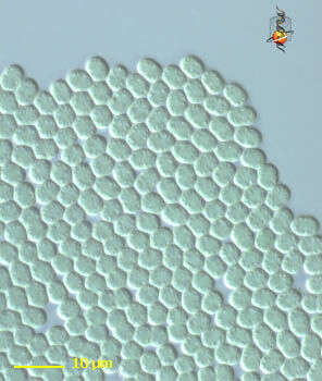
Chroococcus (crow-owe-cock-us) coccoid cyanobacteria, adhering to each other to form extensive flat sheets, no evident mucus sheath or heterocysts. Differential Interference Contrast.
-
Chroococcus (crow-o-cock-us), large cyanobacterium, typically two (but sometimes one) cells enclosed within a mucus sheath. Photosynthetic pigment distributed through cytoplasm, which may have a granular texture, but does not have subcompartments (organelles). Differential interference contrast.
-
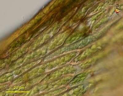
Chroococcus (crow-owe-cock-us) tentative identification. Coccoid blue green algal cells. Found as one of several cyanobacterial epibionts on the leaves of the moss Hygrohypnum, a site which seems to be a focus for nitrogen fixation. In this case the cyanobacterial cells have occupied one of the cortical cells of the plant. Differential interference contrast.
-
Chroococcus (crow-owe-cock-us) tentative identification. Coccoid blue green algal cells. Found as one of several cyanobacterial epibionts on the leaves of the moss Hygrohypnum, a site which seems to be a focus for nitrogen fixation. Differential interference contrast.
-
Chroomonas. Cell observed in freshwater sediments in the vicinity of Broome, Western Australia in September 2003. This image was taken using differential interference contrast optics. Â Â This work was supported by the Australian Biological Resources Study.
-
Variously sized individuals - some species have been reported with at least a five fold size range - so these may be of a single species. Nomarksi, diferential interference contrast optics.
-
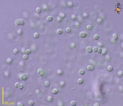
Large blue-green algal (cyanobacterial) cells in mucus sheath. Differential interference contrast optics.
-
Blue green algae in a mucus sheath. Differential interference contrast optics.
-
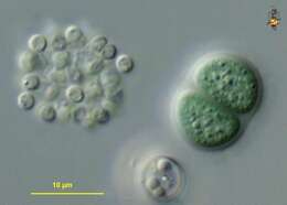
Most Chroococcus cells are quite large. Some empty capsules can be seen to the right of the living cells. Differential interefence contrast optics.
-
The chroococcus cells are to the right, and the cells of a similar size but a brighter green colour to the left are cells of an unidentified chlorophycean green alga (a eukaryote). This image illustrates the blue-green colour from which the blue-green algae get their name. Differential interference contrast optics.
-
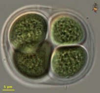
Typical arrangement with paired cells inside a mucus sheath. Differential interference contrast optics.
-
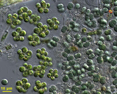
Anacystis (a-na-cyst-is) is a cyanobacterium in which globular cells are located within a gelatinous matrix. Some of the cyanobacteria with this form can be toxic. Differential interference contrast.
-
Anacystis (a-na-cyst-is), a cyanobacterium or blue green algae, in which the cells are clumped together within a mucus material. The clusters have to be compressed so that the individual cells can be observed. This genus adopts a variety of forms. Some Anacystis species are known to produce toxins. Phase contrast.
-
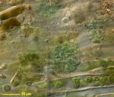
Sampling date 06/2013. Scale bars indicate 10 µm.
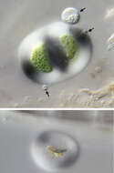
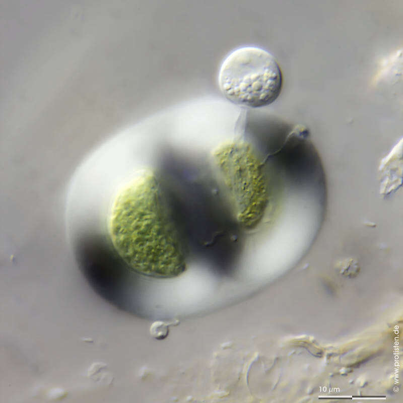
Chroococcus turgidus with chytrid parasite
Chroococcus turgidus, attacked by Chytridiomycota cells. The arrows point to the three chytrid cells; the rhizoids they have drilled through the protective jelly to the host cell are clearly visible in the top two. The cell mass of the cyanobacteria is visibly reduced. In the image attached below, a sucked out
Chroococcus cell. Scale bar indicates 10 µm.Place name: Wetland Schweizeralm near Waidring (Tyrol, Austria) .Latitude: 47.579916 Longitude: 12.575343Microscope Zeiss Standard, camera Canon EOS 600D. DOF image.© Wolfgang Bettighofer,images under Creative Commons License V 3.0 (CC BY-NC-SA).For permission to use of (high resolution) images please contact
postmaster@protisten.de.For further information about the image, please click here:
Link to protisten.de page
-
Sampling date 06/2013. Scale bars indicate 10 µm.
Chroococcus turgidus with chytrid parasite
Chroococcus turgidus, attacked by Chytridiomycota cells. The arrows point to the three chytrid cells; the rhizoids they have drilled through the protective jelly to the host cell are clearly visible in the top two. The cell mass of the cyanobacteria is visibly reduced. In the image attached below, a sucked out
Chroococcus cell. Scale bar indicates 10 µm.Place name: Wetland Schweizeralm near Waidring (Tyrol, Austria) .Latitude: 47.579916 Longitude: 12.575343Microscope Zeiss Standard, camera Canon EOS 600D. DOF image.© Wolfgang Bettighofer,images under Creative Commons License V 3.0 (CC BY-NC-SA).For permission to use of (high resolution) images please contact
postmaster@protisten.de.For further information about the image, please click here:
Link to protisten.de page
-
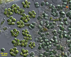
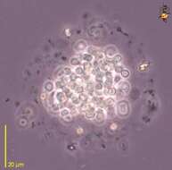
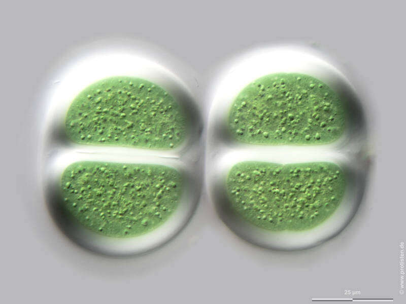
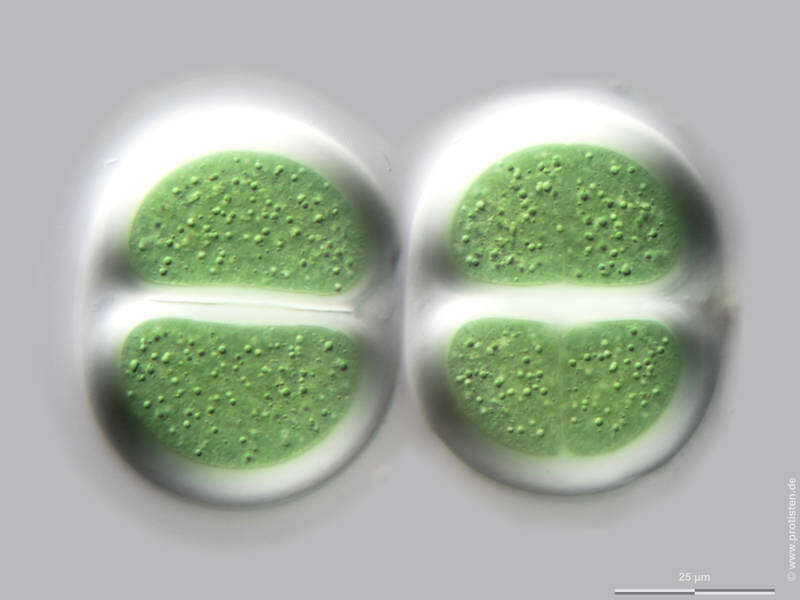
Sampling date 10/2019. Scale bars indicate 25 µm.Several tomographic images (top to bottom) of a colony. Please click on < or > on the image edges or on the dots at the bottom edge of the images to browse through the slides!Place name: Bog Waasenmoos Pass Thurn near Mittersil (Tyrol, Austria).Latitude: 47.30234117 Longitude: 12.41751194Microscope Zeiss Axioplan, camera Olympus OM-D M5 MKII. DOF images.© Wolfgang Bettighofer,images under Creative Commons License V 3.0 (CC BY-NC-SA).For permission to use of (high resolution) images please contact
postmaster@protisten.de.For further information about the image, please click here:
Link to protisten.de page
-
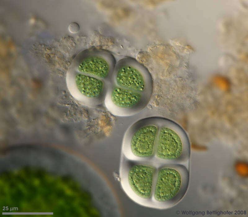
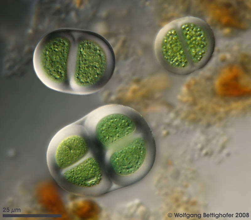
Sampling date 05/2008. Scale bars indicate 25 µm.Place name: Bogs near Salzburg (Austria).Latitude: 48.068516 Longitude: 12.954134Microscope Zeiss Universal, camera Olympus C7070. DOF image.© Wolfgang Bettighofer,images under Creative Commons License V 3.0 (CC BY-NC-SA).For permission to use of (high resolution) images please contact
postmaster@protisten.de.For further information about the image, please click here:
Link to protisten.de page
-
Sampling date 05/2008. Scale bars indicate 25 µm.Place name: Bogs near Salzburg (Austria).Latitude: 48.068516 Longitude: 12.954134Microscope Zeiss Universal, camera Olympus C7070. DOF image.© Wolfgang Bettighofer,images under Creative Commons License V 3.0 (CC BY-NC-SA).For permission to use of (high resolution) images please contact
postmaster@protisten.de.For further information about the image, please click here:
Link to protisten.de page