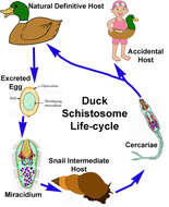
Biology Open Educational Resources|sourceurl=https://flickr.com/photos/71607123@N03/7687096834%7Carchive=%7Creviewdate=2020-11-10 10:27:40|reviewlicense=cc-by-sa-2.0|reviewer=FlickreviewR 2
Wikimedia Commons
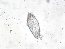
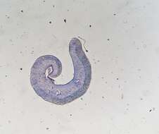
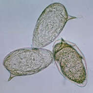
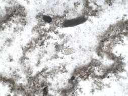
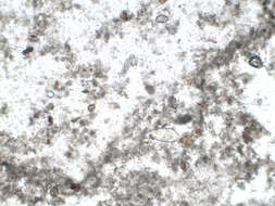
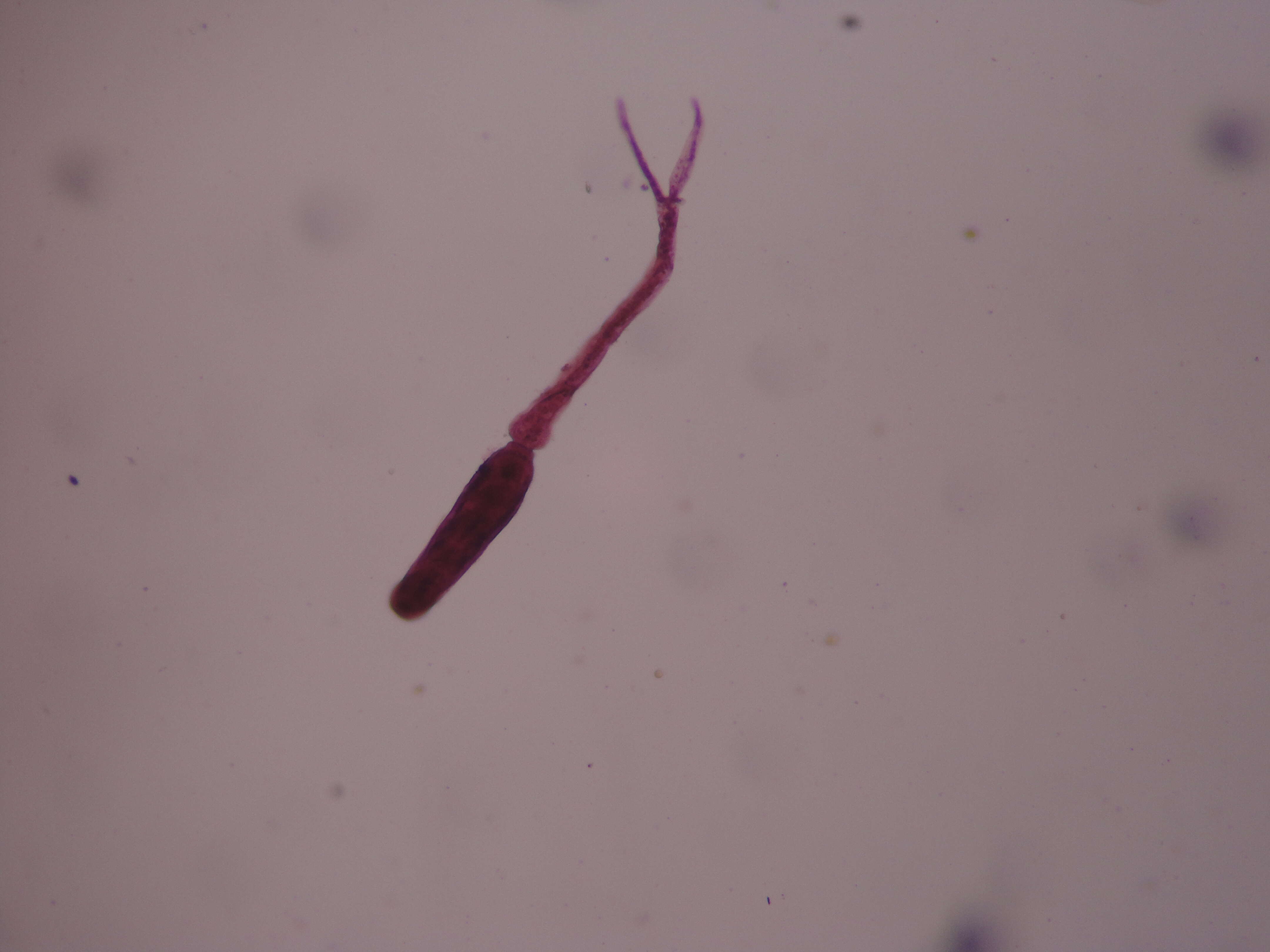
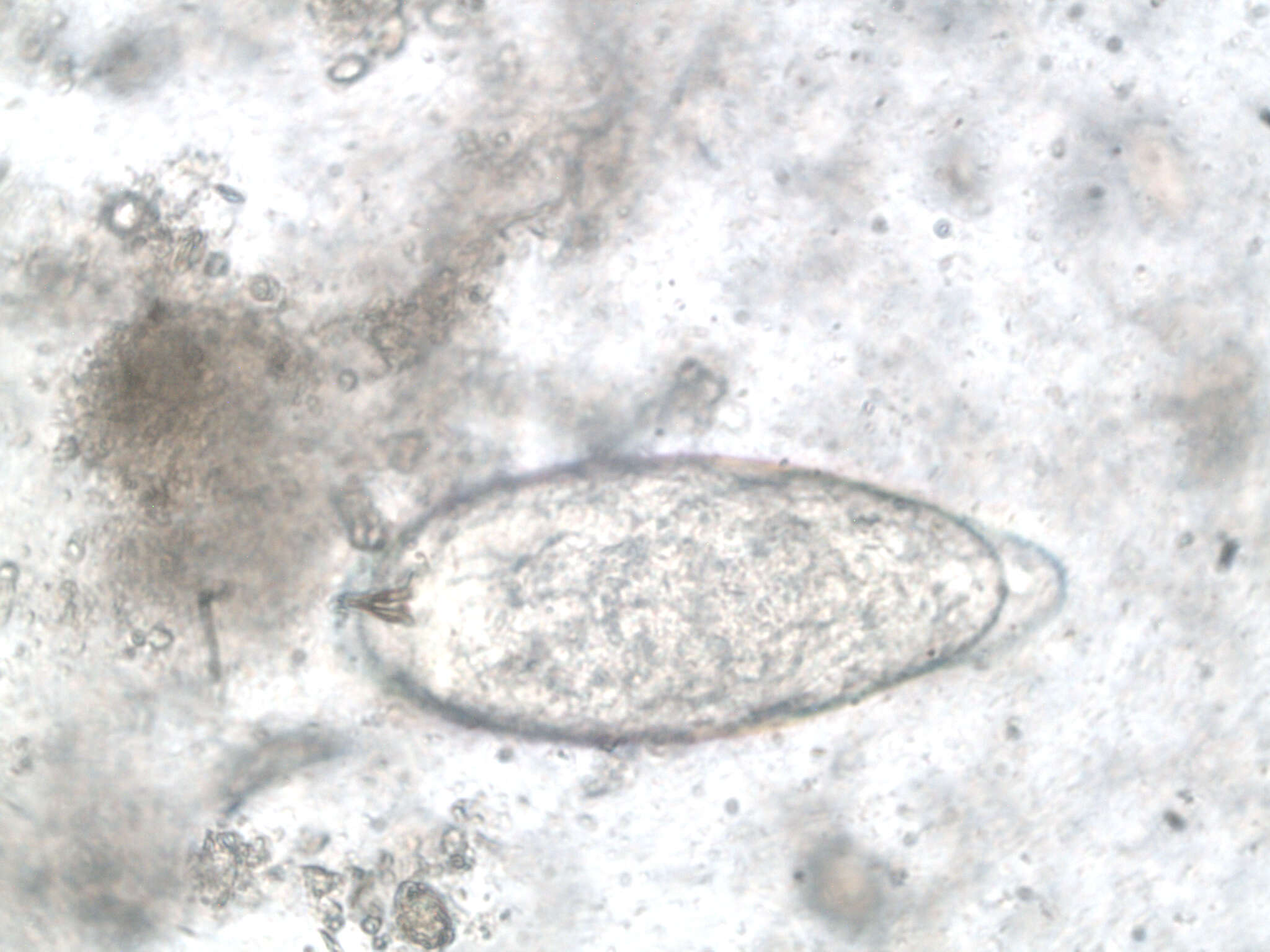
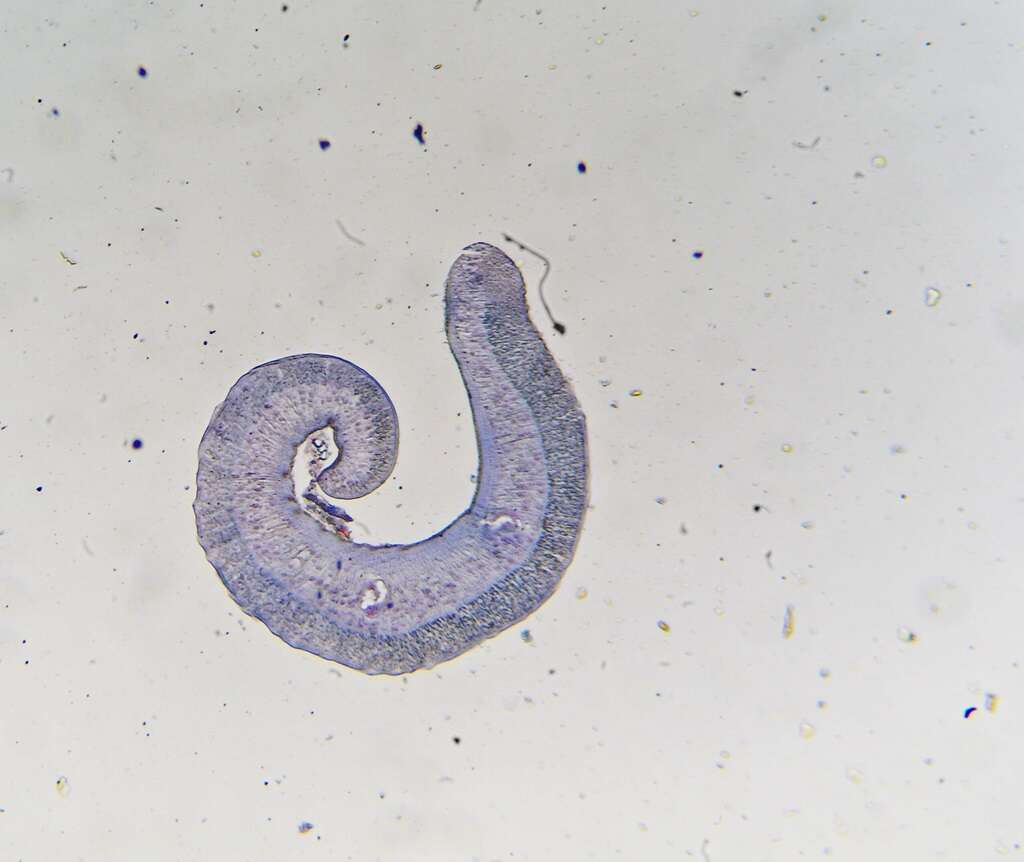
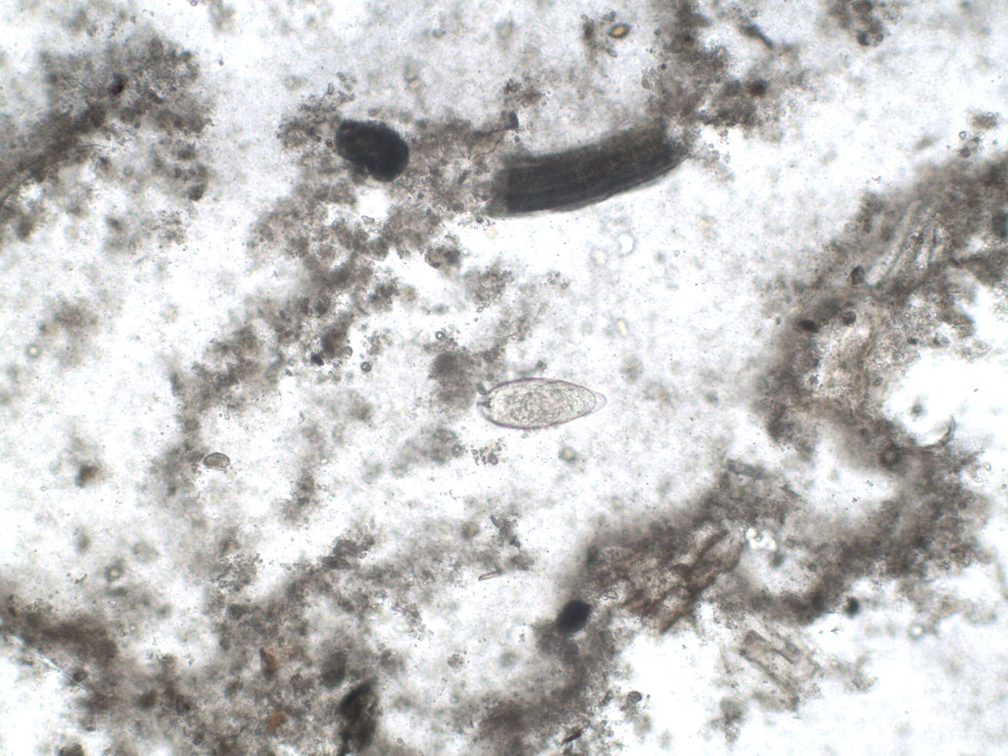
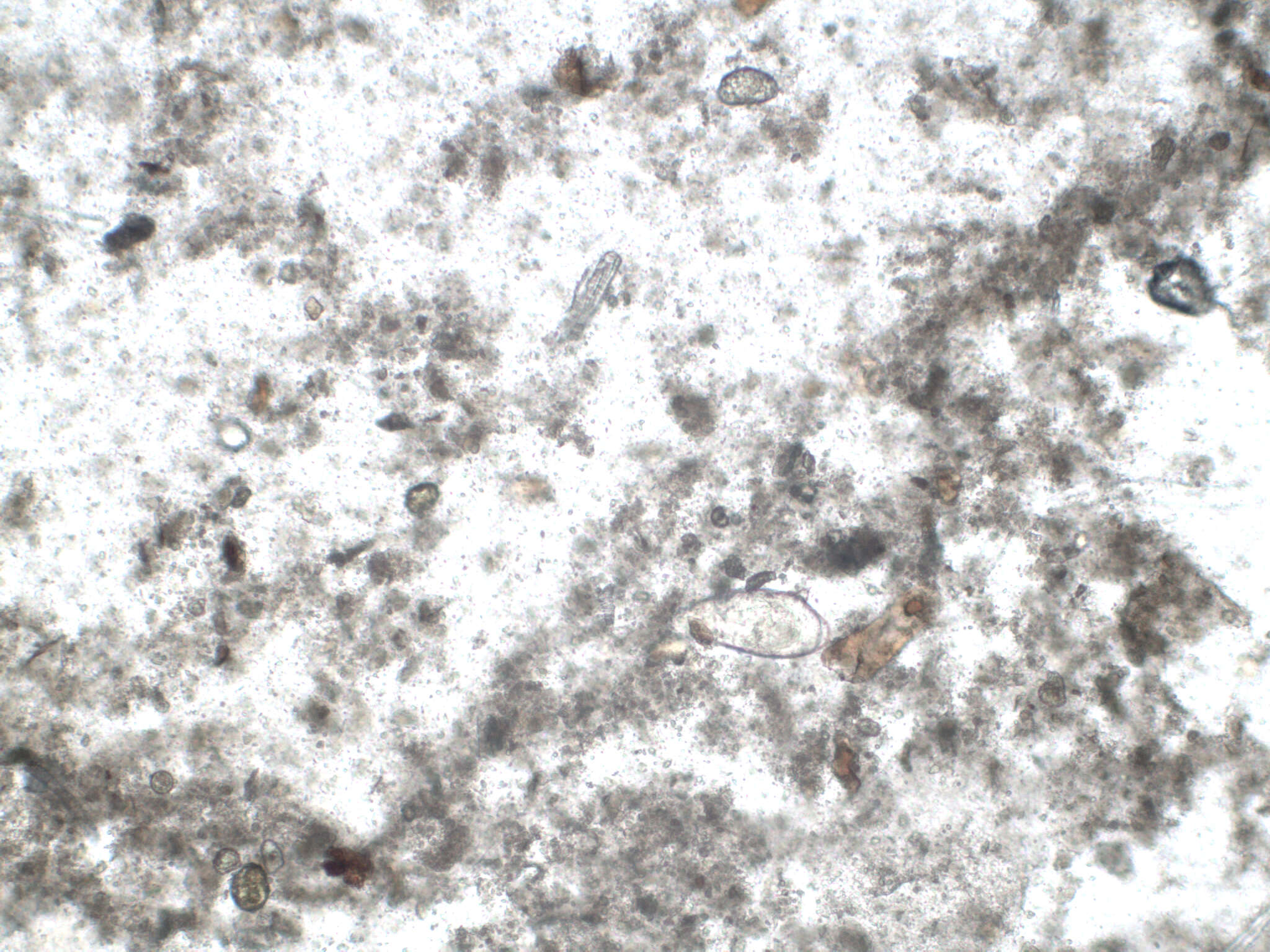
Summary.mw-parser-output table.commons-file-information-table,.mw-parser-output.fileinfotpl-type-information{border:1px solid #a2a9b1;background-color:#f8f9fa;padding:5px;font-size:95%;border-spacing:2px;box-sizing:border-box;margin:0;width:100%}.mw-parser-output table.commons-file-information-table>tbody>tr,.mw-parser-output.fileinfotpl-type-information>tbody>tr{vertical-align:top}.mw-parser-output table.commons-file-information-table>tbody>tr>td,.mw-parser-output table.commons-file-information-table>tbody>tr>th,.mw-parser-output.fileinfotpl-type-information>tbody>tr>td,.mw-parser-output.fileinfotpl-type-information>tbody>tr>th{padding:4px}.mw-parser-output.fileinfo-paramfield{background:#ccf;text-align:right;padding-right:0.4em;width:15%;font-weight:bold}.mw-parser-output.commons-file-information-table+table.commons-file-information-table,.mw-parser-output.commons-file-information-table+div.commons-file-information-table>table{border-top:0;padding-top:0;margin-top:-8px}@media only screen and (max-width:719px){.mw-parser-output table.commons-file-information-table,.mw-parser-output.commons-file-information-table.fileinfotpl-type-information{border-spacing:0;padding:0;word-break:break-word;width:100%!important}.mw-parser-output.commons-file-information-table>tbody,.mw-parser-output.fileinfotpl-type-information>tbody{display:block}.mw-parser-output.commons-file-information-table>tbody>tr>td,.mw-parser-output.commons-file-information-table>tbody>tr>th,.mw-parser-output.fileinfotpl-type-information>tbody>tr>td,.mw-parser-output.fileinfotpl-type-information>tbody>tr>th{padding:0.2em 0.4em;text-align:left;text-align:start}.mw-parser-output.commons-file-information-table>tbody>tr,.mw-parser-output.fileinfotpl-type-information>tbody>tr{display:flex;flex-direction:column}.mw-parser-output.commons-file-information-table+table.commons-file-information-table,.mw-parser-output.commons-file-information-table+div.commons-file-information-table>table{margin-top:-1px}.mw-parser-output.fileinfo-paramfield{box-sizing:border-box;flex:1 0 100%;width:100%}} Description: Set of parasite microscopic images. Date: 29 September 2011, 11:53. Source: Schistosoma haematobium x40mag UK NEQAS. Author: Vivien Rolfe.
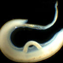
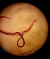
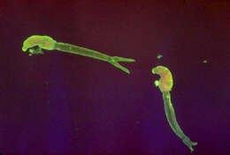
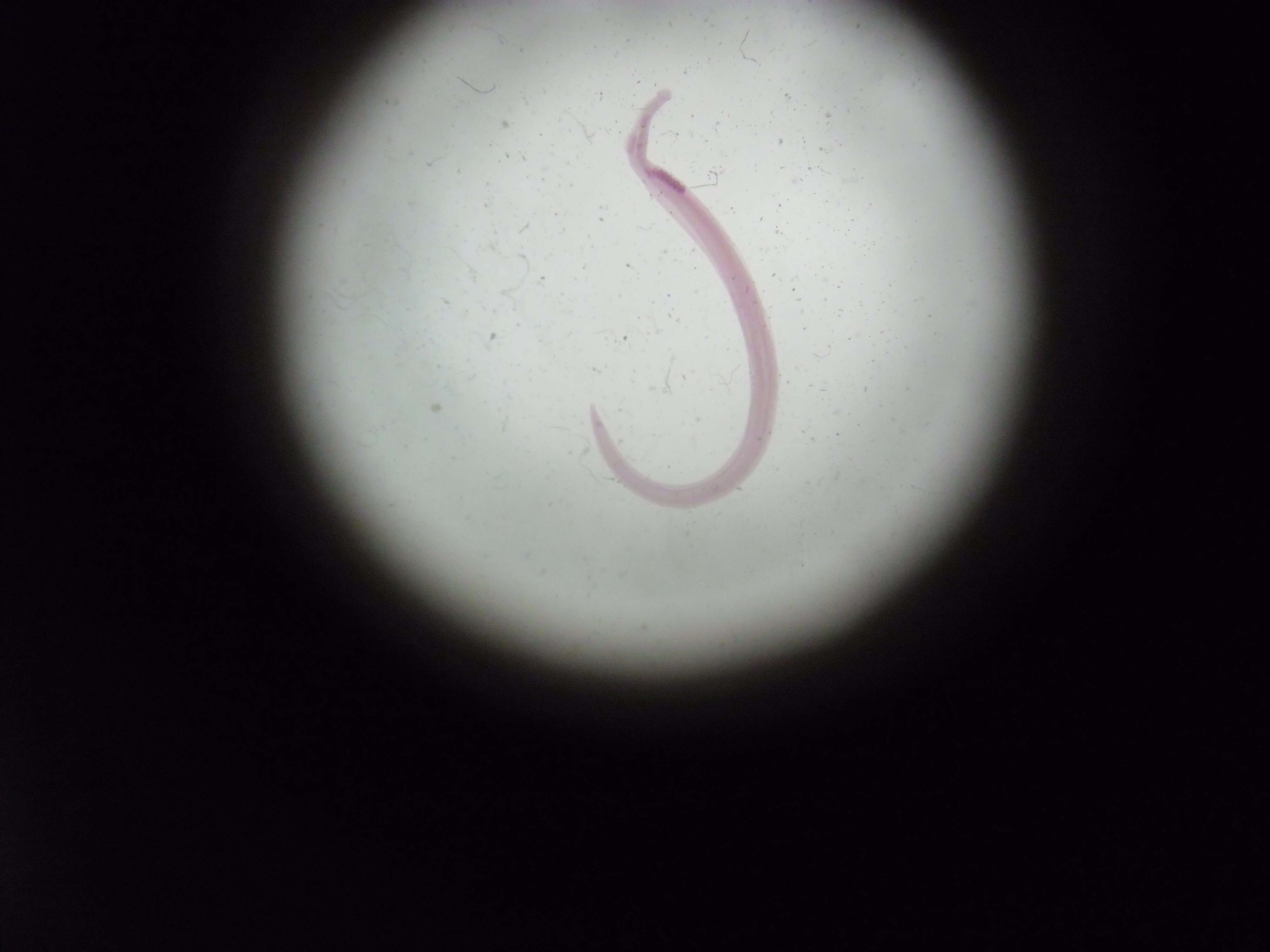
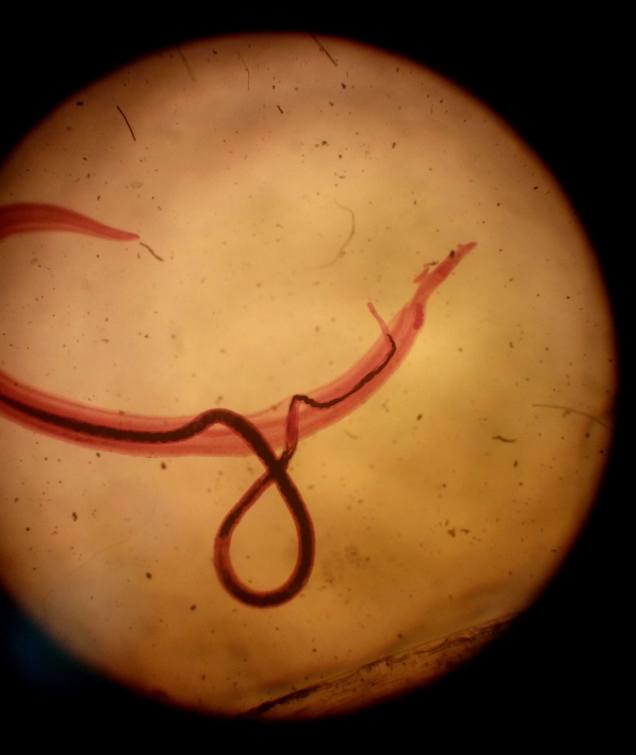
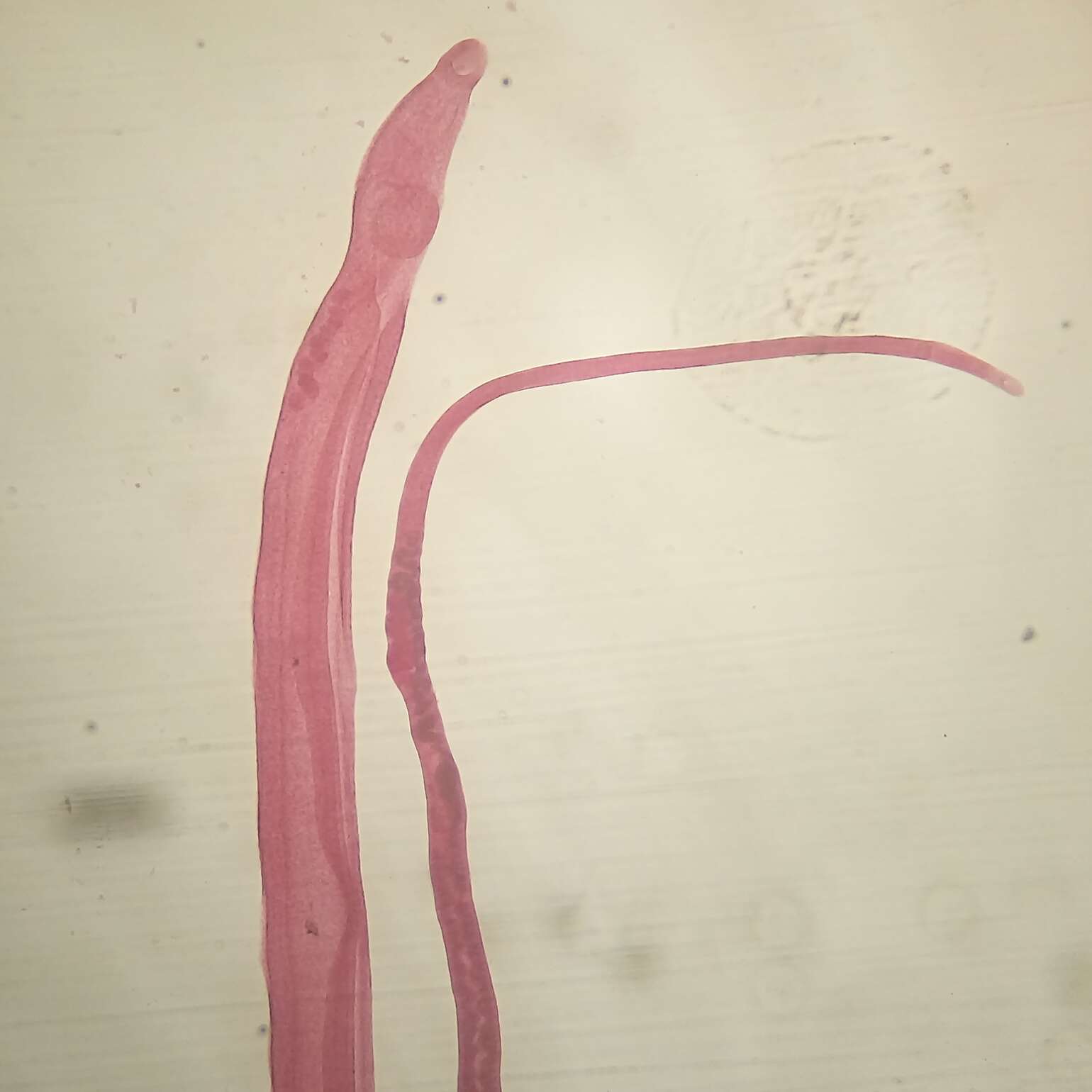
Description: العربية: الصُورة تُظهر زوج من طُفيل البلهارسية المنسونية، حيثُ يظهر الذكر ويمتلك 6-9 خِصي، وتمتلك الأنثى مِبيض غامق الصَبغة ويتواجد بشكل سابق للخط المُنصف. English: Couple of Schistosoma mansoni (Presence of male and female worms together, the male has 6-9 small testes and the female has ovary deeply stained and pre-equatorial). Date: 27 December 2016, 15:04:33. Source: Own work. Author: علاء.
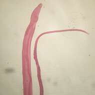
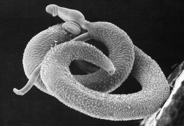
Description: English: Scanning electron micrograph of a pair of Schistosoma mansoni Русский: Пара марит трематоды Schistosoma mansoni. Более мелкая самка заключена в гинекофорный канал самца. Date: 19 February 2006 (original upload date). Source: Transferred from en.wikipedia to Commons by Gliu. Davies Laboratory Uniformed Services University Bethesda, MDdead link Information presented on USUHS web site is considered public information and may be distributed or copied. Use of appropriate byline/photo/image credits is requested. Author: The original uploader was Waisberg at English Wikipedia. Permission (Reusing this file): PD.
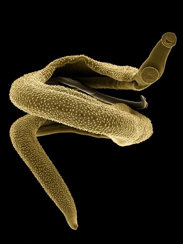
Description: Čeština: Dospělý pár krevniček Schistosoma mansoni. Větší a mohutnější samec přidržuje o mnoho štíhlejší samičku ve své břišní rýze nazývané odborně canalis gynaecophorus. Kolorovaný snímek pochází ze skenovacího elektronového mikroskopu SEM JEOL 6380 LV umístěného na PřF UK. Date: 27 April 2018. Source: Own work. Author: Jana Bulantová.
Description: Português: Casal de Schistosoma mansoni. Macho à esquerda- Fêmea à direita. English: Schistosoma mansoni couple. Male to the left. Female to the right. Date: 2018. Source: Own work. Author: Leonardo M. Lustosa.
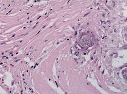
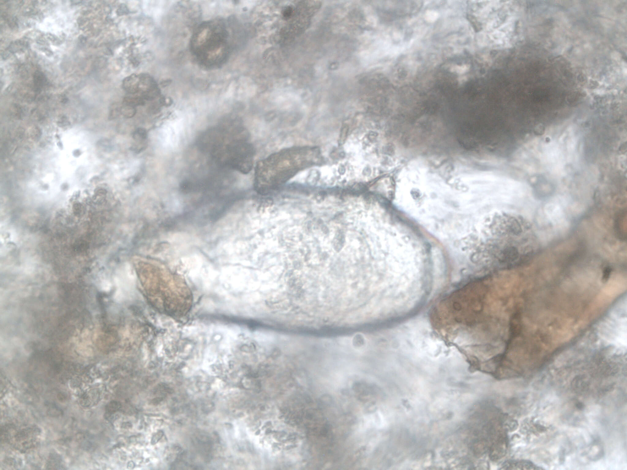
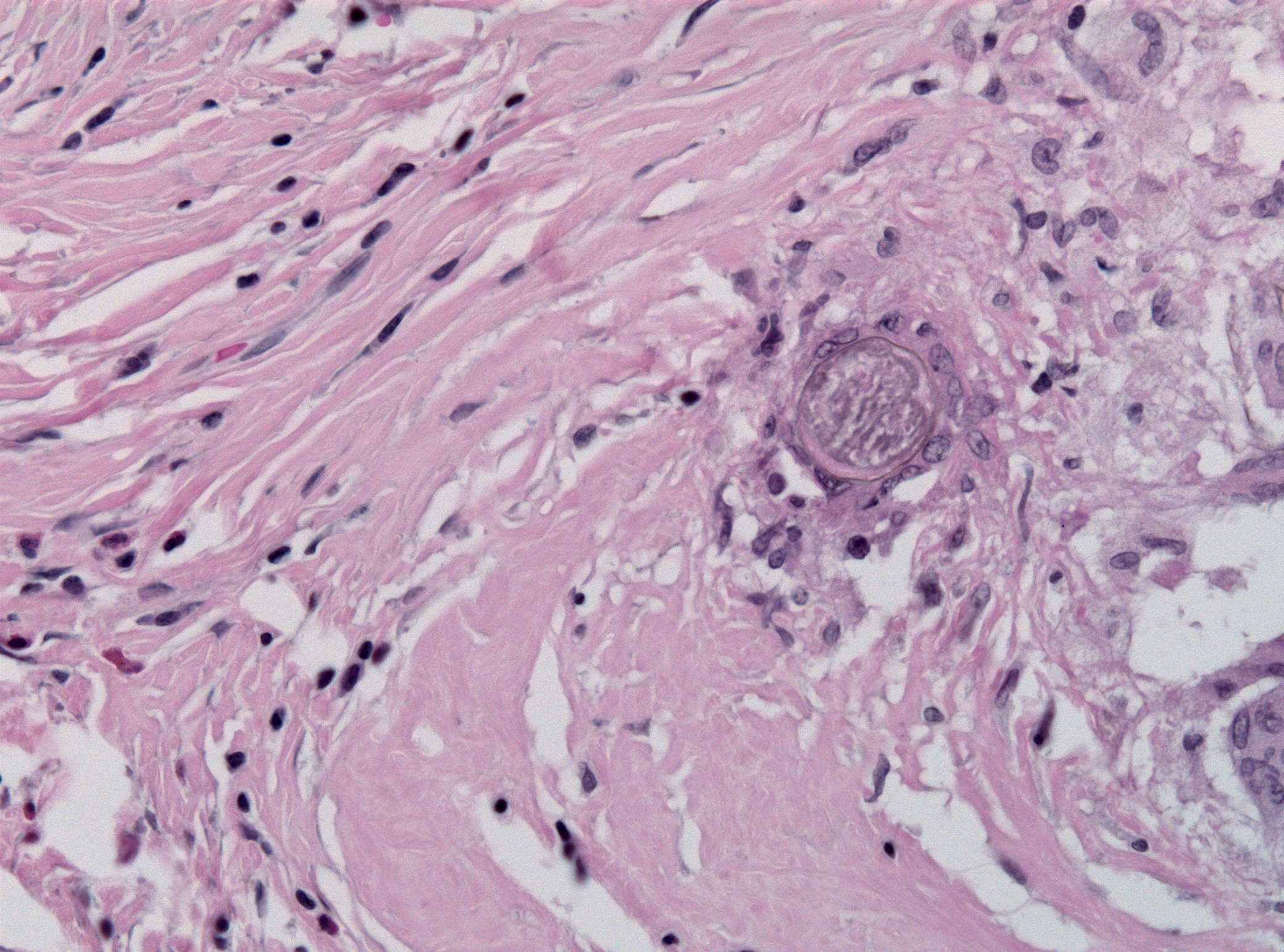
Description: English: Case with cerebral granulomas containing eggs of Schistosoma mansoni (HE stain). Date: 19 October 2010. Source: Own work. Author: Jensflorian.
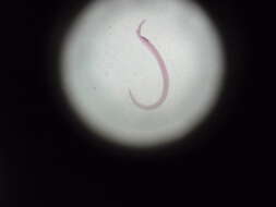
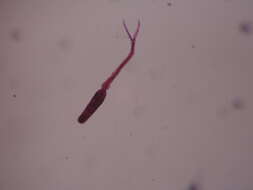
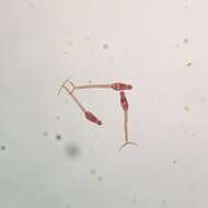
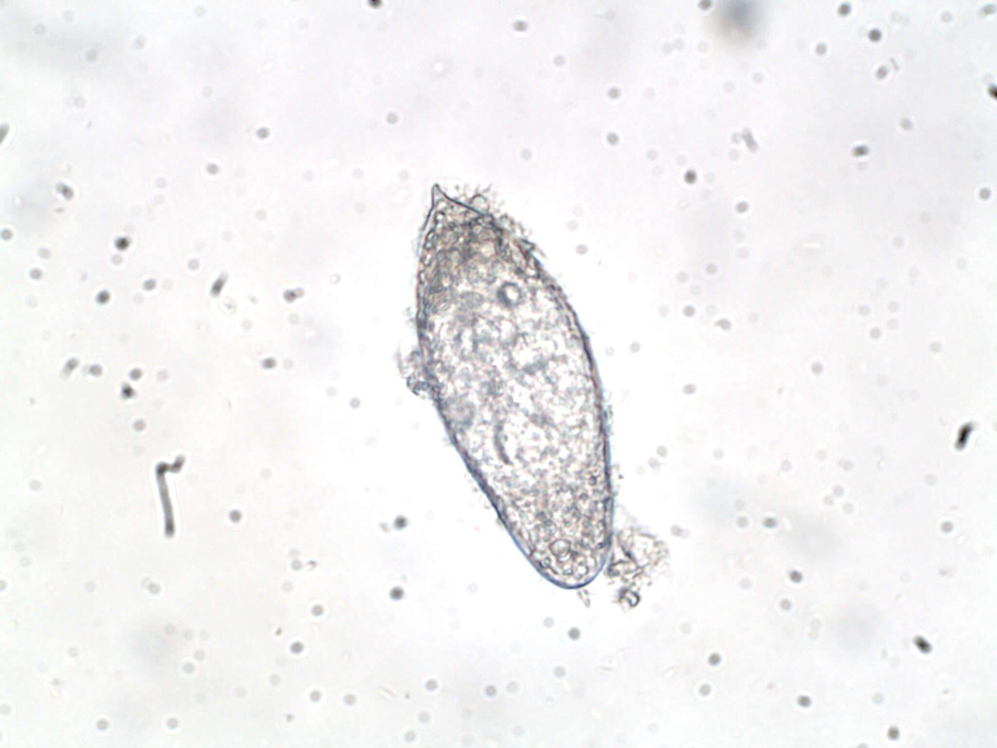
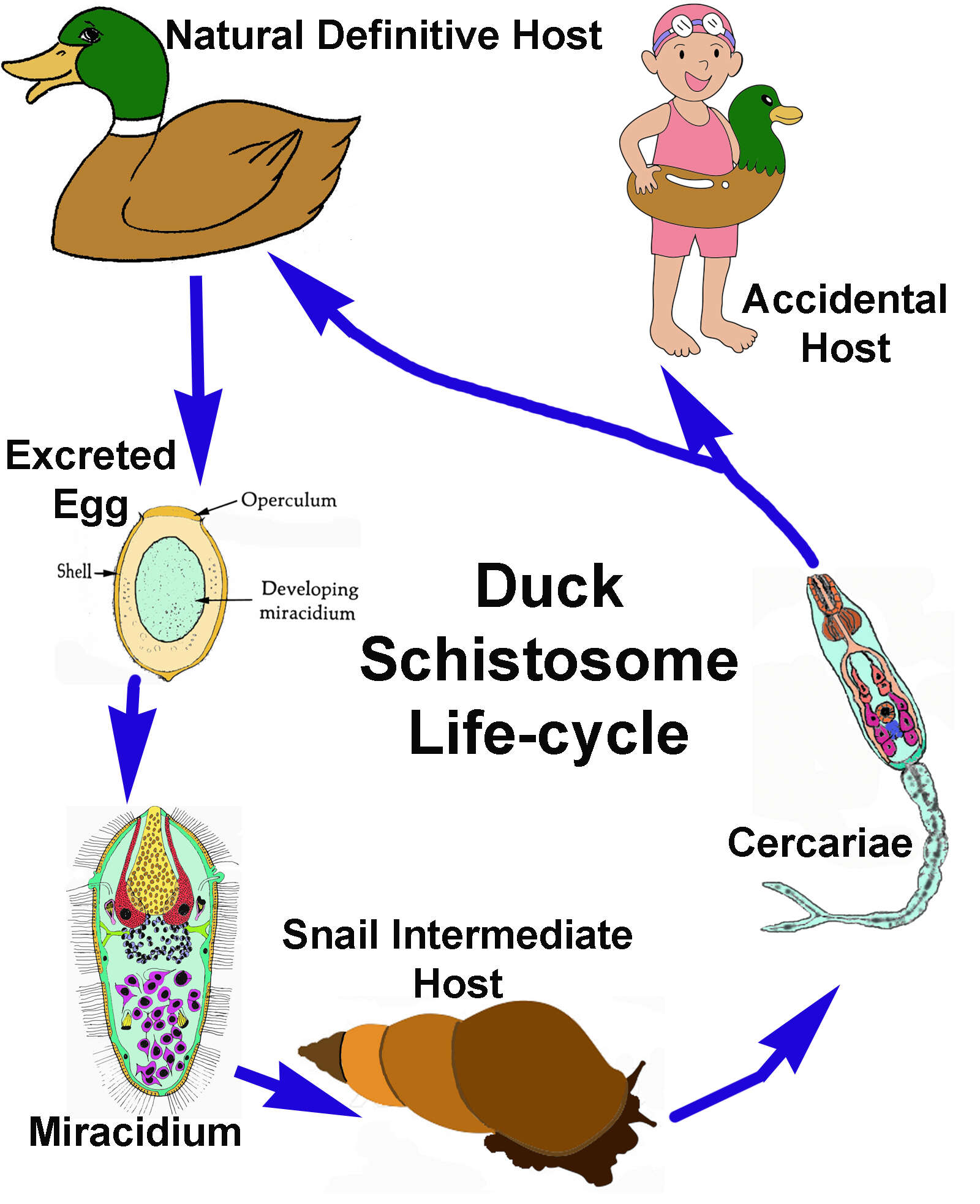
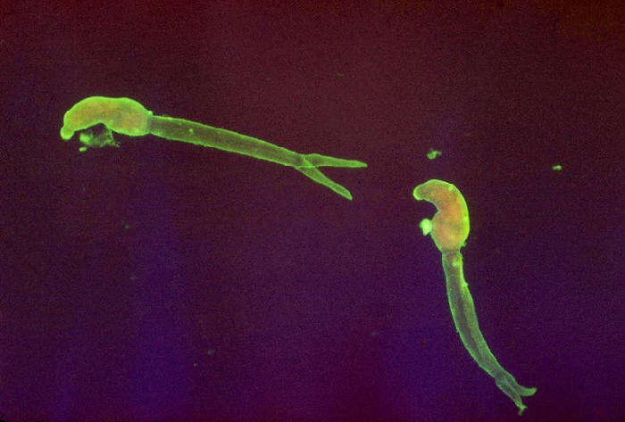
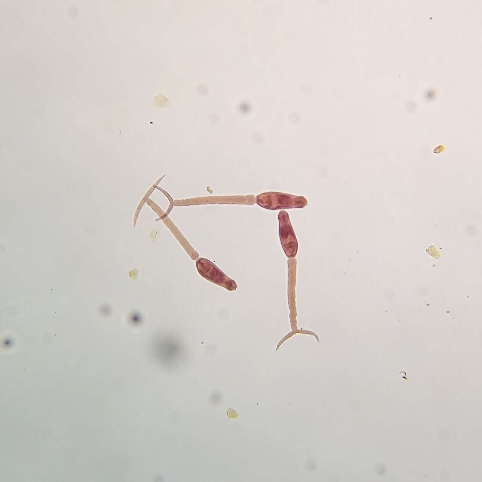
Description: English: Cercariae of Schistosoma mansoni. Indirect fluorescent antibody stain. Parasite. Date: 1963. Source: : This media comes from the Centers for Disease Control and Prevention's Public Health Image Library (PHIL), with identification number #345. Note: Not all PHIL images are public domain; be sure to check copyright status and credit authors and content providers. العربية | Deutsch | English | македонски | slovenščina | +/−. Author: Photo Credit: Content Providers(s): CDC/Dr. Sulzer. Permission(Reusing this file): PD-USGov-HHS-CDC English: None - This image is in the public domain and thus free of any copyright restrictions. As a matter of courtesy we request that the content provider be credited and notified in any public or private usage of this image.