-
-
-
-
-
-
-
-
-
-
-
-
-
-
-
-
-
-
-
-
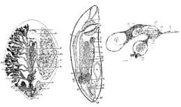
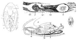
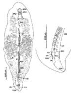
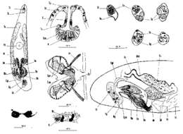
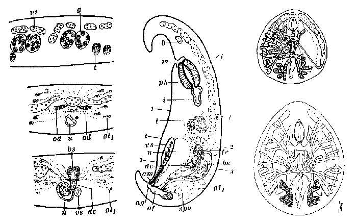
Fig. 1. Freehand drawing of squeezed animal. Fig. 2. Sagital section through proboscis. Fig. 3. Eyes (camera lucida). Fig. 4. Reconstruction of pharynx from a series of sections. Fig. 5. Epithelium (camear lucida)
-
-
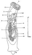
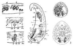
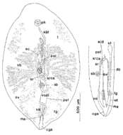
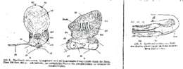
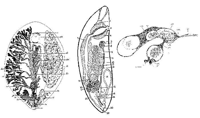
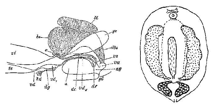
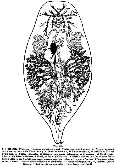
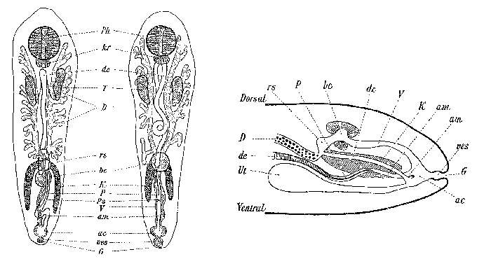
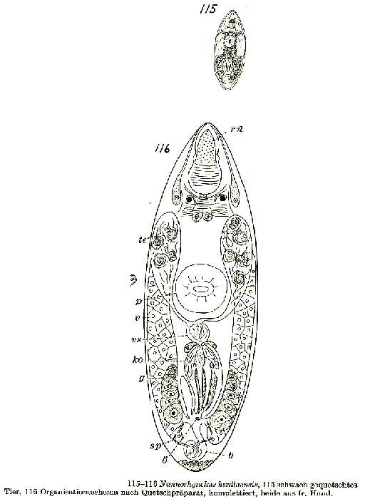
115. Slightly-squeezed animal; 116. Organisation scheme after a squeezed specimen, completed (both free-hand).
-
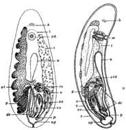
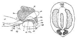
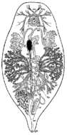
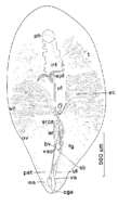
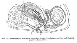
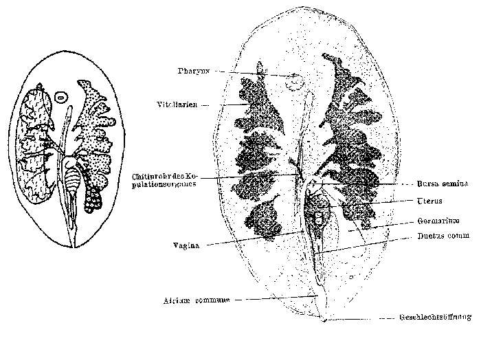
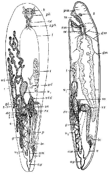
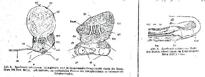
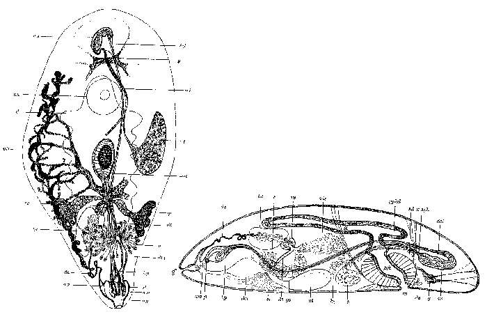
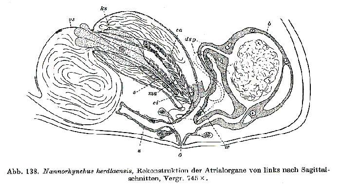
Reconstruction o fthe atrial organs from the left side from sagital sections, 745x.
-
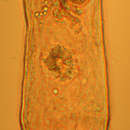
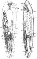
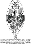
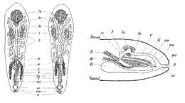
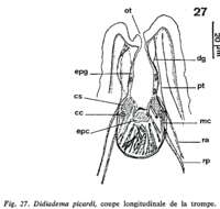
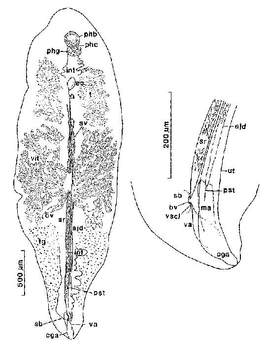
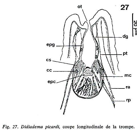
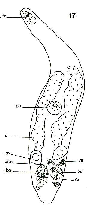
Living animal, squeezed