-
Galende, Castille and Leon, Spain
-
Pera, Faro, Portugal
-
Arbol, Catalunya, Espaa
-
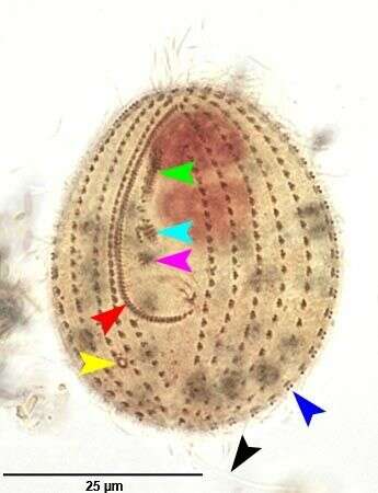
Ventral infraciliature of Calyptotricha pleuronemoides (PHILLIPS,1882). The red arrowhead marks the kinetids of the first adoral membranelle (M1). The light blue and pink arrowheads mark the kinetids of M2 and M3 respectively. the red arrowhead marks the right paraoral (undulating) membrane. The dark blue arrow head marks a dikinetid of a somatic kinety. the black arrowhead marks the single long caudal cilium. The yellow arrowhead marks the excretory pore of the contractile vacuole.Collected from organically enriched stagnant water at the edge of a freshwater stream near Boise, Idaho.Stained by the silver carbonate technique (Foissner,W. Europ. J. Protistol.27:313-330;1991).Brightfield.
-
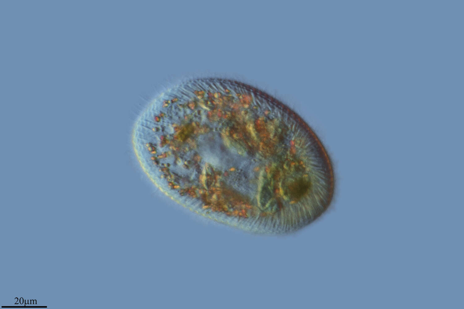
Optical section of the marine frontoniid ciliate, Schistophrya aplanata (Kahl,1933). Schistophrya is a monotypic genus. The cell outline is elongate and bluntly rounded anteriorly and posteriorly. The somatic ciliature is uniform. The pellicle is areolate (marked by uniform rectangular depressions). The slit-like oral aperture is located in mid-body and is bordered by thin slightly serrate lips (not seen in this image). The cytopharyngeal basket of fine trichites is not seen well in these images. A single contractile vacuole is located in the anterior half of the cell. There is a single ovoid macronucleus. A large aggregate of refractile dark granules is present at the anterior end. Fusiform subcortical extrusomes are present (seen in this image). S. aplanata is similar in appearance to the freshwater frontoniid ciliate, Clathrostoma viminale. Collected from a commercial saltwater aquarium in Boise, Idaho February 2004. DIC optics.
-
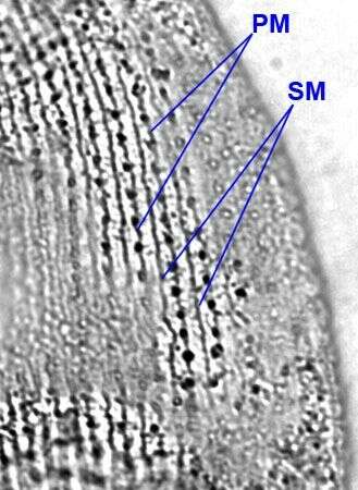
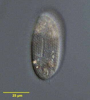
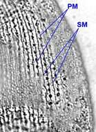
Silverline system of Colpidium kleini (FOISSNER, 1969).There is a single secondary meridian (SM) between each pair of primary meridians (PM).This feature distinguishes C. kleini from the larger C. colpoda whose silverline system shows two secondary meridians between pairs of primary meridians.Stained by the dry silver nitrate technique (see Foissner, W. Europ. J. Protistol., 27:313-330;1991).Brightfield.
-
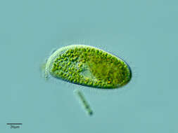
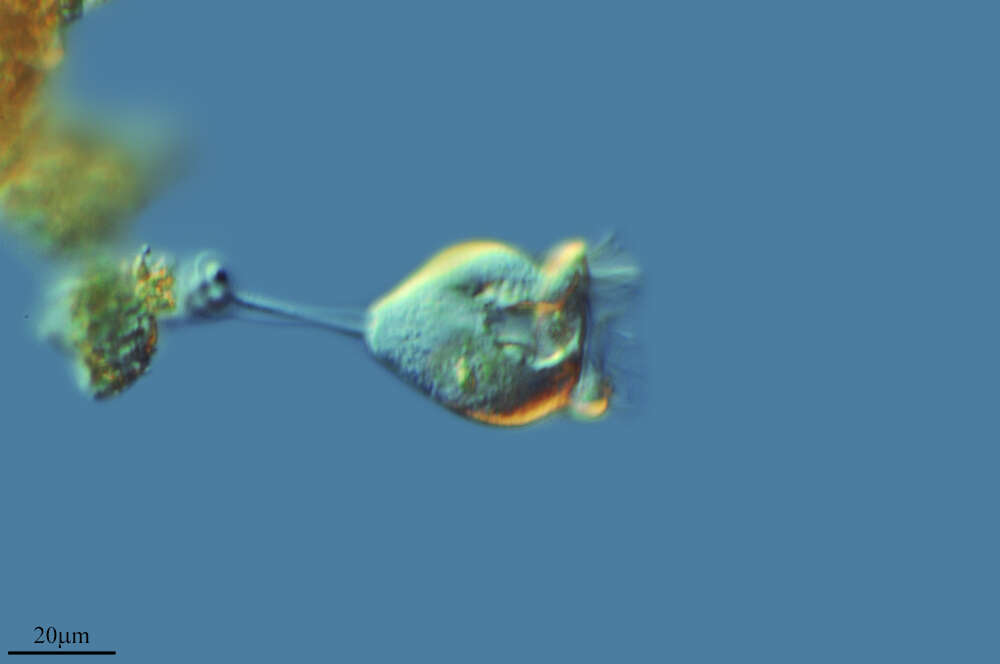
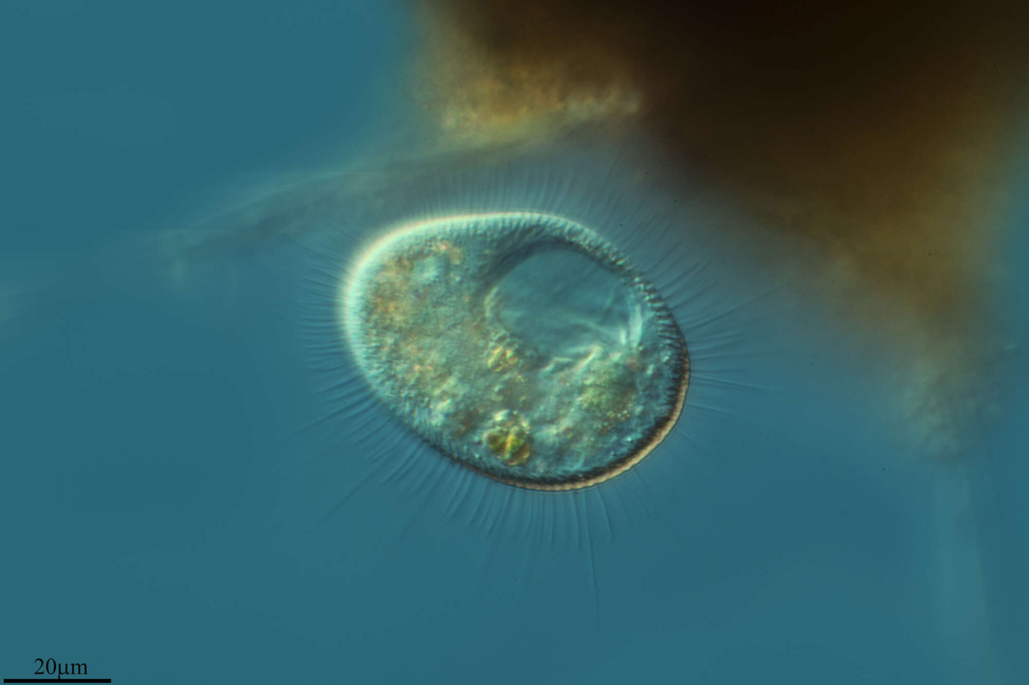
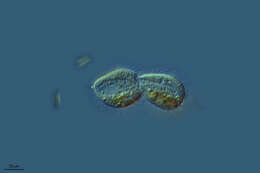
Vaginicola (vadge-in-ee-cola) is a sessile peritrich ciliate. The cells live within a lorica. often found in pairs, the cells attach to the base of the lorica by the posterior ends of the cell. they can contract into the lorica. The oral cilia form a wreath around the anterior end of the cell. No body ciliature. Differential interference contrast.
-
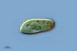
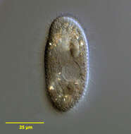
Paramecium (aurelia) (par-a-mee-see-um) is a very familiar genus of ciliates and this (morpho) species is best distinguished by the presence of two small micronuclei pressed up against the macronucleus. This image shows the peniculi or compound ciliary organelles in the mouth. Phase contrast.
-
Hoyo de Manzanares, Madrid, Spain
-
Galende, Castille and Leon, Spain
-
Madrid, Madrid, Spain
-
Gravalos, La Rioja, Spain
-
Ribadelago, Castille and Leon, Spain
-
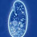
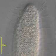
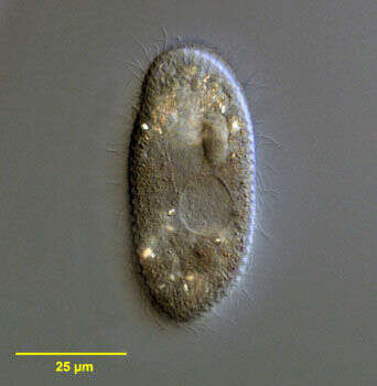
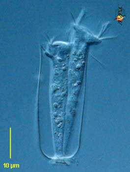
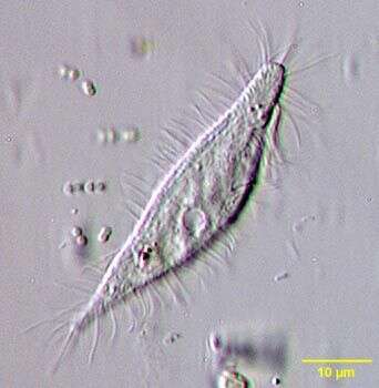
Kahlilembus attenuatus (SMITH, 1897) FOISSNER, BERGER & KOHMANN, 1994, a hymenostome ciliate. Body shape is elongate and fusiform, the anterior end rounded and the posterior end strongly tapered. The oral apparatus is seen in these images on the organism's left extending posteriorly from the anterior end about one quarter of the body length. 3 adoral membranelles occupy the area just anterior to the cytostome and a prominent flag-like undulating membrane lines the right side of the cytostome. Somatic cilia are arranged in 8 to 10 longitudinal kineties. A spherical macronucleus is located centrally. The contractile vacuole (seen well here) is located just posterior to the midbody. Primarily bactiverous. From freshwater pond near Boise, Idaho. Oblique illumination.
-
Portrait of the marine frontoniid ciliate, Schistophrya aplanata (Kahl, 1933). Schistophrya is a monotypic genus. The cell outline is elongate and bluntly rounded anteriorly and posteriorly. The somatic ciliature is uniform. The pellicle is areolate (marked by uniform rectangular depressions). The slit-like oral aperture is located in mid-body and is bordered by thin slightly serrate lips (seen well in this image). The cytopharyngeal basket of fine trichites is not seen well in these images. A single contractile vacuole is located in the anterior half of the cell. There is a single ovoid macronucleus. A large aggregate of refractile dark granules is present at the anterior end. Fusiform subcortical extrusomes are present. S. aplanata is similar in appearance to the freshwater frontoniid ciliate, Clathrostoma viminale. Collected from a commercial saltwater aquarium in Boise, Idaho February 2004. DIC optics.
-
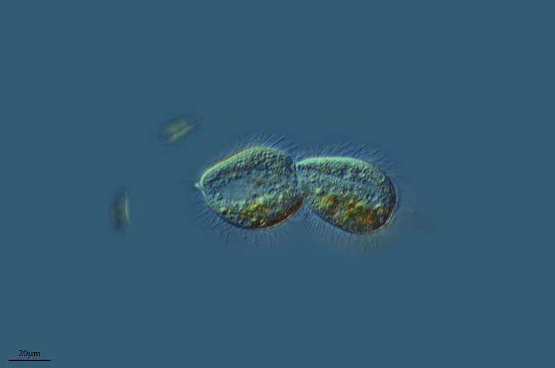
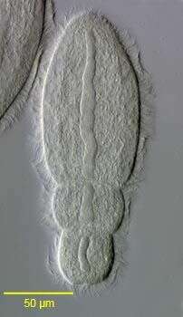
Group portrait of the endocommensal astomatid ciliate, Anoplophrya maupasi(Cépède,1910) in the digestive tract of the oligochaete, Aelosoma hemprichi (see description of characteristics in: Lom, J., 1956. Arch. Protistenk., 101: 286). Collected from a freshwater pond near Boise, Idaho. June 2005. DIC.
-
Vaginicola (vadge-in-ee-cola) is a sessile peritrich ciliate. The cells live within a lorica. often found in pairs, the cells attach to the base of the lorica by the posterior ends of the cell. they can contract into the lorica. The oral cilia form a wreath around the anterior end of the cell. No body ciliature. Phase contrast.
-
Paramecium (aurelia) (par-a-mee-see-um) is a very familiar genus of ciliates and this (morpho) species is best distinguished by the presence of two small micronuclei pressed up against the macronucleus. They can be seen here to the north of the nucleus. Phase contrast.
-
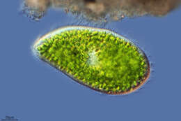
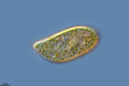
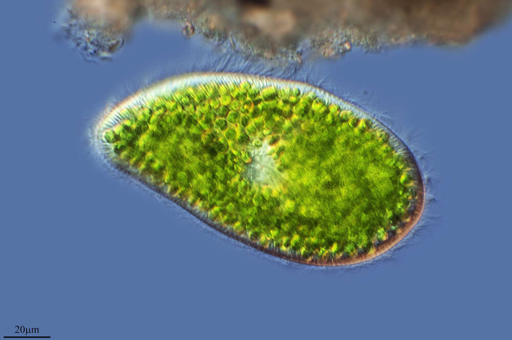
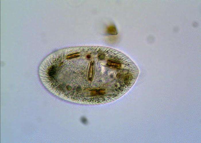
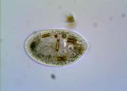
Ovoid, 60-100 micron long. Numerous large trichocysts.
-
Villoslada de Cameros, La Rioja, Spain
-
Detail view of Dexiotricha granulosa (Kent, 1881) Foissner, 1994 showing the prominent ring-shaped cytoplasmic glycogen granules. From freshwater pond near Boise, Idaho. DIC.
-
Portrait of the marine frontoniid ciliate, Schistophrya aplanata (Kahl,1933). Schistophrya is a monotypic genus. The cell outline is elongate and bluntly rounded anteriorly and posteriorly. The somatic ciliature is uniform. The pellicle is areolate (marked by uniform rectangular depressions). The slit-like oral aperture is located in mid-body and is bordered by thin slightly serrate lips (seen well in this image). The cytopharyngeal basket of fine trichites is not seen well in these images. A single contractile vacuole is located in the anterior half of the cell. There is a single ovoid macronucleus. A large aggregate of refractile dark granules is present at the anterior end. Fusiform subcortical extrusomes are present. S. aplanata is similar in appearance to the freshwater frontoniid ciliate, Clathrostoma viminale. Collected from a commercial saltwater aquarium in Boise, Idaho February 2004. DIC optics.
-
View of the bandform macronucleus of the endocommensal astomatid ciliate, Anoplophrya maupasi (Cépède,1910) from the digestive tract of the oligochaete,Aelosoma hemprichi (see description of characteristics in: Lom, J., Arch. Protistenk.,101:p.286,1956).There are two smaller opisthes seen here. Collected from a freshwater pond near Boise, Idaho. June 2005. DIC.
-