-
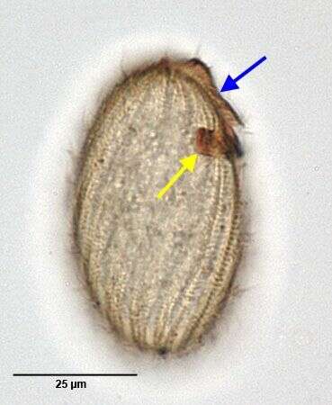
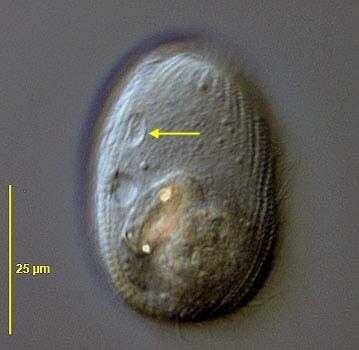
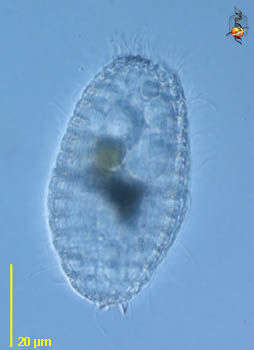
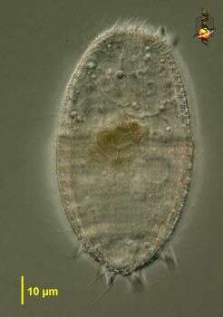
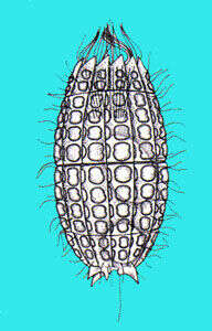
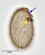
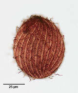
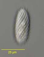
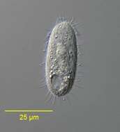
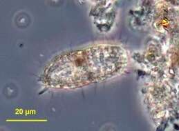
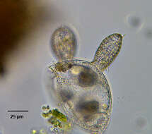
Infraciliature (right lateral view) of the prorodontid ciliate, Placus luciae (Kahl,1926) Kahl,1930. The cell is ovoid and slightly laterally compressed. The pellicle has broad slightly spiral ridges separated by narrow furrows. The small oval oral aperture is apical anterior, surrounded by a ring of unciliated kinetosomes. The cytopharynx is supported by fine trichites. The somatic ciliature uniform, with cilia. There is a "dorsal brush" (blue arrow), about 1/4 to 1/3 cell length, composed of a double parallel row of kinetosomes bearing longer cilia. It arises adjacent to the oral aperture and terminates at a ventral circular "pit" or "fossette" which has a supporting ring of transversely oriented trichites (yellow arrow). The function of this structure is unknown. The ovoid macronucleus is centrally located. The contractile vacuole is posterolateral. Collected from a slow-flowing freshwater stream near Boise, Idaho.Stained by the silver carbonate technic (see Foissner, W.Europ. J. Protistol.27,313-330;1991). Brightfield.
-
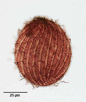
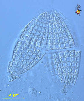
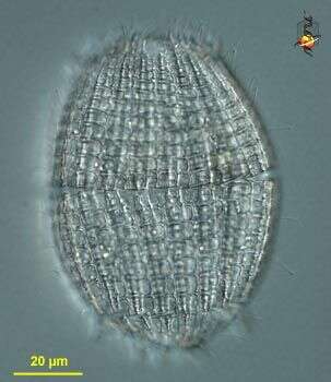
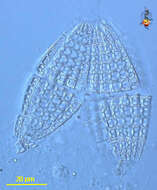
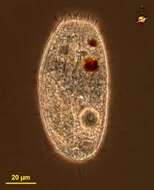
Infraciliature (dorsal view) of the prorodontid ciliate, Placus luciae (Kahl,1926) Kahl,1930. The cell is ovoid and slightly laterally compressed. The pellicle has broad slightly spiral ridges separated by narrow furrows.In this preparation the reticulate pattern of the ridges is seen. Fine transverse striations cross the narrow furrows between the somatic kineties to a darkly stained longitudinal fibril on the right margin of the ridges. The small oval oral aperture is apical anterior, surrounded by a ring of unciliated kinetosomes. The cytopharynx is supported by fine trichites. The somatic ciliature uniform, with cilia. There is a "brush" , about 1/4 to 1/3 cell length, composed of a double parallel row of kinetosomes bearing longer cilia. This arises adjacent to the oral aperture and terminates at a ventral circular "pit" which has a supporting ring of transversely oriented trichites. The function of this structure is unknown . The ovoid macronucleus is centrally located. The contractile vacuole is posterolateral. Collected from a slow-flowing freshwater stream near Boise, Idaho.Stained by the silver carbonate technique (see Foissner, W.Europ. J. Protistol.27,313-330;1991) . Brightfield.
-
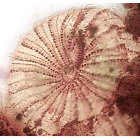
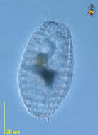
Anterior view of the infraciliature of the prorodontid ciliate, Placus luciae (Kahl,1926) Kahl,1930. The cell is ovoid and slightly laterally compressed. The pellicle has broad slightly spiral ridges separated by narrow furrows. The small oval oral aperture is apical anterior, surrounded by a ring of unciliated kinetosomes (seen well here). The cytopharynx is supported by fine trichites. The somatic ciliature uniform, with cilia. There is a "brush" (seen here at 12 0'clock) composed of a double parallel row of kinetosomes bearing longer cilia. This is about 1/4 to 1/3 cell length arising adjacent to the oral aperture. It terminates at a ventral circular "pit" which has a supporting ring of transversely oriented trichites. The function of this structure is unknown . Collected from a slow-flowing freshwater stream near Boise, Idaho.Stained by the silver carbonate technic (see Foissner, W.Europ. J. Protistol.27,313-330;1991). Brightfield.
-
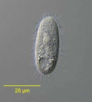
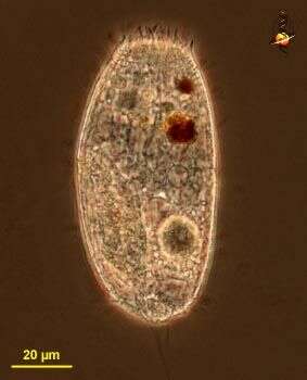
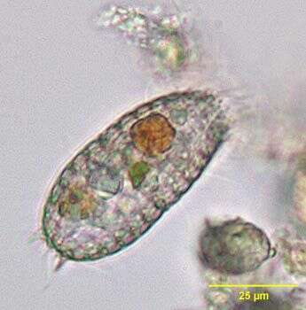
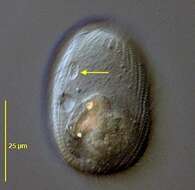
Portrait (left lateral view) of the prorodontid ciliate, Placus luciae (Kahl,1926) Kahl,1930. The cell is ovoid and slightly laterally compressed. The pellicle has broad slightly spiral ridges separated by narrow furrows. The small oval oral aperture is apical anterior, surrounded by a ring of unciliated kinetosomes. The cytopharynx is supported by fine trichites. The somatic ciliature uniform, with cilia.There is a "brush" composed of a double parallel row of kinetosomes bearing longer cilia. This is about 1/4 to 1/3 cell length arising adjacent to the oral aperture. It terminates at a ventral circular "pit" (arrow) which has a supporting ring of transversely oriented trichites. The function of this structure is unknown. The ovoid macronucleus is centrally located. The contractile vacuole is poster lateral. Collected from a slow-flowing freshwater stream near Boise, Idaho. DIC.
-
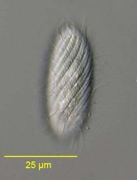
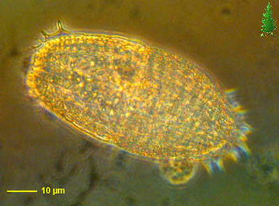
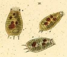
Surface detail of the prorodontid ciliate, Placus striatus (Cohn, 1866). The somatic kineties of P. striatus are distinctly more spiralled than those of P. luciae.DIC.
-
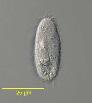
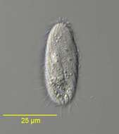
Portrait of the prorodontid ciliate, Placus striatus (Cohn,1866).DIC.
-
Portrait of the prorodontid ciliate, Placus striatus (Cohn,1866).DIC.
-
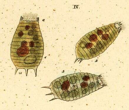
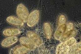
Coleps (coal-eps) - the pirranha or hyaena of the ciliate world. Eats detritus but definitely has a penchant for bits of animals - and is not squeamish about attacking living but damaged organisms. Shed exoskeletons of crustacea are attacked, the Coleps then gnaws on the tissue (and is thus referred to as histophagous). Distinguished by having calcareous plates around the outside of the body, shown here dissociated from a cell that was killed by compression. Differential interference contrast.
-
Coleps (coal-eps) - the pirranha or hyaena of the ciliate world. Eats detritus but definitely has a penchant for bits of animals - and is not squeamish about attacking living but damaged organisms. Shed exoskeletons of crustacea are attacked, the Coleps then gnaws on the tissue (and is thus referred to as histophagous). Distinguished by having calcareous plates around the outside of the body. Differential interference contrast.
-
Coleps (coal-eps) - the pirranha or hyaena of the ciliate world. Eats detritus but definitely has a penchant for bits of animals - and is not squeamish about attacking living but damaged organisms. Shed exoskeletons of crustacea are attacked, the Coleps then gnaws on the tissue (and is thus referred to as histophagous). This cell seems to have been eating algae. Also eats other detrital material. Distinguished by having calcareous plates around the outside of the body. Phase contrast.
-
Coleps (coal-eps) - the pirranha or hyaena of the ciliate world. Eats detritus but definitely has a penchant for bits of animals - and is not squeamish about attacking living but damaged organisms. Shed exoskeletons of crustacea are attacked, the Coleps then gnaws on the tissue (and is thus referred to as histophagous). Distinguished by having calcareous plates around the outside of the body. This is a recent product of cell division, it is derived from the posterior half of the parent cell - because the back half of the cell contains thickened cortical plates, whereas the front part has weakly developed plates. Differential interference contrast.
-
Coleps (coal-eps) - the pirranha or hyaena of the ciliate world. Eats detritus but definitely has a penchant for bits of animals - and is not squeamish about attacking living but damaged organisms. Shed exoskeletons of crustacea are attacked, the Coleps then gnaws on the tissue (and is thus referred to as histophagous). Distinguished by having calcareous plates around the outside of the body, and with posterior spines. Differential interference contrast.
-
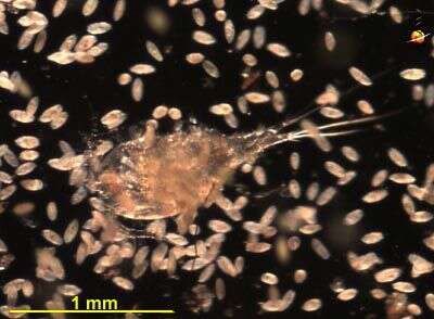
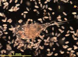
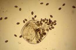
Coleps (coal-eps) - the pirranha or hyaena of the ciliate world. Eats detritus but definitely has a penchant for bits of animals - and is not squeamish about attacking living but damaged organisms. Shed exoskeletons of crustacea are attacked, the Coleps then gnaws on the tissue (and is thus referred to as histophagous). Here Coleps is seen attacking a damaged copepod. Dark ground.
-
Coleps (coe-leps) is a predatory ciliate, made distinctive by the calcareous plates which enclose the cell. anterior end usually flattened, posterior end typically with a number of short spines. Most usually consumed dead and decaying animal matter. Not common. Phase contrast. Material from Nymph Creek and Nymph Lake, thermal sites within Yellowstone National Park, photograph by Kathy Sheehan and David Patterson.
-
-
-
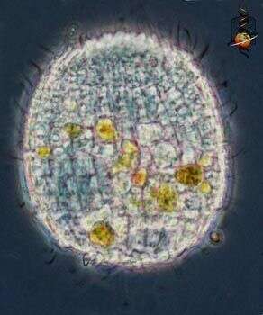
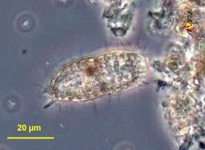
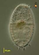
Portrait of Coleps (Nitzsch,1827) a common barrel shaped ciliate with intricate ectoplasmic plates composed of calcium carbonate and variable numbers of posterior and, less commonly, anterior spines. Fine structure of plates is used in species identification. Histophagous, they feed voraciously on dead protists and metazoans. From freshwater pond near Boise, Idaho. Brightfield.
-
Coleps observed in freshwater sediments in the vicinity of Broome, Western Australia in September 2003. This image was taken using phase contrast optics. This work was supported by the Australian Biological Resources Study.
-
Collected from Cumloden Swamp on September 9, 2002.
-
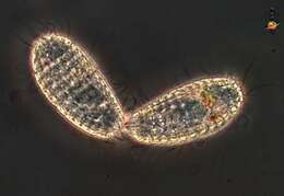
Portrait of Coleps (Nitzsch,1827), a common barrel shaped ciliate with intricate ectoplasmic plates composed of calcium carbonate and variable numbers of posterior and, less commonly, anterior spines. Fine structure of plates is used in species identification. Histophagous, they feed voraciously on dead protists and metazoans. These individuals are devouring a deceased Lembadion. From freshwater pond near Boise, Idaho. Brightfield.
-
-
Coleps is one of nature's little scavengers. It will attach dead and decaying metazoa, eating residual pieces of tissue on cast off exoskeletons or attaching organisms that are wounded. It will also eat detrital material. This species retains chloroplasts from algal food - a phenomenon referred to as kleptoplasty. Chloroplasts are derived from blue-green algae that have survived symbiotically within other cells, and kleptoplasty exploits aspects of the autonomy of these organelles. Phase contrast microscopy.
-
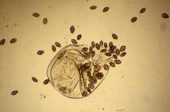
Coleps is a ciliate and one of nature's little scavengers. In nature it can eat remanent tissue attached to cast off exoskeletons of arthropods, or will attack wounded organisms. Here about 50 cells crowd round the remains of a Daphnia.
-
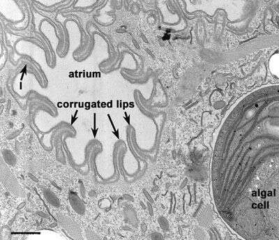
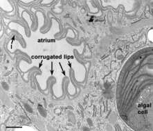
Opening to oral region at the anterior end of Coleps hirtus. Corrugated lips containing lamellae (l) surround the atrium. The oral region is bordered by cilia. Rods extend inward (see Figs. 12 and 13) from these cilia. Toxicysts and mucocysts lie in the cytosol. An algal cell, enclosed in a smooth membrane is also present. EM taken on 3/24/69 by R. Allen with Philips 300 TEM. Neg. 14,800X. Bar = 0.5µm.
This image is available in Richard Allen's collection.