-
-
-
-
-
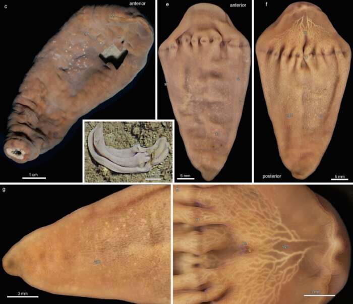
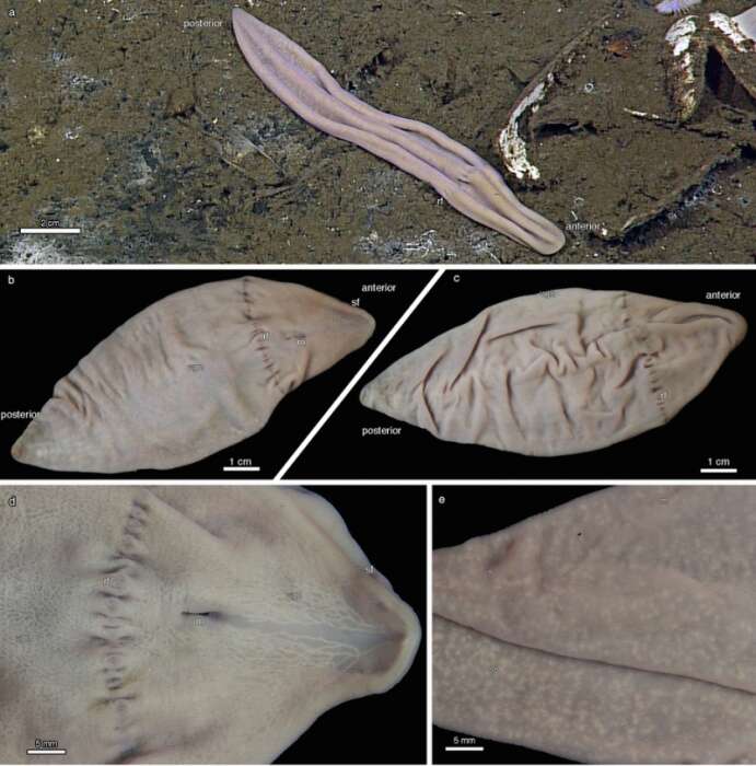
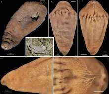
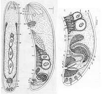
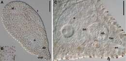
c, Ventral view of the holotype SIO-BIC BI1037. Although highly contracted and incomplete (the posterior end was removed and frozen and the ventral area removed for histology), the specimen is still over 10?cm long. The mouth (m), ring furrow (rf) and side furrow (sf) are visible. d, Frame grab of paratype SIO-BIC BI1039, a female in situ. e, Dorsal view of paratype SIO-BIC BI1039 (relaxed) showing ring furrow (rf), side furrow (sf), oocytes in body wall (oo) and part of the ventral surface (v). f, Ventral view of paratype SIO-BIC BI1039 showing the mouth (m), ring furrow (rf), oocytes in body wall (oo) and part of the epidermal ventral glandular network (vgn). g, Close-up of the ventral posterior of paratype SIO-BIC BI1039 showing the trailing off of the ventral glandular network (vgn) and oocytes clearly visible in the body wall. h, Close-up of the ventral anterior of paratype SIO-BIC BI1039 showing the mouth (m), ring furrow (rf) and the beginning of the ventral glandular network (vgn) near the anterior tip of the animal.
-
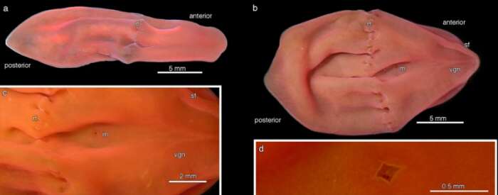
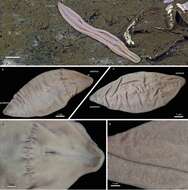
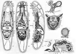
b, Ventral view of the holotype SIO-BIC BI1041, a male (relaxed) showing mouth (m), ring furrow (rf), side furrow (sf) and epidermal ventral glandular network (vgn). c, Dorsal view of the holotype showing the ring furrow (rf). The ventral glandular network (vgn) is visible where part of the ventral side is exposed. d, Close-up of the ventral anterior end of female paratype SIO-BIC BI1044, showing mouth (m), ring furrow (rf), side furrow (sf) and beginning of ventral glandular network (vgn) near the anterior tip of the animal. e, Close-up of the dorsal posterior of female paratype SIO-BIC BI1044, showing oocytes of different sizes distributed in the parenchyma
-
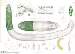
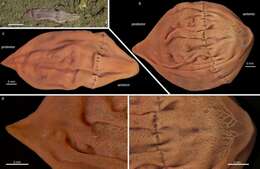
a, Frame grab of holotype SIO-BIC BI1040, a female, from the Guaymas Transform Fault, Gulf of California, Mexico, at ~1,700?m depth in situ. The ring furrow (rf) is visible towards the anterior end. b, Ventral view of the holotype SIO-BIC BI1040, (relaxed) showing mouth (m), ring furrow (rf), oocytes (oo), side furrow (sf) and epidermal ventral glandular network (vgn). Part of the dorsal side (d) is visible. c, Dorsal view of paratype SIO-BIC BI1039 showing ring furrow (rf) and oocytes (oo). d, Close-up of the ventral posterior of the holotype, showing the trailing off of the ventral glandular network (vgn), oocytes and the distinctively tapering posterior tip. e, Close-up of the anterior end of the holotype, showing the mouth (m), ring furrow (rf) and the beginning of the ventral glandular network (vgn) near the anterior tip of the animal.
-
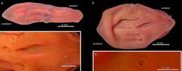
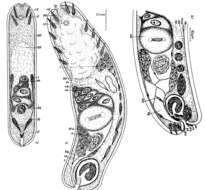
a, Dorsal view of the live unrelaxed holotype SIO-BIC BI1036, showing ring furrow (rf). b, Ventral view of the holotype (relaxed) showing mouth (m), ring furrow (rf) side furrow (sf) and epidermal ventral glandular network (vgn). c, Close-up of the anterior end of the holotype, showing mouth (m), ring furrow (rf), side furrow (sf) and the ventral glandular network (vgn) near the anterior tip of the animal. d, Close-up of the diamond-shaped mouth.
-
-
-
-
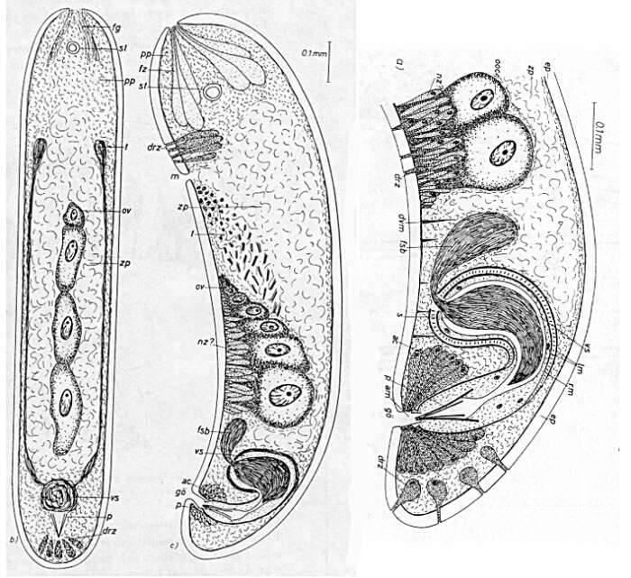
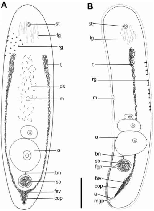
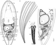
Philactinoposthia brevis sp.nov. Reconstructions show arrangement of organs. A. Dorsal reconstruction of whole living animal. B. Sagittal reconstruction of the whole animal. Scale bar: 80 μm.
-
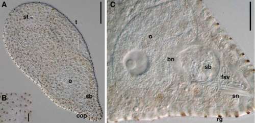
Philactinoposthia brevis sp.nov. Photomicrographs of living specimens ?C. A. Dorsal view of whole specimen. Scale bar: 60 μm B. Dorsal view of posterior part of body. Scale bar: 40 μm.
-
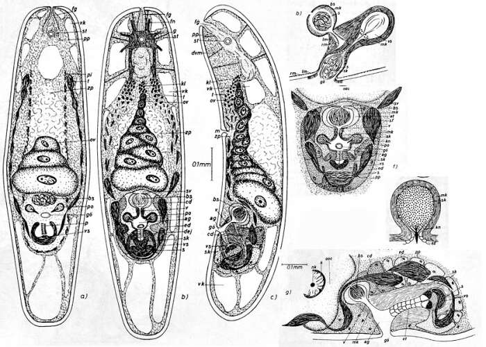
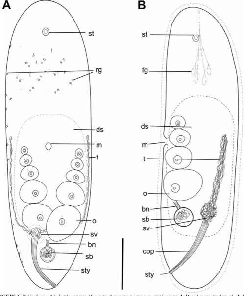
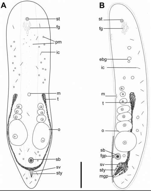
Philactinoposthia ischiae sp.nov. Reconstructions show arrangement of organs. A. Dorsal reconstruction of whole living animal. B. Sagittal reconstruction of the whole animal. Scale bar: 90 μm.
-
Philactinoposthia ischiae sp.nov. Photomicrographs of living specimens. A. Dorsal view of whole specimen. Scale bar: 50 μm. B. View of posterior part of body. Scale bar: 70 μm. C. Dorsal view of anterior part of body. Scale bar: 45 ?m.
-
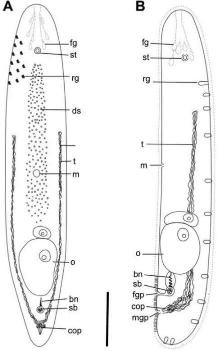
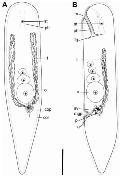
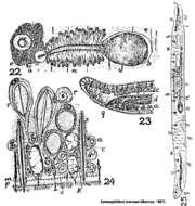
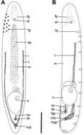
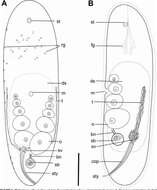
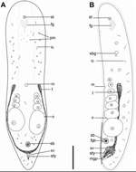
A. Dorsal reconstruction of whole living animal. B. Sagittal reconstruction of the whole animal. Scale bar: 500 μm.
-
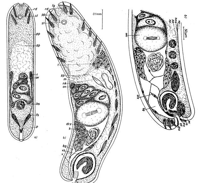
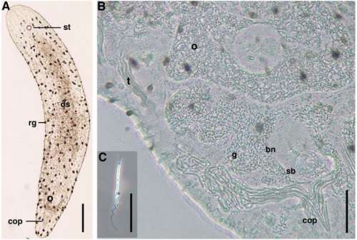
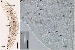
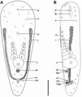
A. Dorsal view of whole squeezed specimen. Scale bar: 300 μm. B. View of posterior part of body. Scale bar: 90 ?m. C. Female copulatory organ and rhabdoid glands. Scale bar: 110 ?m.
-
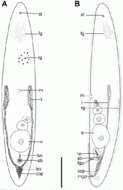
A. Dorsal reconstruction of whole living animal. B. Sagittal reconstruction of the whole animal. Scale bar: 80 μm.
-
A. Dorsal view of whole squeezed specimen. Scale bar: 80 μm. B. View of posterior part of body. Scale bar: 15 μm. C. Rhabdoid glands. Scale bar: 10 μm.
-
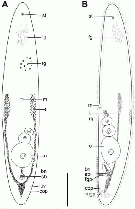
Childia aculifera sp.nov. Reconstructions show arrangement of organs. A. Dorsal reconstruction of whole living animal. B. Sagittal reconstruction of the whole animal. Scale bar: 220 μm.
-
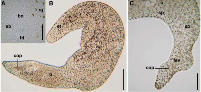
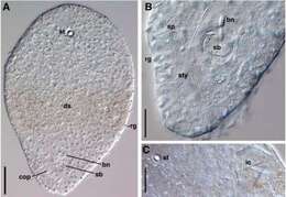
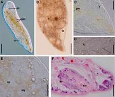
Childia aculifera sp.nov. ?E photomicrographs of living specimens. A. Dorsal view of whole specimen. Scale bar: 250 μm. B. View of posterior part of body. Scale bar: 100 μm. C. View of posterior end. Scale bar: 75 ?m. D. Inclusions (arrowhead). Scale bar: 30 μm. E. View of posterior part of body. Scale bar: 40 μm. F. Photomicrograph of sagittal histological sections stained with hematoxylin-eosin. Scale bar: 30 μm.
-
A. Dorsal view of whole specimen. Scale bar: 500 μm. B. View of mid-body. Scale bar: 60 μm. C. Male copulatory organ and inclusions. Scale bar: 30 μm. D. View of posterior end of body. Scale bar: 80 μm.
-
A. Dorsal reconstruction of whole living animal. B. Sagittal reconstruction of the whole animal. Scale bar: 500 μm.
-
Pharyngia furva sp.nov. Reconstructions show arrangement of organs. A. Dorsal reconstruction of whole living animal. B. Sagittal reconstruction of the whole animal. Scale bar: 300 μm