-
Sydney, New South Wales, Australia
-
Xin Qi, Xiaolong Lin, Xinhua Wang
Zookeys
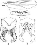
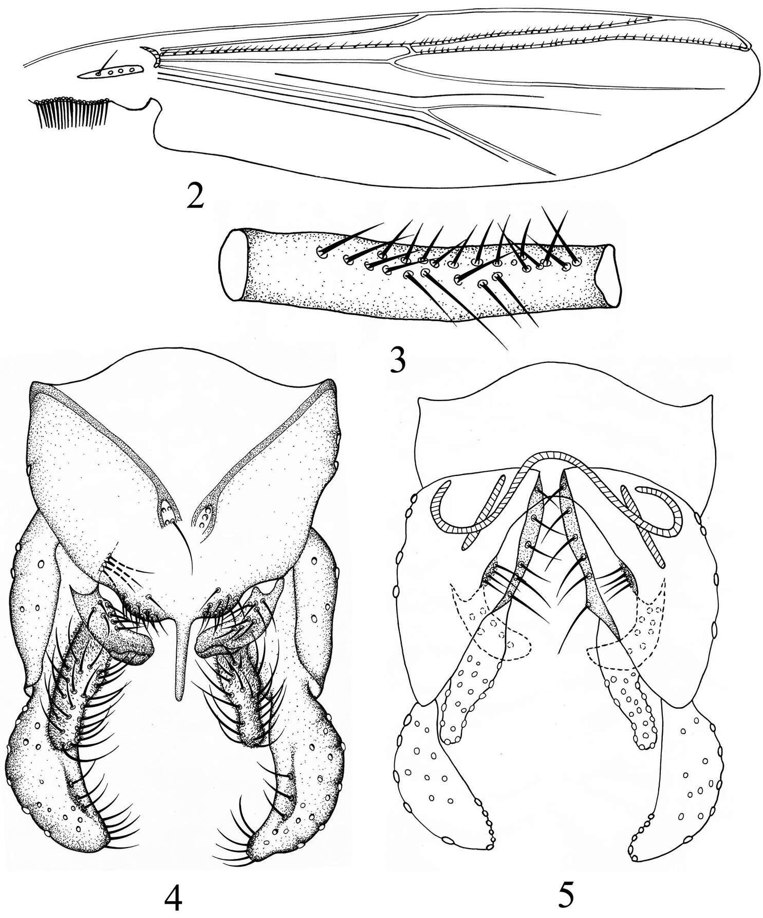
Figures 2–5.Microtendipes zhejiangensis sp.n., male. 2 wing 3 two rows of directed setae in front femur 4 hypopygium (dorsal view) 5 hypopygium (ventral view).
-
Xiaolong Lin, Xin Qi, Ruilei Zhang, Xinhua Wang
Zookeys
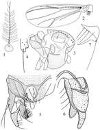
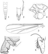
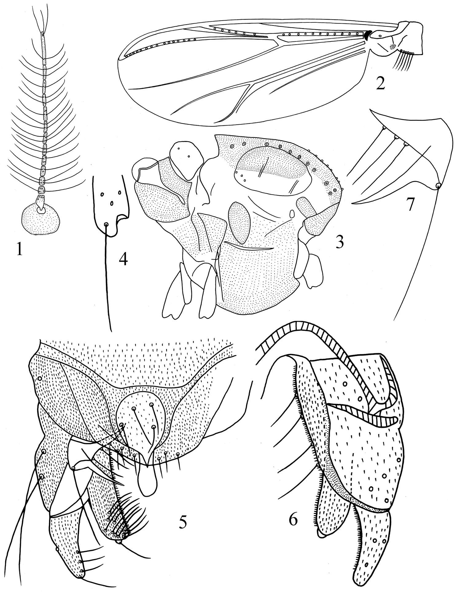
Figures 1–7.Polypedilum (Uresipedilum) minimum sp. n. 1 Antenna. 2 Wing. 3 Thorax 4 Fore tibia scale 5 Dorsal view of hypopygium 6 Ventral view of hypopygium 7 Superior volsella.
-
Jing Ren, Xiaolong Lin, Xinhua Wang
Zookeys
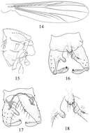
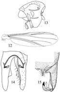
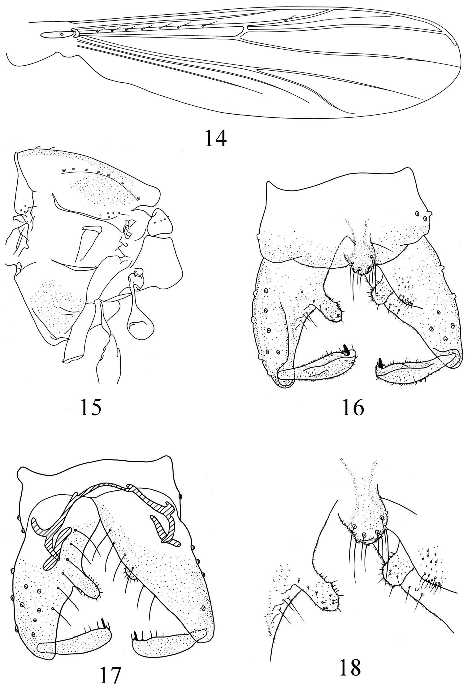
Figures 14–18.Pseudorthocladius (Pseudorthocladius) digitus sp. n., male. 14 wing 15 thorax 16 hypopygium (dorsal view) 17 hypopygium (ventral view) 18 anal point and inferior volsella.
-
Washington, District of Columbia, United States
-
Sydney, New South Wales, Australia
-
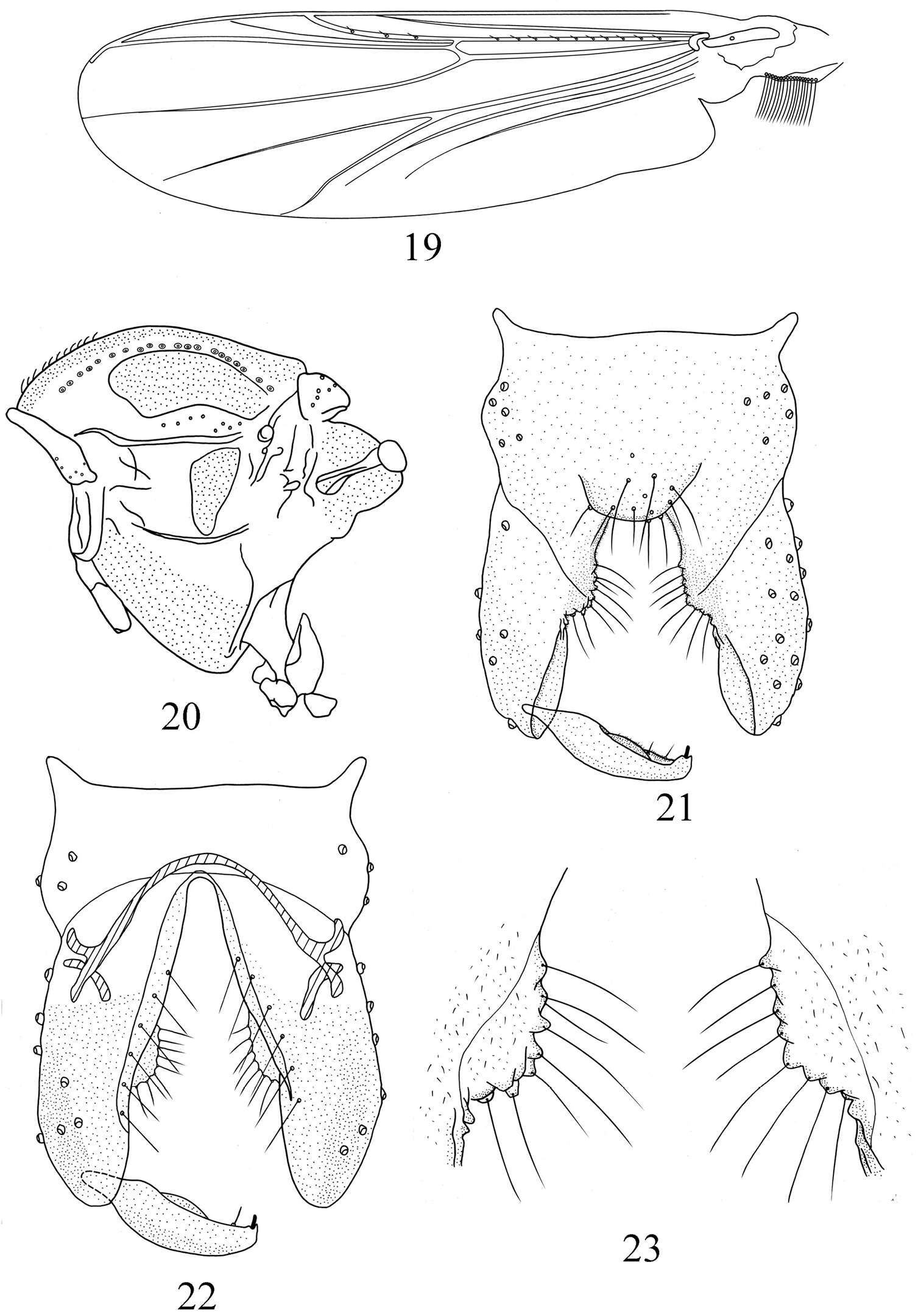
Jing Ren, Xiaolong Lin, Xinhua Wang
Zookeys
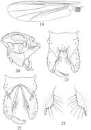
Figures 19–23.Pseudorthocladius (Pseudorthocladius) ovatus sp. n., male. 19 wing 20 thorax 21 hypopygium (dorsal view) 22 hypopygium (ventral view) 23 inferior volsella.
-
Sydney, New South Wales, Australia
-
Sydney, New South Wales, Australia
-
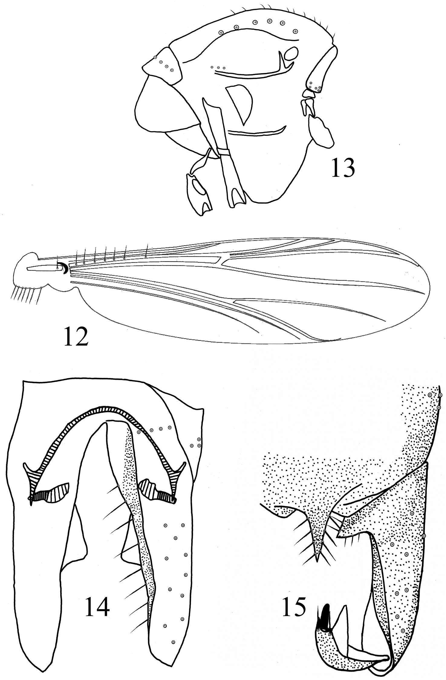
Wenbin Liu, Xiaolong Lin, Xinhua Wang
Zookeys
Figures 12–15.Rheocricotopus (Psilocricotopus) serratus sp. n., male. 12 wing 13 thorax 14 hypopygium (ventral view) 15 hypopygium (dorsal view).
-
Sydney, New South Wales, Australia
-
Wenbin Liu, Xiaolong Lin, Xinhua Wang
Zookeys
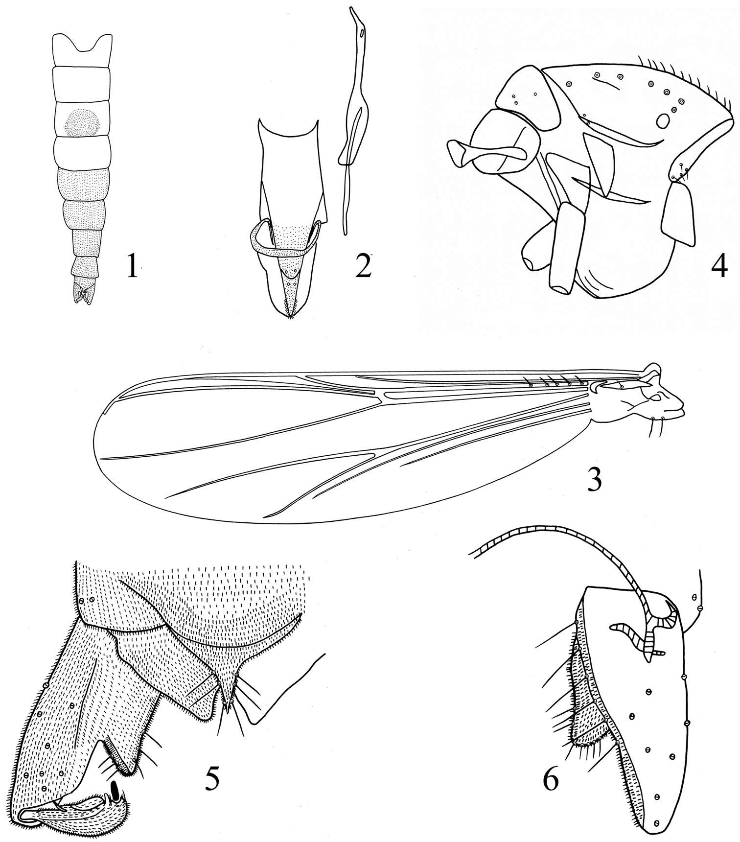
Figures 1–6.Rheocricotopus (Psilocricotopus) brochus sp. n., male. 1 abdomen tergites coloration 2 cibarial pump, tentorium and stipes 3 wing 4 thorax 5 hypopygium (dorsal view) 6 hypopygium (ventral view).
-
Sydney, New South Wales, Australia
-
Sydney, New South Wales, Australia
-
Wenbin Liu, Xiaolong Lin, Xinhua Wang
Zookeys
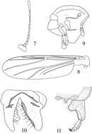
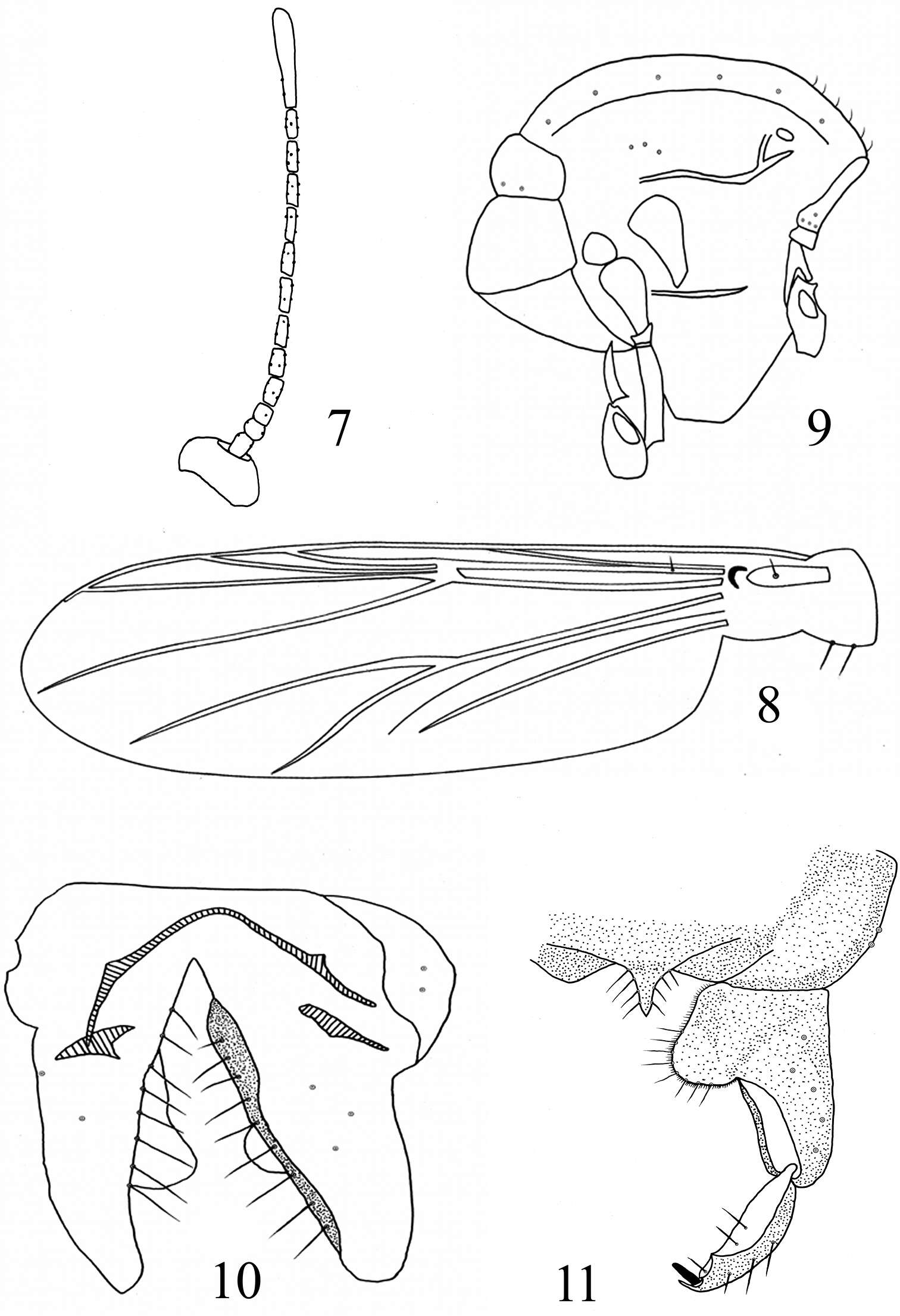
Figures 7–11.Rheocricotopus (Psilocricotopus) rotundus sp. n., male. 7 antenna 8 wing 9 thorax 10 hypopygium (ventral view) 11 hypopygium (dorsal view).
-
Sydney, New South Wales, Australia
-
Sydney, New South Wales, Australia
-
Sydney, New South Wales, Australia
-
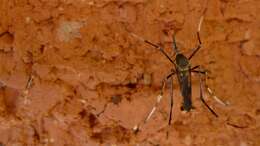
Grande Baie, Riviere du Rempart, Mauritius
-
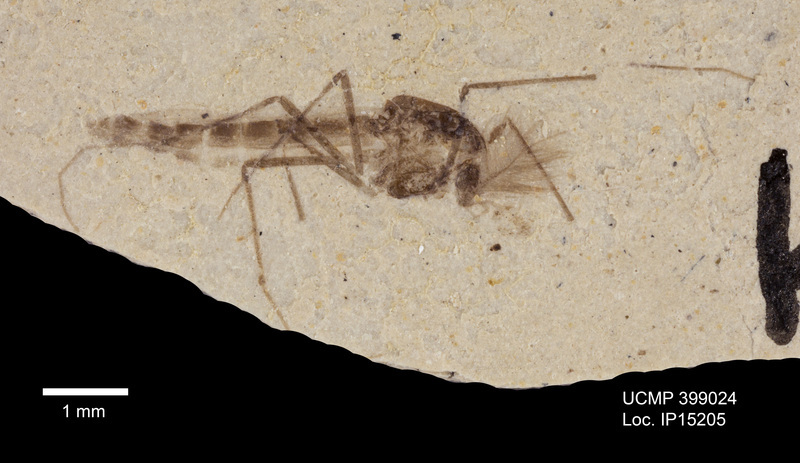
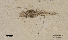
2015 University of California Museum of Paleontology
CalPhotos
-
2015 University of California Museum of Paleontology
CalPhotos
-
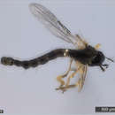
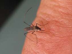
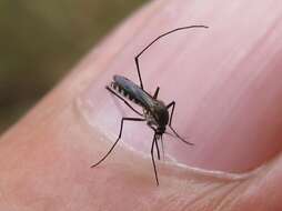
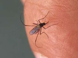
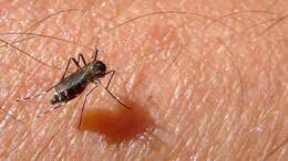
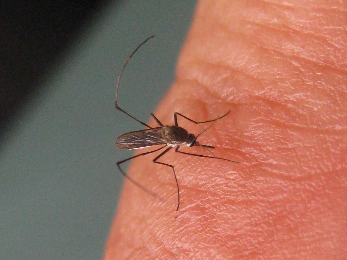
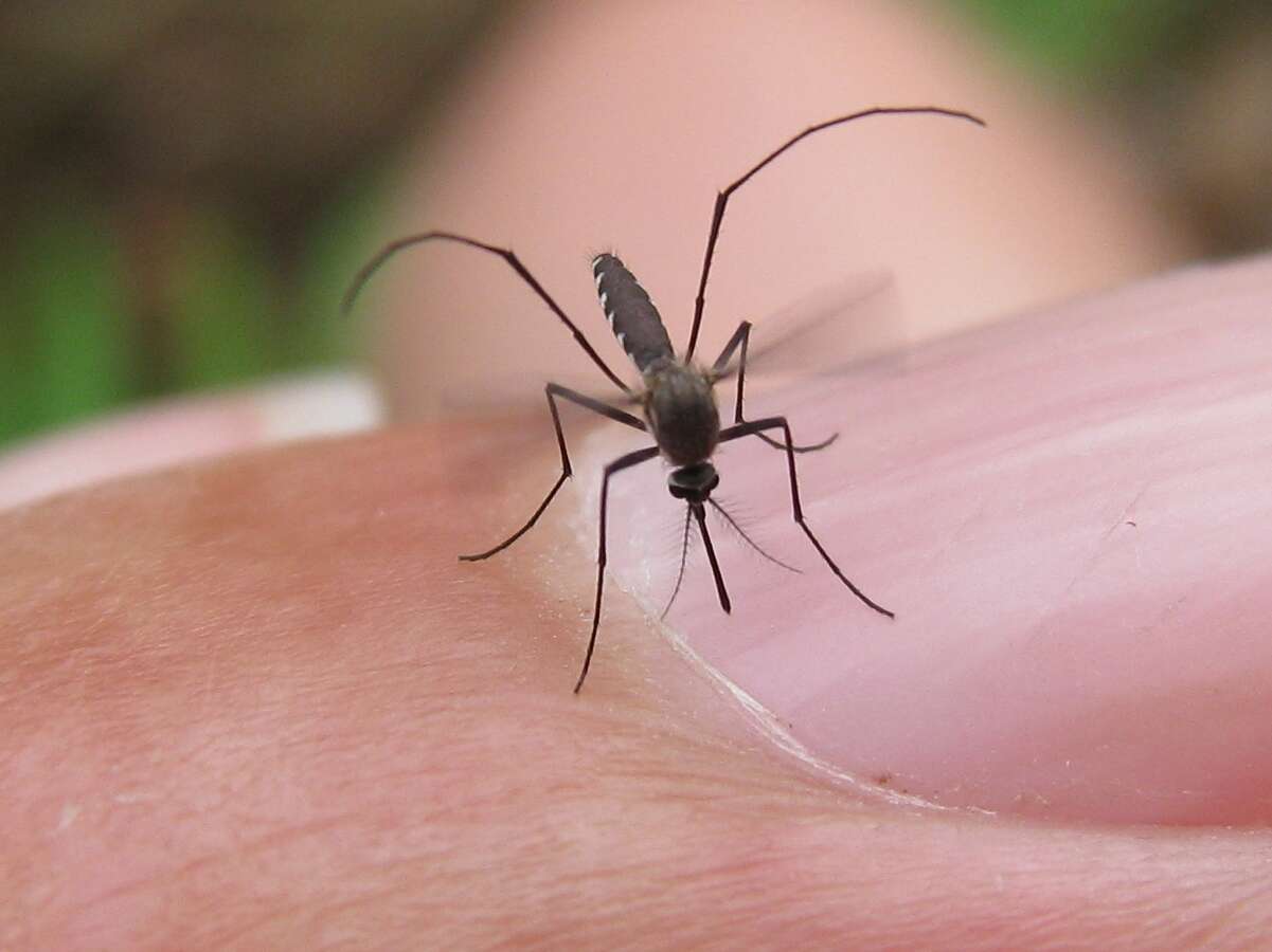
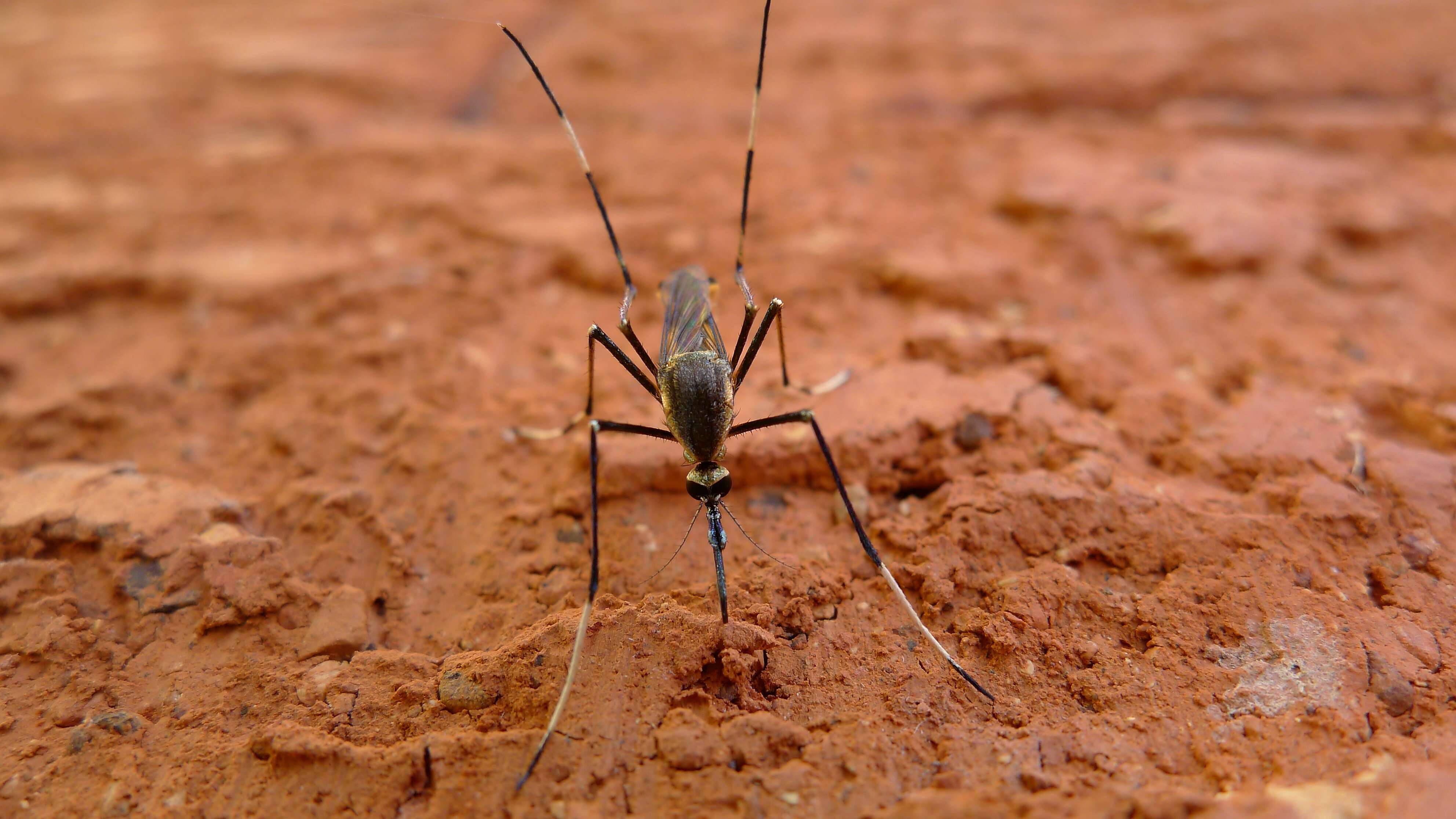
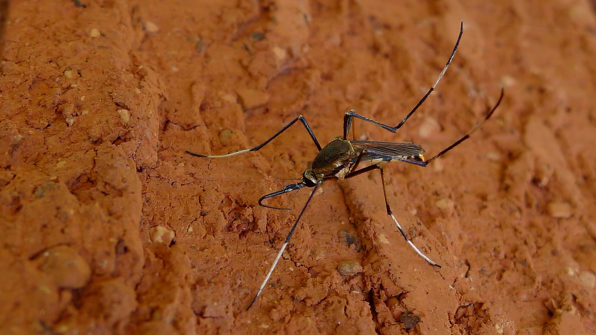
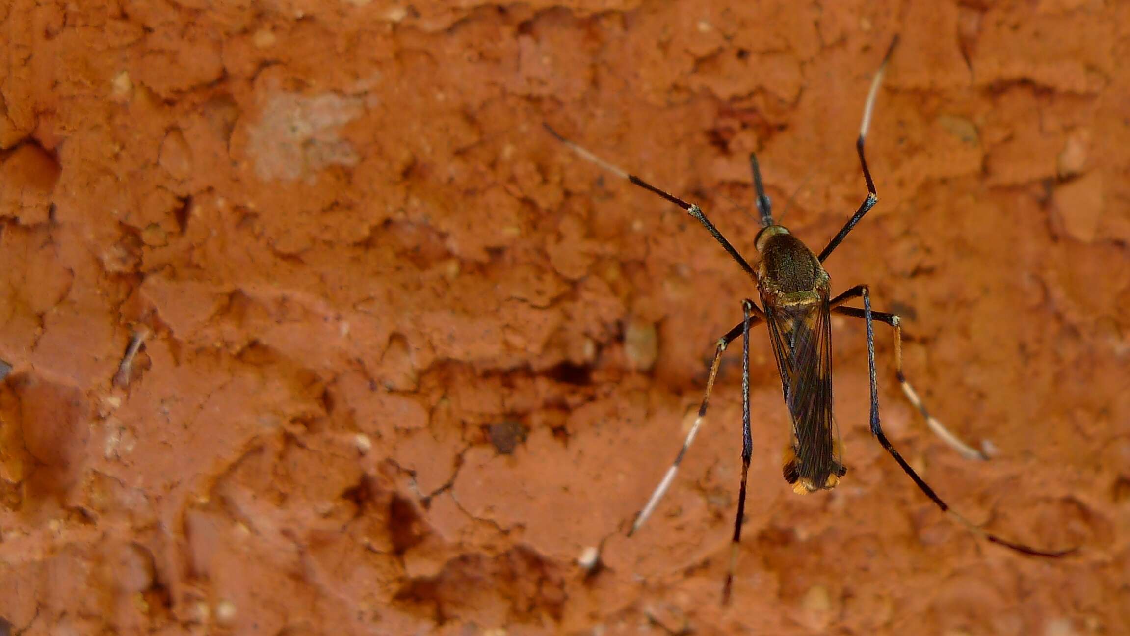
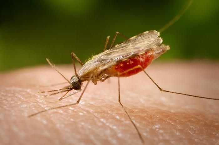
This close-up photograph shows an Anopheles minimus mosquito, a malaria vector of the Orient, as she was feeding on a human host. Note the blood meal that this mosquito had ingested, as it collected inside its stomach within its abdominal segment, extracting it from the host though its proboscis, which it had used to penetrate the skin, much like a straw.Created: 2005
-
This close-up 2005 photograph shows an Anopheles minimus, a malaria vector of the Orient mosquito, from a lateral perspective as she was feeding on a human host. Note the blood meal that this mosquito had ingested, as it collected inside its stomach within its abdominal segment, extracting it from the host though its proboscis, which it had used to penetrate the skin, much like a straw.Created: 2005
-
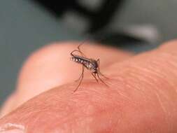
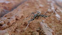
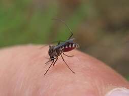
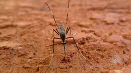
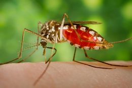
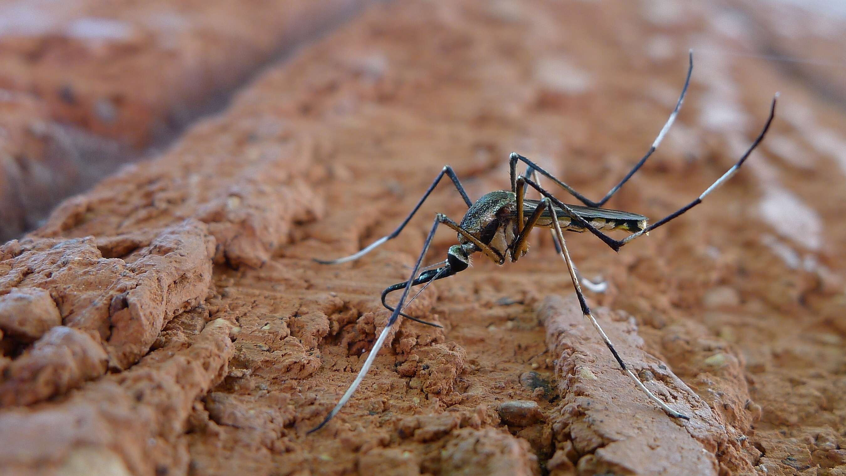
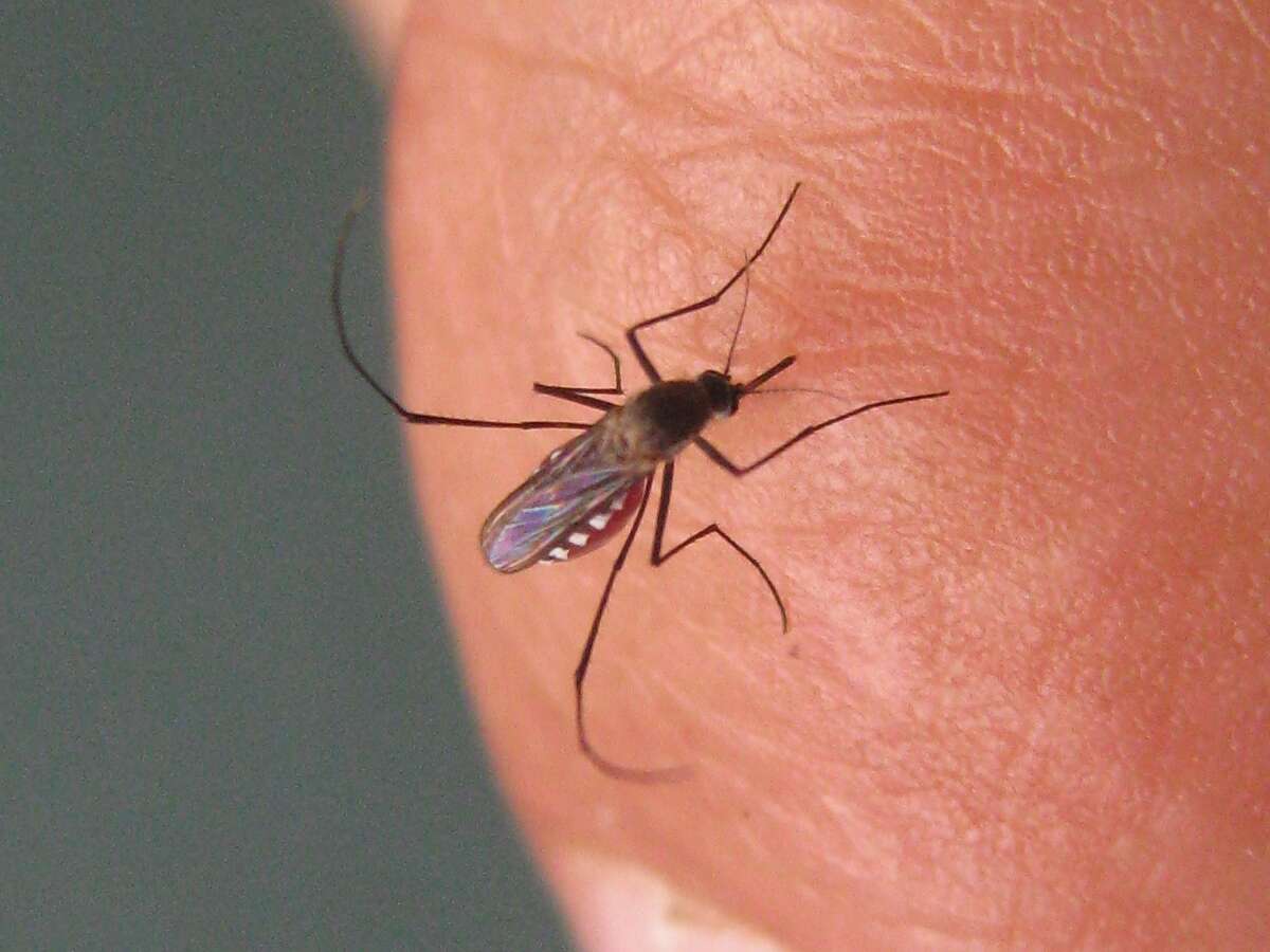
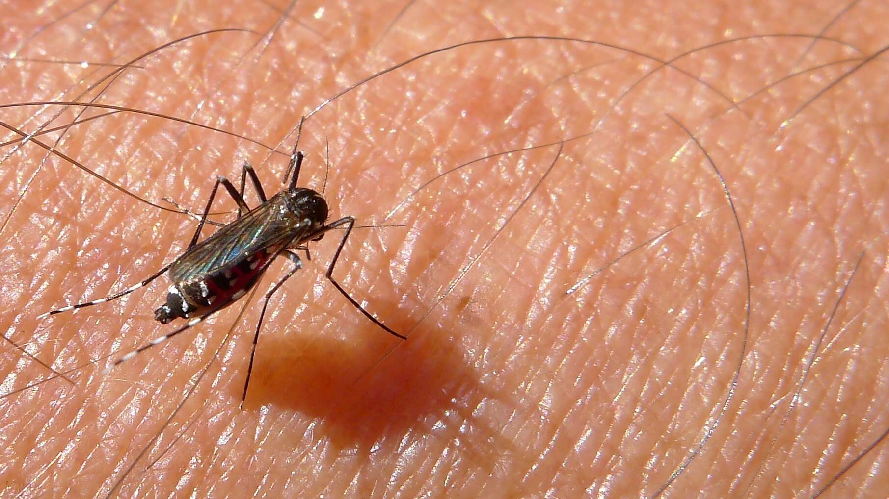
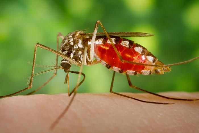
An Armigeres subalbatus mosquito of the Nagasaki colony was depicted in this 2005 photograph, as she was ingesting a blood meal after having lighted on a human finger. Note the pooling of the blood inside the mosquitos abdomen as it fills its stomach. The blood was being suctioned through the insects proboscis, which is its straw-like mouth that is used to penetrate the hosts skin much like a syringe.Created: 2005