-
Amager Fælled
-
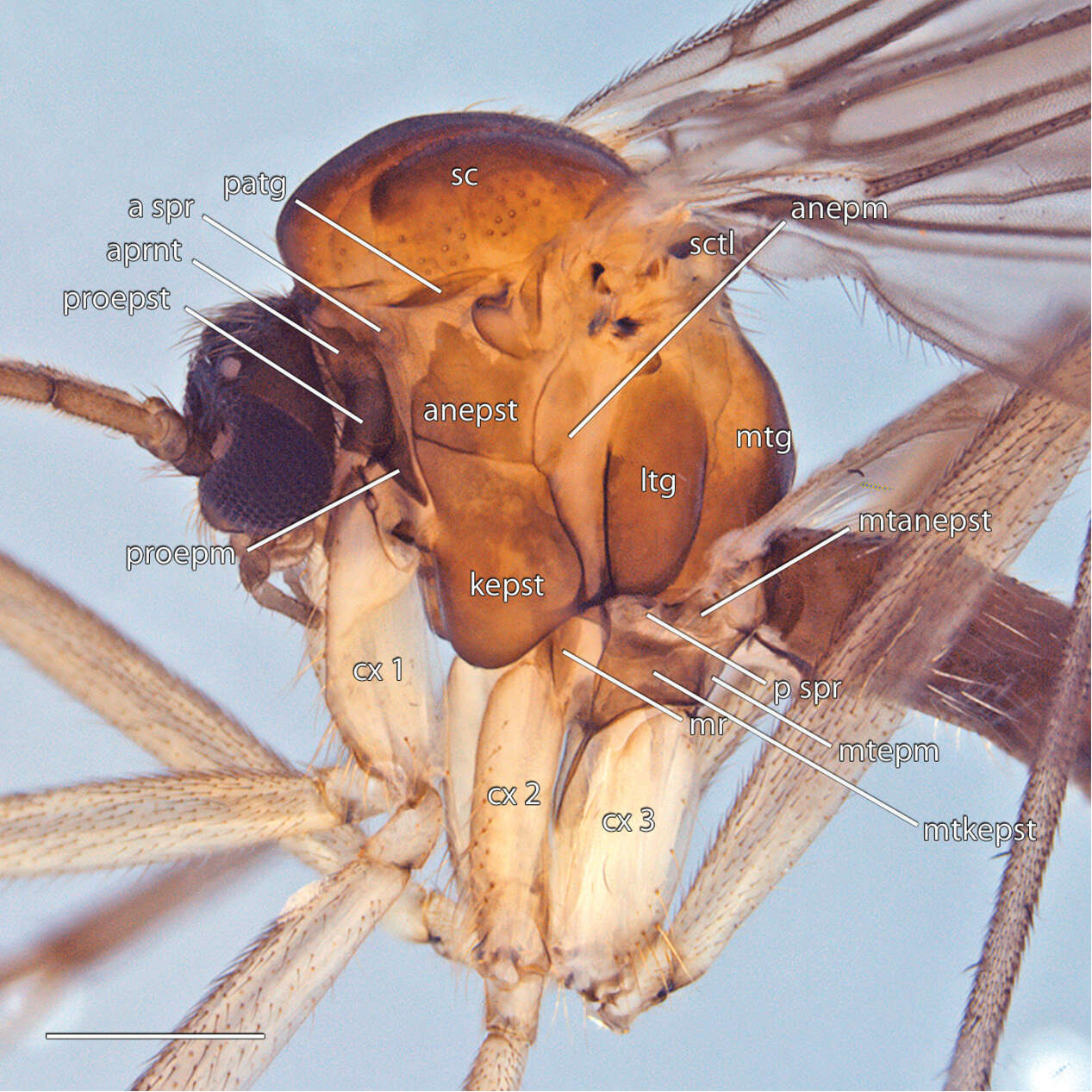
Figure 7. Acomoptera digitata sp. n., thorax, lateral view [692363]. Scale line = 0.5 mm. Abbreviations: anepm anepimeron anepst anepisternum aprnt antepronotum a spr anterior spiracle cx coxa kepst katepisternum ltg laterotergite mr meron mtg mediotergite mtanepst metanepisternum mtepm metepimeron mtkepst metakatepisternum p spr posterior spiracle patg paratergite proepm proepimeron proepst proepisternum sc scutum sctl scutellum.
-
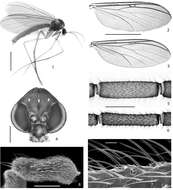
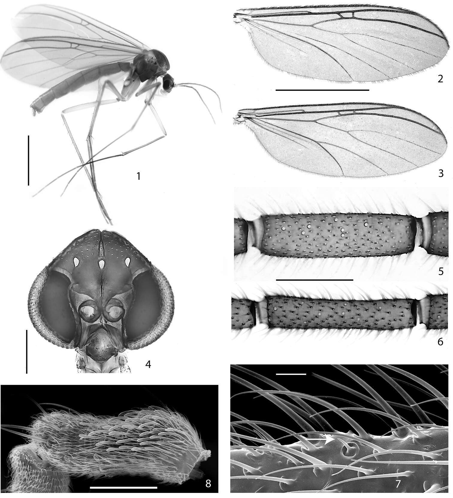
Figures 1–8.1–2, 4–5, 7–8. Acomopterella martinovskyi. 1 Female habitus 2 Male wing 4 Male head, frontal view 5 Flagellomere 4, male. Sensilla chaetica visible as pale spots. 7 Sensillum chaeticum (at arrowhead) on flagellomere 10, male 8 Sensilla on palpomere 3, male 3, 6 Acomopterella yoshiwaesp. n., male. 3 Wing 6 Flagellomere 4. Length of scale bar = 2 mm (for 1–3), 200 µm (for 4), 100 µm (for 5–6), 10 µm (for 7), 50 µm (for 8).
-
Olavi Kurina, Sarah Siqueira Oliveira
Zookeys
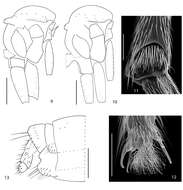
Figures 12–14.Cordyla australica sp. n. female terminalia. 12 lateral view 13 dorsal view 14 ventral view. Scale bar = 0.1 mm. Abbreviations: cerc= cercus, gp= gonapophysis, st= sternite, tg= tergite.
-
Kai Shi, Junhao Huang, Sujiong Zhang, Hong Wu
Zookeys
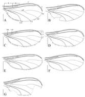
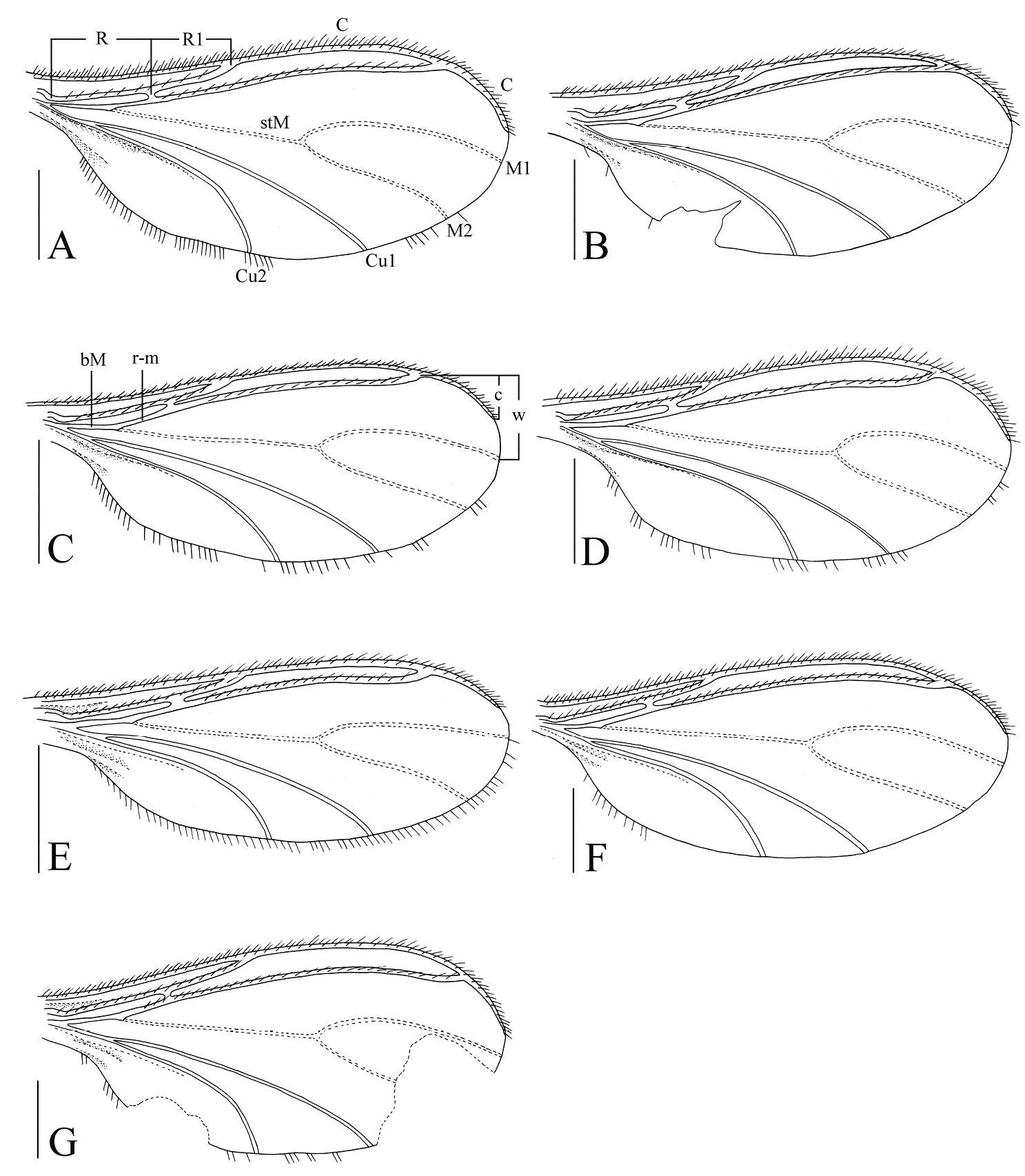
Figure 8.Wings, dorsal view. A Peyerimhoffia hamata sp. n. (holotype) B Peyerimhoffia obesa sp. n. (holotype) C Peyerimhoffia sparsula sp. n. (holotype) D Peyerimhoffia longiprojecta sp. n. (holotype) E Peyerimhoffia brachypoda sp. n. (holotype) F Peyerimhoffia yunnana sp. n. (holotype) G Peyerimhoffia shennongjiana sp. n. (holotype). Scale, 0.50 mm.
-
Jan Ševčík, Heikki Hippa, Rodzay Abdul Wahab
Zookeys
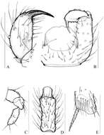
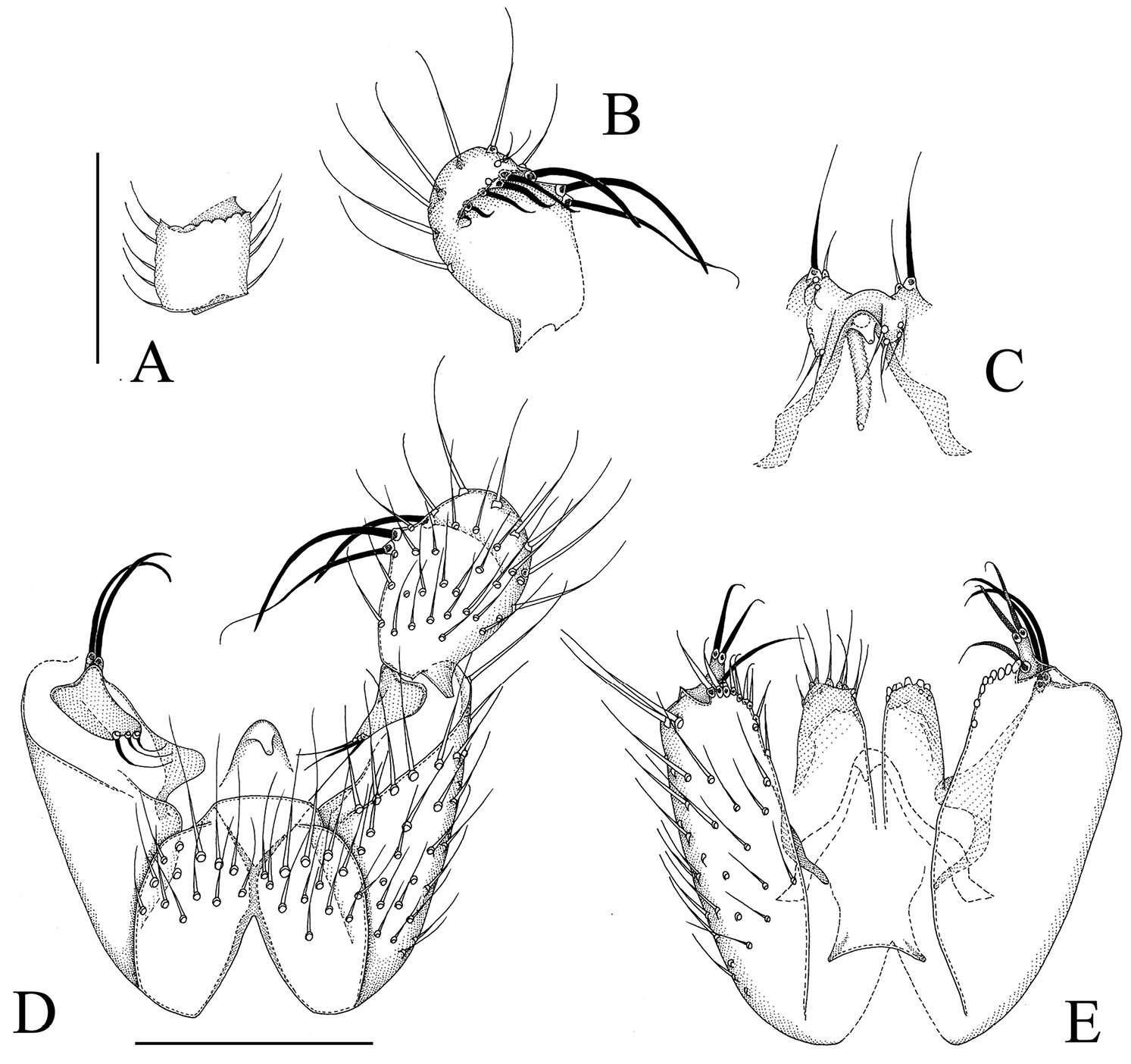
Figure 6.Manota megachaeta sp. n. (holotype). A Antennal flagellomere 4, lateral view B Gonostylus, dorsal view C Aedeagus and hypoproct, ventral view D Hypopygium, ventral view E Hypopygium, dorsal view. Scale 0.1 mm.
-
All Biocode files are based on field identifications to the best of the researcher’s ability at the time.
-
Småland, Sweden
-
Amager Fælled
-
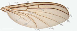
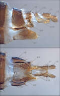
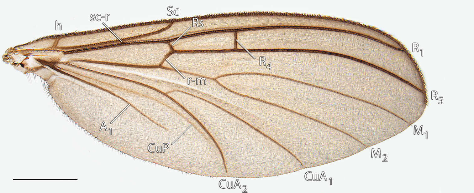
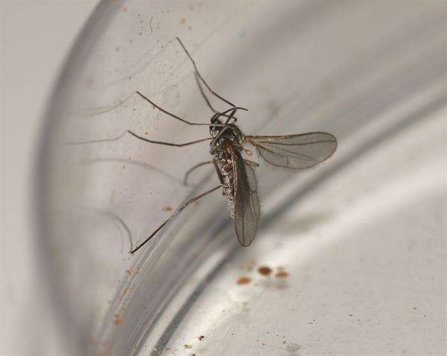
Figure 8. Acomoptera digitata sp. n., wing, dorsal view [692360]. Scale line = 1 mm.
-
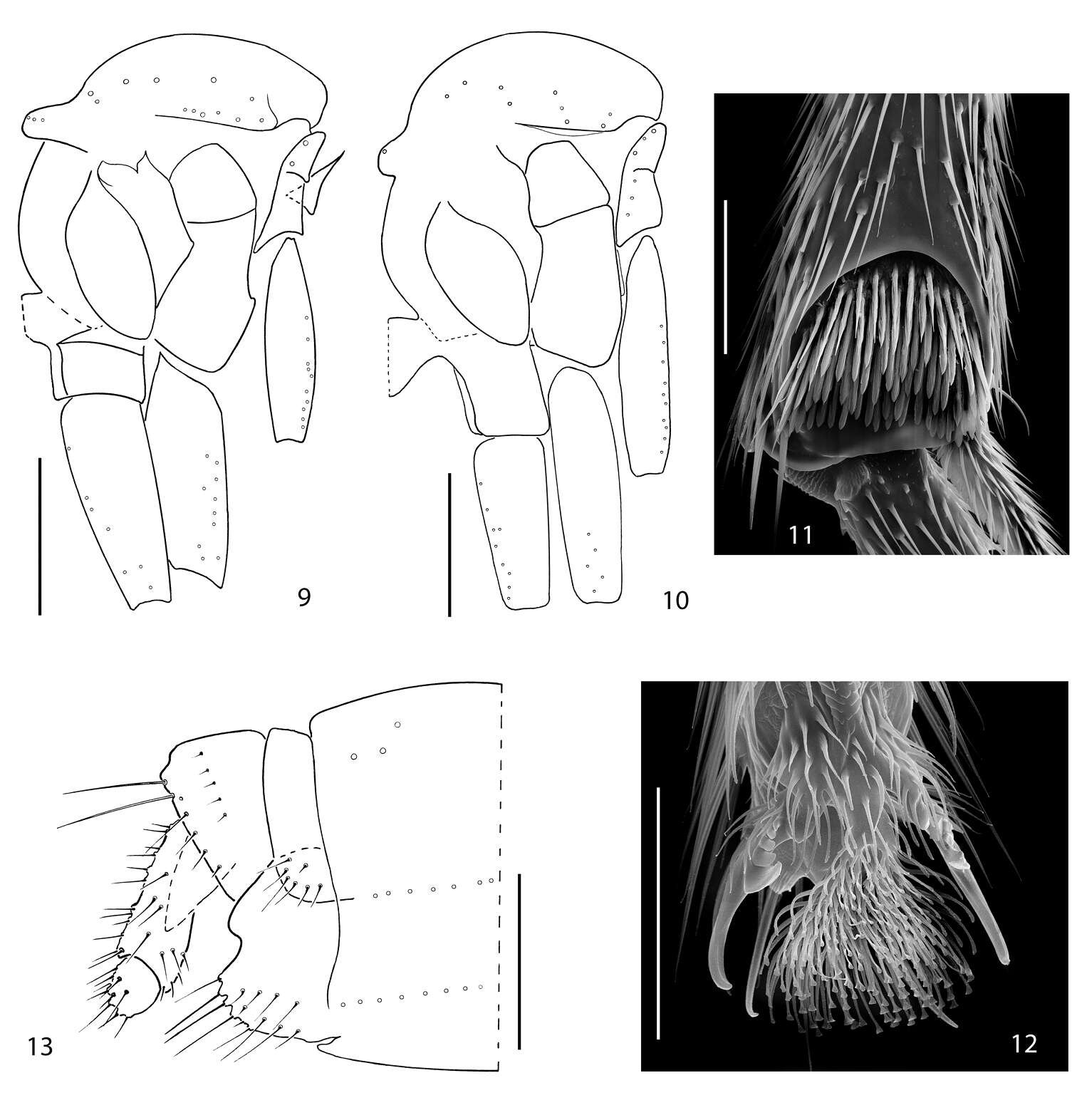
Figures 9–13.9 Acomopterella yoshiwaesp. n., thorax in lateral view. 10–13 Acomopterella martinovskyi. 10 Thorax in lateral view 11 Fore tibial organ, male 12 Fore claw, male 13 Female terminalia in lateral view. Length of scale bar = 0,5 mm (for 9–10), 50 µm (for 11–12), 250 µm (for 13).
-
Kai Shi, Junhao Huang, Sujiong Zhang, Hong Wu
Zookeys
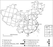
Figure 9.Geographical distribution of Peyerimhoffia from China.
-
Jan Ševčík, Heikki Hippa, Rodzay Abdul Wahab
Zookeys
Figure 7.Manota pileata sp. n. A Antennal flagellomere 4, lateral view B Aedeagus and hypoproct, ventral view C Hypopygium, ventral view D Hypopygium, dorsal view. Scale 0.1 mm.
-
All Biocode files are based on field identifications to the best of the researcher’s ability at the time.
-
Småland, Sweden
-
Amager Fælled
-
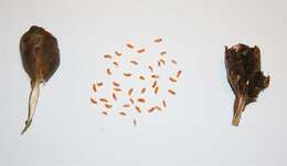
Figure 9. Acomoptera digitata sp. n., female genitalia: A lateral view [692365] B ventral view [692366]. Scale line = 0.1 mm. Abbreviations: c cercus st sternite t tergite.
-
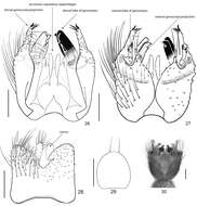
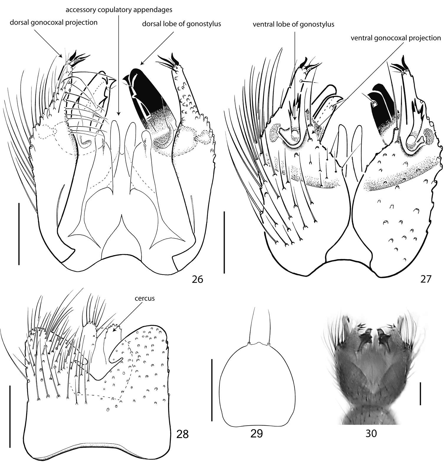
Figures 26–30.Terminalia of Acomopterella yoshiwae sp. n., male. 26 Terminalia in dorsal view, tergite IX removed 27 Terminalia in ventral view 28 Tergite IX, dorsal view 29 Hypoproct, ventral view 30 Terminalia incl. tergite IX in dorsal view. Length of scale bar = 100 µm.
-
Kai Shi, Junhao Huang, Sujiong Zhang, Hong Wu
Zookeys
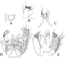
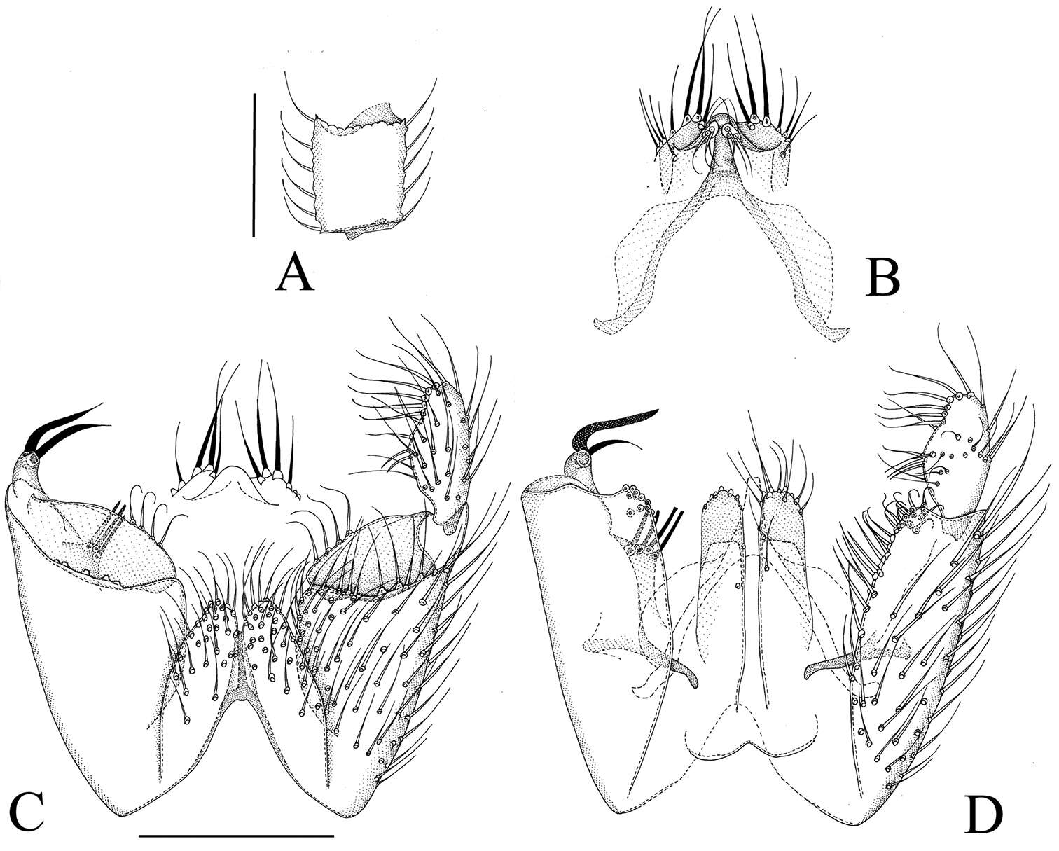
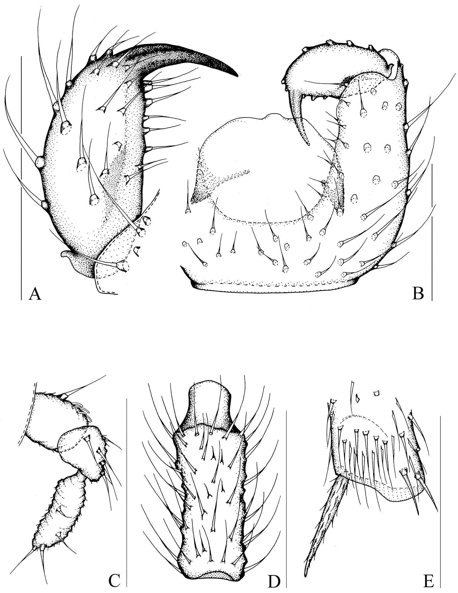
Figure 3.Peyerimhoffia sparsula Shi & Huang, sp. n., male, holotype. A Left gonostylus, ventral view B Part of hypopygium, ventral view C Palp, lateral view D Fourth flagellomere, lateral view E Apex of foretibia, prolateral view. Scale, 0.10 mm.
-
Jan Ševčík, Heikki Hippa, Rodzay Abdul Wahab
Zookeys
Figure 8.Manota ricina sp. n. (holotype). A Antennal flagellomere 4, lateral view. B Gonostylus, dorsal view C Aedeagus and hypoproct, ventral view D Hypopygium, ventral view E Hypopygium, dorsal view. Scale 0.1 mm.
-
Kielstrup Sø, Jylland, Danmark
-
Amager Fælled
-
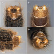
Figure 10. Acomoptera digitata sp. n., male genitalia, images: A dorsal view [692367] B ventral view [692368] C lateral view [692369] D posterior view [692370]. Scale line = 0.1 mm.
-
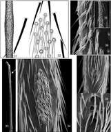
Figures 31–38.Mid tibial organ. Not on the same scale. 31–32 Acomopterella yoshiwae sp. n. 33–34 Acomopterella martinovskyi 33 General outer view of mid tibia, the arrowhead points to the tibial organ 35–36 Speolepta leptogaster 37–38 Ectrepesthoneura hirta.