-
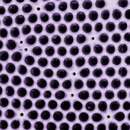
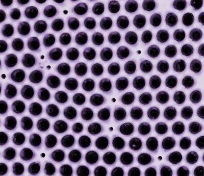
This image was made from samples taken during a scientific cruise in the Pacific. Water was filtered to concentrate the organisms that were present, then dried onto a thin sheet of plastic and then shadowed with a fine layer of metal to provide contrast. The preparation was then observed with an electron-microscope. This technique has been used to document the diversity of marine microbes, especially, protists in the oceans.
-
Note that the delicate spines are chitinous. Focus on valve surface. Scale bar indicates 50 µm. The image was built up using several photomicrographic frames with manual stacking technique. Sample from North Sea near Heligoland (spring diatom bloom). Images were taken using Zeiss Universal with Olympus C7070 CCD camera.
-
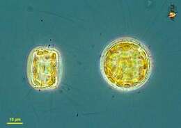
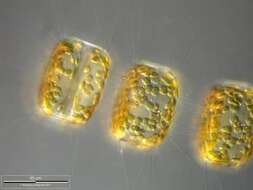
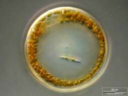
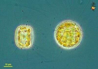
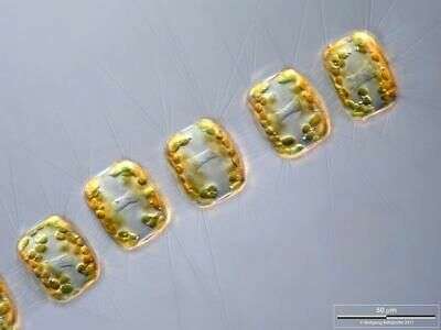
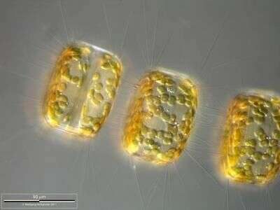
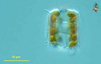
Thalassiosira (tha-lassy-owe-sire-a) eccentrica is one of the hundred or so species in the diverse and common genus of centric diatoms frequently found in marine waters. Some species can be very large. Species are distinguished primarily by the pattern of sculpting in the valve elements of the shell or frustule. The cell to the left is seen in girdle view, the one to the right in valve view. The margins of the valves give rise to a small number of fine chitinous filaments which are believed to function in flotation. These filaments are evident in arising from the cell to the right. With many small golden plastids. Phase contrast microscopy.
data on this strain.
-
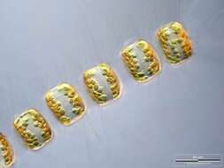
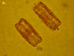
Chain of Porosira glacialis. Note that the delicate spines are chitinous. Focus on frustule surface. Scale bar indicates 50 µm. The image was built up using several photomicrographic frames with manual stacking technique. Sample from North Sea near Heligoland (spring diatom bloom). Images were taken using Zeiss Universal with Olympus C7070 CCD camera.
-
Marginal silica processes are visible. Scale bar indicates 25 µm. The image was built up using several photomicrographic frames with manual stacking technique. Sample from North Sea near Heligoland (spring diatom bloom). Images were taken using Zeiss Universal with Olympus C7070 CCD camera.
-
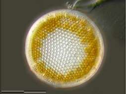
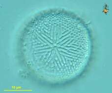
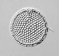
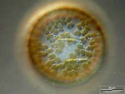
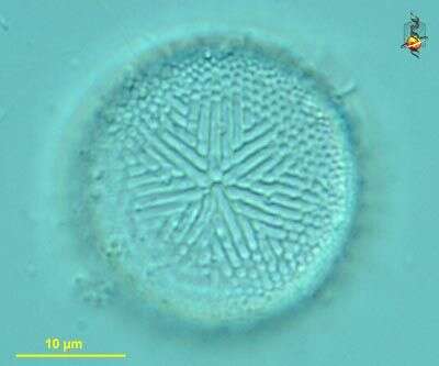
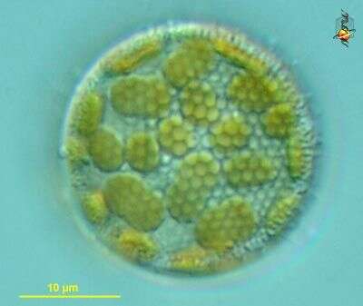
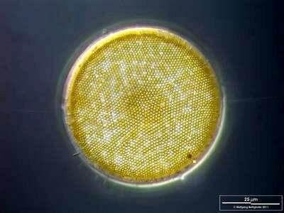
Thalassiosira (tha-lassy-owe-sire-a) eccentrica is one of the hundred or so species in the diverse and common genus of centric diatoms often found in marine waters. Some species can be very large. Species are distinguished primarily by the pattern of sculpting in the valve elements of the shell or frustule. This species has a seven-fold symmetry in the strutting which lies immediately below the surface of the valve, and can be seen in this isolated valve. Differential interference microscopy.
data on this strain.
-
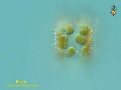
Chain of Porosira glacialis. Note that the delicate spines are chitinous. Focus on cell center showing cytoplasmic accumulation around the nucleus. Scale bar indicates 50 µm. The image was built up using several photomicrographic frames with manual stacking technique. Sample from North Sea near Heligoland (spring diatom bloom). Images were taken using Zeiss Universal with Olympus C7070 CCD camera.
-
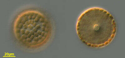
Cells at two focal levels. Material from a plankton tow off Martha's Vineyard, Massachusetts. Image by Jeff Cole.
-
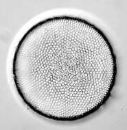
Thalassiosira (tha-lassy-owe-sire-a) eccentrica is one of the hundred or so species in the diverse and common genus of centric diatoms often found in marine waters. Some species can be very large. Species are distinguished primarily by the pattern of sculpting in the valve elements of the shell or frustule, and this shows the surface of an isolated valve. Differential interference microscopy.
data on this strain.
-
Closeup of Porosira glacialis chain. Note that the delicate spines are chitinous. Focus on frustule surface. Scale bar indicates 50 µm. The image was built up using several photomicrographic frames with manual stacking technique. Sample from North Sea near Heligoland (spring diatom bloom). Images were taken using Zeiss Universal with Olympus C7070 CCD camera.
-
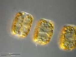
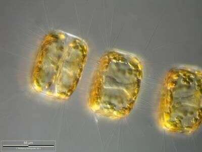
Thalassiosira (tha-lassy-owe-sire-a) eccentrica is one of the hundred or so species in the diverse and common genus of centric diatoms frequently found in marine waters. Some species can be very large. Species are distinguished primarily by the pattern of sculpting in the valve elements of the shell or frustule. The margins of the valve have a number of stout processes and these give rise to the chitinous threads. This is a girdle view and shows the processes as well as the plastids.
data on this strain.
-
Closeup of Porosira glacialis chain. Note that the delicate spines are chitinous. Focus on cell center showing cytoplasmic accumulation around the nucleus. Scale bar indicates 50 µm. The image was built up using several photomicrographic frames with manual stacking technique. Sample from North Sea near Heligoland (spring diatom bloom). Images were taken using Zeiss Universal with Olympus C7070 CCD camera.
-
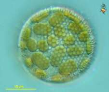
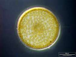
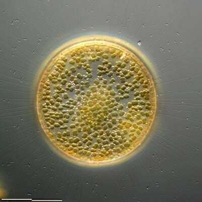
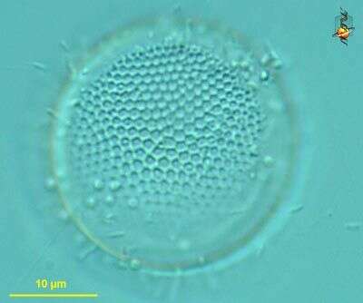
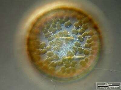
Thalassiosira (tha-lassy-owe-sire-a) eccentrica is one of the hundred or so species in the diverse and common genus of centric diatoms commonly found in marine waters. Some species can be very large. Species are distinguished primarily by the pattern of sculpting in the valve elements of the shell or frustule. This is the valve view showing the sculpting of the valve and the underlying plastids. Differential interference microscopy.
data on this strain.
-
-
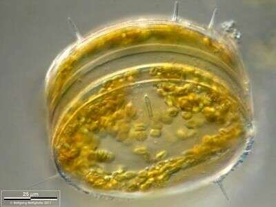
Thalassiosira (tha-lassy-owe-sire-a) eccentrica , is one of the hundred or so species in the diverse and common genus of centric diatoms commonly found in marine waters. Some species can be very large. Species are distinguished primarily by the pattern of sculpting in the valve elements of the shell or frustule. This is the girdle view showing the plastids located near the valves and a central nucleus. Differential interference microscopy.
data on this strain.
-
Some specimen of this centric diatom carried naviculoid ones on the valve(s). Scale bar indicates 25 µm. The image was built up using several photomicrographic frames with manual stacking technique. Sample from North Sea near Heligoland (spring diatom bloom). Images were taken using Zeiss Universal with Olympus C7070 CCD camera.
-
-
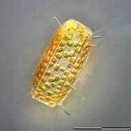
The oblique view exhibits short silicous spines, the so called occluded processes. On the lower left near the scale bar a chitinous spine is visible. Scale bar indicates 25 µm. The image was built up using several photomicrographic frames with manual stacking technique. Sample from North Sea near Heligoland (spring diatom bloom). Images were taken using Zeiss Universal with Olympus C7070 CCD camera.
-
One labiate process is visible together with some chitinous spines. Scale bar indicates 25 µm. The image was built up using several photomicrographic frames with manual stacking technique. Sample from North Sea near Heligoland (spring diatom bloom). Images were taken using Zeiss Universal with Olympus C7070 CCD camera.
-
Some specimen of this centric diatom carried naviculoid ones on the valve(s). Scale bar indicates 25 µm. The image was built up using several photomicrographic frames with manual stacking technique. Sample from North Sea near Heligoland (spring diatom bloom). Images were taken using Zeiss Universal with Olympus C7070 CCD camera.
-
-
The oblique view exhibits short silicous spines, the so called occluded processes. On the lower left, lower right and central above chitinous spines are visible. Scale bar indicates 50 µm. The image was built up using several photomicrographic frames with manual stacking technique. Sample from North Sea near Heligoland (spring diatom bloom). Images were taken using Zeiss Universal with Olympus C7070 CCD camera.
-
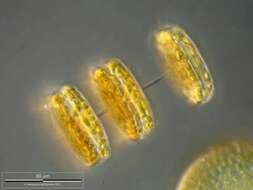
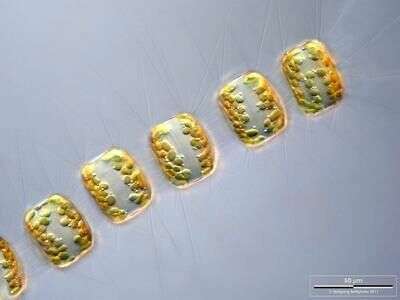
The members of the colony are interconnected with a bundle of threads. Numerous delicate spines protruding from the valve's margin are visible. Scale bar indicates 50 µm. The image was built up using several photomicrographic frames with manual stacking technique. Sample from North Sea near Heligoland (spring diatom bloom). Images were taken using Zeiss Universal with Olympus C7070 CCD camera.
-
The oblique view exhibits short silicous spines, the so called occluded processes. Some chitinous spines protruding from the fultoportulae (also called strutted processes) along the dotted valve margin are also visible. Scale bar indicates 50 µm. The image was built up using several photomicrographic frames with manual stacking technique. Sample from North Sea near Heligoland (spring diatom bloom). Images were taken using Zeiss Universal with Olympus C7070 CCD camera.