-
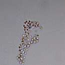
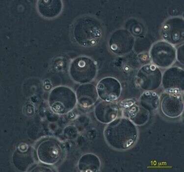
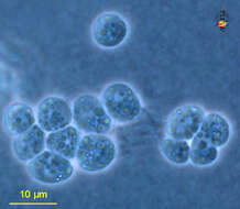
Aplanochytrium (a-plan-o-kit-ree-um), a labyrinthulid. This image from a culture on agar and does not show any ectoplasmic network. Phase contrast.
-
Aplanochytrium (a-plan-o-kit-ree-um), a labyrinthulid. This image from a culture on agar and shows a few filaments of the ectoplasmic network. Phase contrast.
-
Aplanochytrium (a-plan-o-kit-ree-um), a labyrinthulid. This image from a culture on agar and does not show any ectoplasmic network. Detail of cell bodies. Phase contrast.
-
Aplanochytrium (a-plan-o-kit-ree-um), a labyrinthulid. This image from a culture on agar and shows some of the ectoplasmic network. Phase contrast.
-
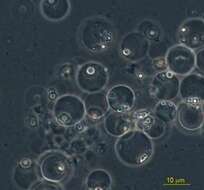
Aplanochytrium (a-plan-o-kit-ree-um), a labyrinthulid. This image from a culture on agar and does not show any ectoplasmic network. Phase contrast.
-
-
-
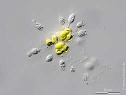
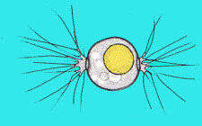
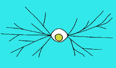
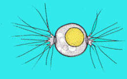
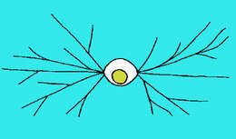
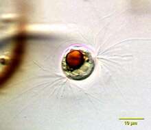
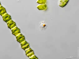
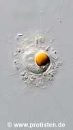
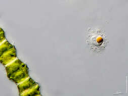
Diplophys, small spherical cell with a clear, yellow or orange lipid drop (or drops) in the centre of the cell, and two tufts of fine pseudopodia extending from the opposing poles of the cell. Phase contrast micrograph.
-
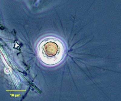
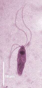
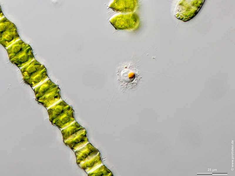
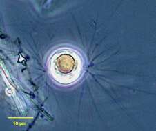
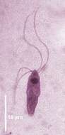
Portrait of Diplophrys archeri (Barker,1868), a small amoeba within a round organic shell composed of small plates not visible with light microscope. One distinctive orange cytoplasmic droplet thought to be composed of lipid is seen in this image. Two tufts of fine branching pseudopodia emerge from either pole of the shell. From freshwater pond with abundant decaying leaves near Boise, Idaho. Phase contrast
-
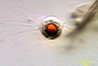
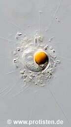
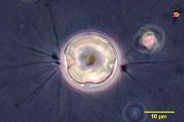
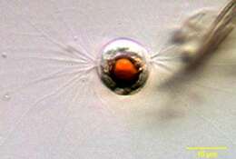
Portrait of Diplophrys archeri (Barker,1868, a small amoeba within a round organic shell composed of small plates not visible with light microscope. One distinctive orange cytoplasmic droplet thought to be composed of lipid is seen in this image. Two tufts of fine branching pseudopodia emerge from either pole of the shell. From freshwater pond with abundant decaying leaves near Boise, Idaho. Oblique illumination.
-
-
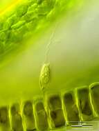
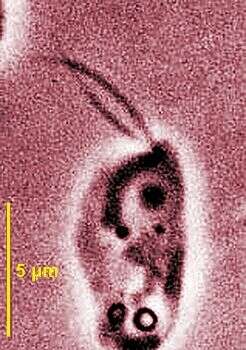
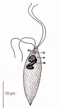
Phase contrast micrograph showing the two short anterior flagella, the nucleus and the mitochondrion close to the base of the flagella.
-
ATCC 50177. This parasite was recently placed within stramenopiles.
-
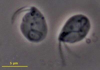
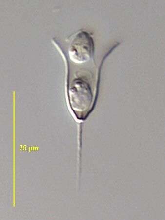
Karotomorpha cells are small (12-16 µm) flagellates with four (two pairs) long flagella inserted subapically. Anterior nucleus, body surface ridged. Dictyosomes situated at the base of the flagella. Pinocytic nutrition, occurring in the gut of amphibia. Karotomorpha bufonis from Bufo bufo (Giemsa staining).
-
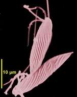
Scanning EM showing the corrugated cell surface and the four anterior flagella.
-
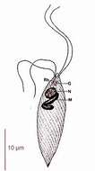
Diagram showing the two pairs of flagella, the rhizoplast, Golgi, nucleus, mitochondrion and the corrugated cell surface
-
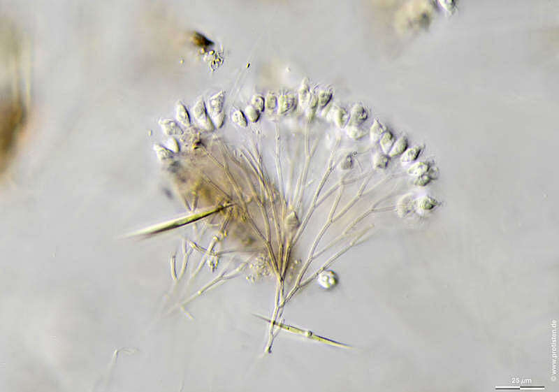
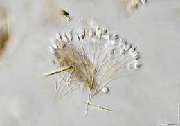
The coiling of the long anterior flagellum when it retracts (seen in the lowermost cell here) is typical of this genus. There is a daughter cell adherent to the inner surface of the chitinous lorica by its posterior flagellum in this image. DIC.
-
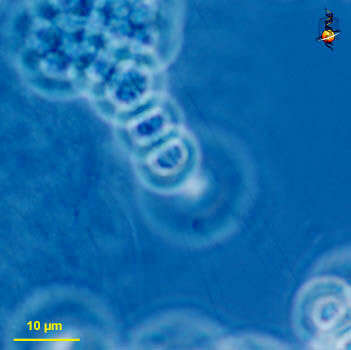
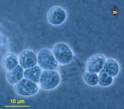
Sampling date 09/2018. Scale bars indicate 25 µm (1), 10 µm (2).Two images.Please click on < or > on the image edges or on the dots at the bottom edge of the images to browse through the slides!Place name: Bog Kaltenhof near Kiel (Schleswig-Holstein, Germany) Latitude: 54.42102744 Longitude: 10.07686615Microscope Zeiss Axioplan, camera Olympus OM-D M5 MKII. DOF images.© Wolfgang Bettighofer,images under Creative Commons License V 3.0 (CC BY-NC-SA).For permission to use of (high resolution) images please contact
postmaster@protisten.de.For further information about the image, please click here:
Link to protisten.de page
-
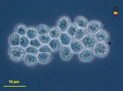
Sampling date 08/2018. Scale bar indicates 25 µm.Place name: Lake Vollstedter See near Kiel (Germany) Latitude: 54.24105528 Longitude: 9.859339Microscope Zeiss Axioplan, camera Olympus OM-D M5 MKII. DOF image.© Wolfgang Bettighofer,images under Creative Commons License V 3.0 (CC BY-NC-SA).For permission to use of (high resolution) images please contact
postmaster@protisten.de.For further information about the image, please click here:
Link to protisten.de page
-
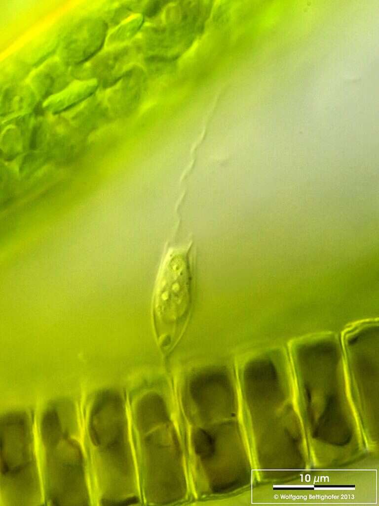
Scale bar indicates 10 µm.Bicosoecida lacustris living on diatom Ellerbeckia spec.Place name: Tropical freshwater aquarium Latitude: 54.3018013 Longitude: 10.07120132Microscope Zeiss Axioplan, camera Canon EOS 600D. DOF image.© Wolfgang Bettighofer,images under Creative Commons License V 3.0 (CC BY-NC-SA).For permission to use of (high resolution) images please contact
postmaster@protisten.de.For further information about the image, please click here:
Link to protisten.de page
-
Sampling date 10/2018. Scale bar indicates 25 µm.Place name: Pond Suploch, Hiddensee (Germany) Latitude: 54.538638 Longitude: 13.097802Microscope Zeiss Universal, camera Olympus OM-D M5 MKII. DOF image.© Wolfgang Bettighofer,images under Creative Commons License V 3.0 (CC BY-NC-SA).For permission to use of (high resolution) images please contact
postmaster@protisten.de.For further information about the image, please click here:
Link to protisten.de page