-
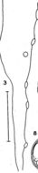
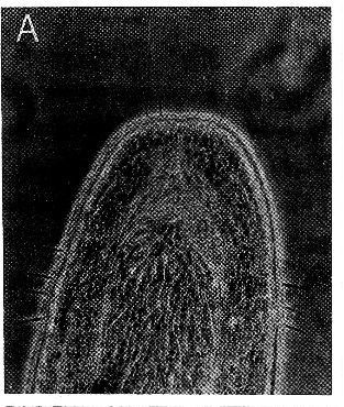
Teilweise evertierter Russel
-
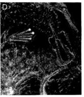
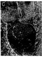
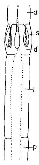
Diaphragma mit Haupt- und Reservestiletten.
-
Bulbus globosus
-
Russelpapillen
-
Vorderende mit Tastcirren
-
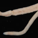
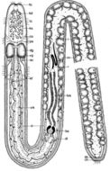
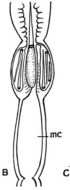
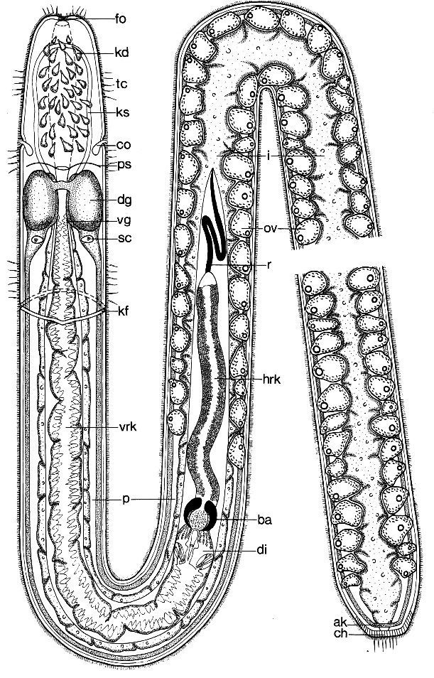
Ototyphlonemertes pallida. Habitus und Organisation eines geschlechtsreifen Weibchens
-
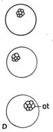
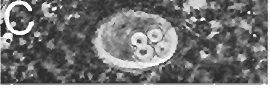
Statocysts. showing variation in composition of statoliths
-
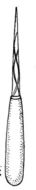
Stylet and basis more highly enlarged to show delicate spiral groans on stylet
-
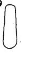
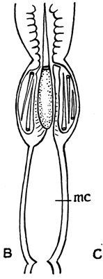
Stylet apparatus with slender basis and stylet and nanow middle chamber
-
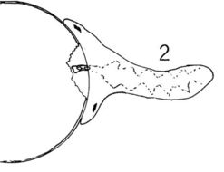
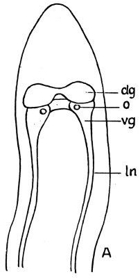
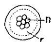
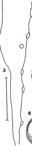
Outline of anterior portion of body, showing statocysts
-
Statolith with several globules
-
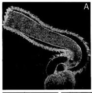
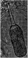
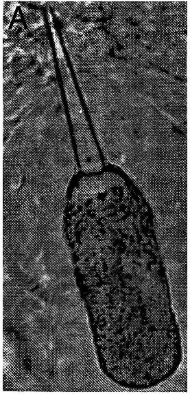
Long proboscis with proboscis canal
-
Statocyst
-
Russelausschnitt mit Papillen, Diaphragma und BalIan
-
Hauptstilett
-
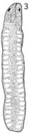
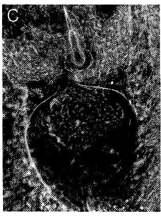
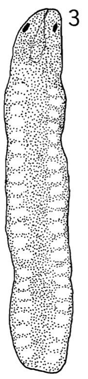
Dorsal view of live C. errans. Ovaries becoming visible as rows of transparent spots on both sides. Scale = 1.0 mm.
-
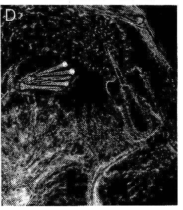
C. errans feeding on egg of C. magister. Esophagus used in sucking particles of egg yolk into intestine.
-
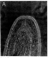
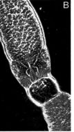
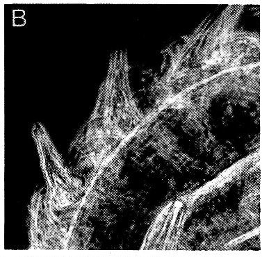
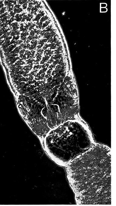
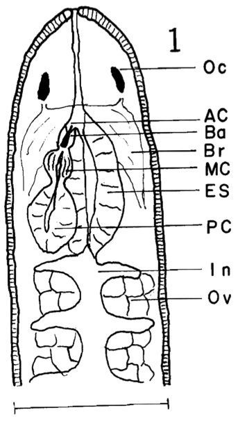
Anterior portion of body of living worm from dorsal surface. Oc, ocellus; Ac, anterior proboscis chamber; Ba, basis and stylet..Anterior portion of body of living worm from dorsal surface. Oc, ocellus; Ac, anterior proboscis chamber; Ba, basis and stylet; Br, brain; Me, middle proboscis chamber; Es, esophagus; Pc, posterior proboscis chamber; In, intestine; Ov, ovary. Scale .5 mm.
-
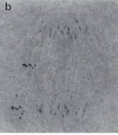
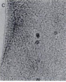
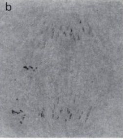
Chromosomes in mitotic anaphase during division from two to four cell stage in haploid embryo of experimental Carcinonemertes...Chromosomes in mitotic anaphase duringdivision from two to four cell stage in haploid embryo of experimental Carcinonemertes errans. Chromosome number is one-half that of Figure 1b. Magnification: l000x.
-
Chromosomes in mitotic anaphase during division from one to two cell stage in embryo of control Carcinonemertes errans...Chromosomes in mitotic anaphase during division from one to two cell stage in embryo ofcontrol Carcinonemertes errans.The small darkspots to the left of the chromosomes are remains of chromosomes in the two polar bodies. Magnification: l000X
-
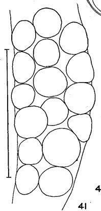
Part of a mucous sheath of C. mitsukurii from Charybdis erythrodactyla from the Society Islands. Scale 0.5 mm.
-
Outline of basis of C. mitsukurii as seen in section. Scale 0.02 mm.
-
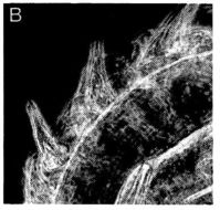
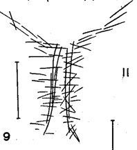
Sketch of cephalic muscle fibers in region of rhynchodaetim in C. mitsukurii. The anterior end is directed toward the bottom
-
Fragment of mucous sheath of C . mitsukurii from egg mass of Charybdis erythrodactyla from Society Islands. Scale 1 mm .