-
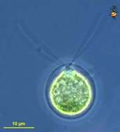
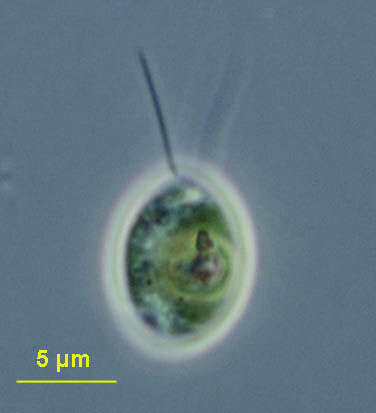
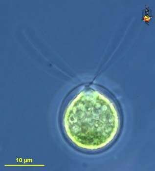
Chlamydomonas (clam-ee-doe-moan-ass), a solitary volvocid (flagellated green algal cell). Cell surrounded by a cellulosic wall, with two similar flagella emerging from near the apex. The photosynthetic pigments are located within a cup-shpaed chloroplast which has a large pyrenoid with associated polysaccharide materials. Many taxa described (there are books on this genus). Eyespot located within plastid. Flagella beat with a breast-stroke pattern. Phase contrast.
-
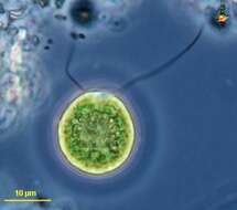
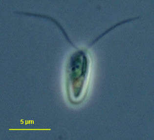
Chlamydomonas (clam-ee-doe-moan-ass), a solitary volvocid (flagellated green algal cell). Cell surrounded by a cellulosic wall, with two similar flagella emerging from near the apex. The photosynthetic pigments are located within a cup-shaped chloroplast which has a large pyrenoid with associated polysaccharide materials located posteriorly. The nucleus is located within the cup. This image shows one anterior contractile vacuole. Animations by Rosemary Arbur of flagellar beat patterns are available
here.Phase contrast.
-
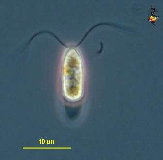
Chlamydomonas (clam-ee-doe-moan-ass), a solitary volvocid (flagellated green algal cell). Cell surrounded by a cellulosic wall. Cell damaged, no flagella. The photosynthetic pigments are located within a cup-shaped chloroplast which has a large pyrenoid with associated polysaccharide materials located posteriorly. The nucleus is located within the cup. This image shows the red eyespot to the right and two anterior contractile vacuoles. Phase contrast.
-
Chlamydomonas (clam-ee-doe-moan-ass), a solitary volvocid (flagellated green algal cell). Cell surrounded by a cellulosic wall, with two similar flagella emerging from near the apex. Elongate species. Animations by Rosemary Arbur of flagellar beat patterns are available
here. Phase contrast.
-
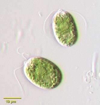
Chlamydomonas (clam-ee-doe-moan-ass), a solitary volvocid (flagellated green algal cell). Cell surrounded by a cellulosic wall. With plastid containing chlorophyll B giving the bright green colour, two similar flagella emerge from the anterior of the cell. Differential interference contrast.
-
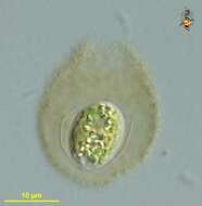
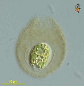
Chlamydomonas (clam-ee-doe-moan-ass), a solitary volvocid (flagellated green algal cell). Cell surrounded by a cellulosic wall and enclosed in mucilagenous sheath - cells in this form were inactive as if encysted. Differential interference contrast.
-
Chlamydomonas (clam-ee-doe-moan-ass), a solitary volvocid (flagellated green algal cell). Cell surrounded by a cellulosic wall. With plastid containing chlorophyll B giving the bright green colour, two similar flagella emerge from the anterior of the cell. Phase contrast.
-
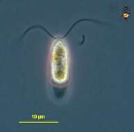
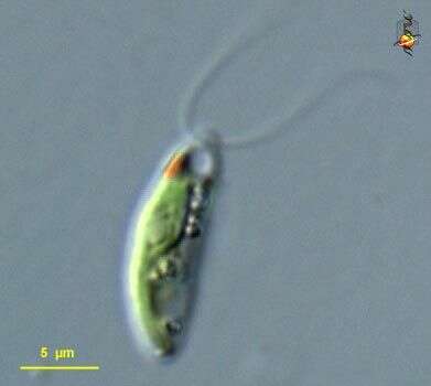
Chlamydomonas (clam-ee-doe-moan-ass) a common volvocid (green alga) flagellate. Cells vary in shape from elongate to rounded, this being one of the more elongate cells. With a cell wall, a cup-shaped chloroplasts with chlorophyll B, a red eyespot located external to the plastid, and two equal flagella emerging from the anterior pole of the cell. Differential interference contrast. Animations by Rosemary Arbur of flagellar beat patterns are available
here. Material from Nymph Creek and Nymph Lake, thermal sites within Yellowstone Park, photograph by Kathy Sheehan and David Patterson.
-
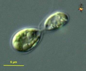
Chlamydomonas (clam-ee-dough-moan-ass) a common volvocid (green alga) flagellate. Cells vary in shape from elongate to rounded, this being one of the more elongate cells. With a cell wall, a cup-shaped chloroplasts with chlorophyll B, a red eyespot located external to the plastid, and two equal flagella emerging from the anterior pole of the cell. These cells undergo a form of sexual reproduction referred to as conjugation in which two similar to near similar cells fuse and exchange genetic information. Animations by Rosemary Arbur of flagellar beat patterns are available
here. Differential interference contrast. Material from Nymph Creek and Nymph Lake, thermal sites within Yellowstone National Park, photograph by Kathy Sheehan and David Patterson.
-
-
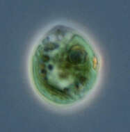
Chlamydomonas (clam-ee-dough-moan-ass) iconic volvocid motile green alga, with two similar flagella inserting into the anterior end of the cell. Photosynthetic pigments include chlorophyll B which gives the cells their bright green colour. Phase contrast micrograph.
-
Phase contrast microscopy.
-
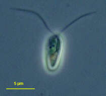
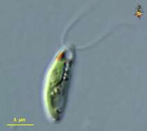
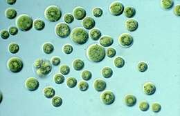
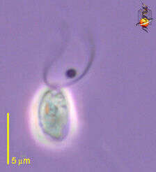
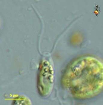
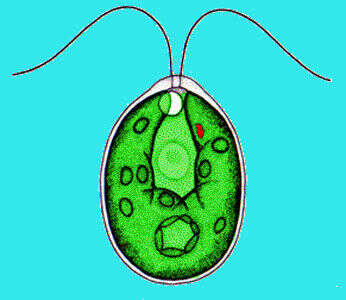
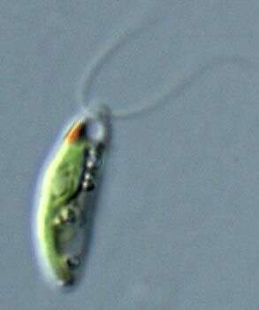
Chlamydomonas, a volvocid flagellate. The genus is very large probably with many synonymous species. Two equal length flagella emerge from a prominent anterior papilla in this species. A small contractile vacuole can be seen just posterior to the flagellar insertion site. A well-demarcated central nucleus can be seen in these images. A very small stigma is present. There is a single large cup shaped chloroplast in this species. A large pyrenoid is present in the posterior half of the cell. Some species have a gelatinous sheath although the cell wall is closely applied to the protoplast in this species. From freshwater pond near Boise, Idaho. Oblique illumination.
-
Chlamydomonas is a green alga, common in freshwater habitats like Swan Lake. The cells are small with two flagella used for locomotion. A single red eyespot (stigma) is used to sense light levels and control the direction of 'swimming'. Animations by Rosemary Arbur of flagellar beat patterns are available
here.
-
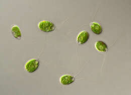
Differential interference contrast image of a cluster of cells attached to the coversl;ip by their flagella.
-
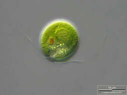
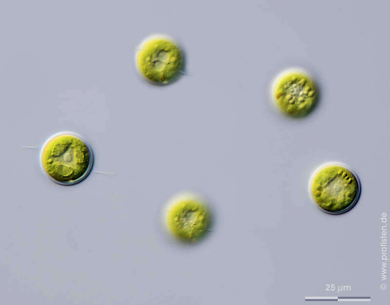
Place name: Ponds near Hausen (Hessisch Lichtenau , Germany) Latitude: 51.21453 Longitude: 9.868894Microscope Zeiss Axioskop, camera DSLR type.Copyright Winfried Hölz, Hausen, Germany.© Wolfgang Bettighofer,images under Creative Commons License V 3.0 (CC BY-NC-SA).For permission to use of (high resolution) images please contact
postmaster@protisten.de.For further information about the image, please click here:
Link to protisten.de page
-
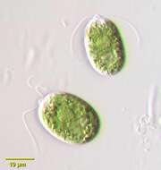
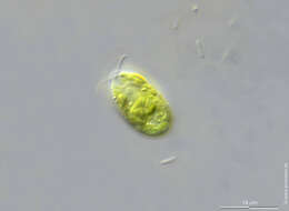
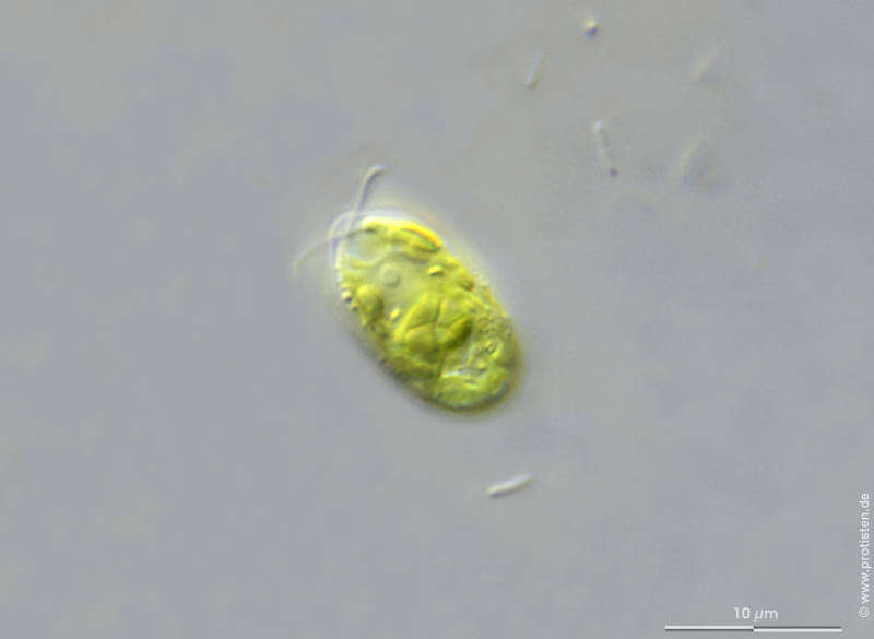
Sampling date 04/2016. Scale bar indicates 25 µm.Place name: Bog Dosenmoor near Neumuenster (Schleswig-Holstein, Germany)Latitude: 54.136219 Longitude: 10.026433Microscope Zeiss Axioplan, camera Olympus OM-D M5 MKII.© Wolfgang Bettighofer,images under Creative Commons License V 3.0 (CC BY-NC-SA).For permission to use of (high resolution) images please contact
postmaster@protisten.de.For further information about the image, please click here:
Link to protisten.de page
-
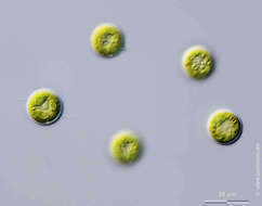
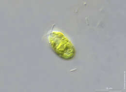
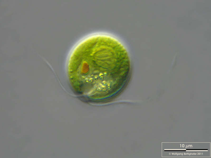
Sampling date 05/2011. Scale bar indicates 10 µm.Place name: Creek in Oder valley 100 km north east of Berlin (Germany)Latitude: 53.135032 Longitude: 14.348738Microscope Zeiss Universal, camera Olympus C7070WZ.© Wolfgang Bettighofer,images under Creative Commons License V 3.0 (CC BY-NC-SA).For permission to use of (high resolution) images please contact
postmaster@protisten.de.For further information about the image, please click here:
Link to protisten.de page
-
Sampling date 06/2013. Scale bars indicate 10 µm.Two images.First:Central pyrenoid with starch cap.Second:Optical section through the starch-enveloped pyrenoid.Please click on < or > on the image edges or on the dots at the bottom edge of the images to browse through the slides!Place name: Wetland Mittermoos near Fieberbrunn (Tyrol, Austria)Latitude: 47.47998695 Longitude: 12.5240922Microscope Zeiss Universal, camera Canon EOS 600D. DOF images.© Wolfgang Bettighofer,images under Creative Commons License V 3.0 (CC BY-NC-SA).For permission to use of (high resolution) images please contact
postmaster@protisten.de.For further information about the image, please click here:
Link to protisten.de page