-
Jeremy A. Miller, Charles E. Griswold, Nikolaj Scharff, Milan Řezáč, Tamás Szűts, Mohammad Marhabaie
Zookeys
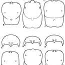
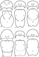
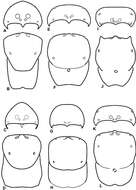
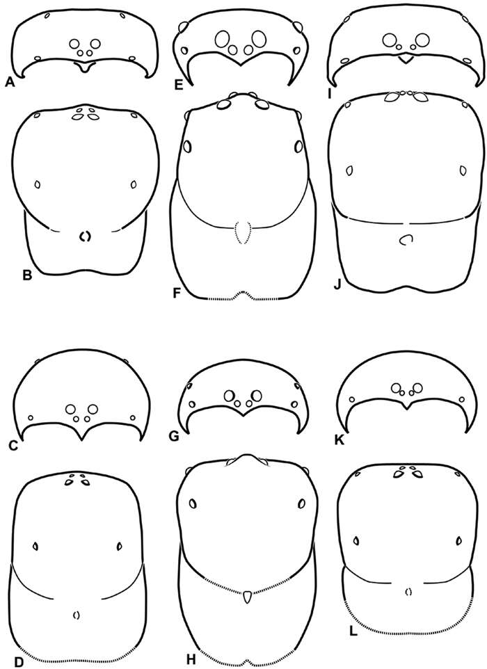
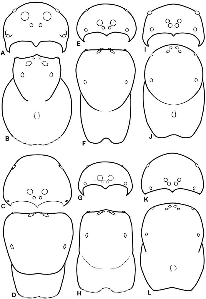
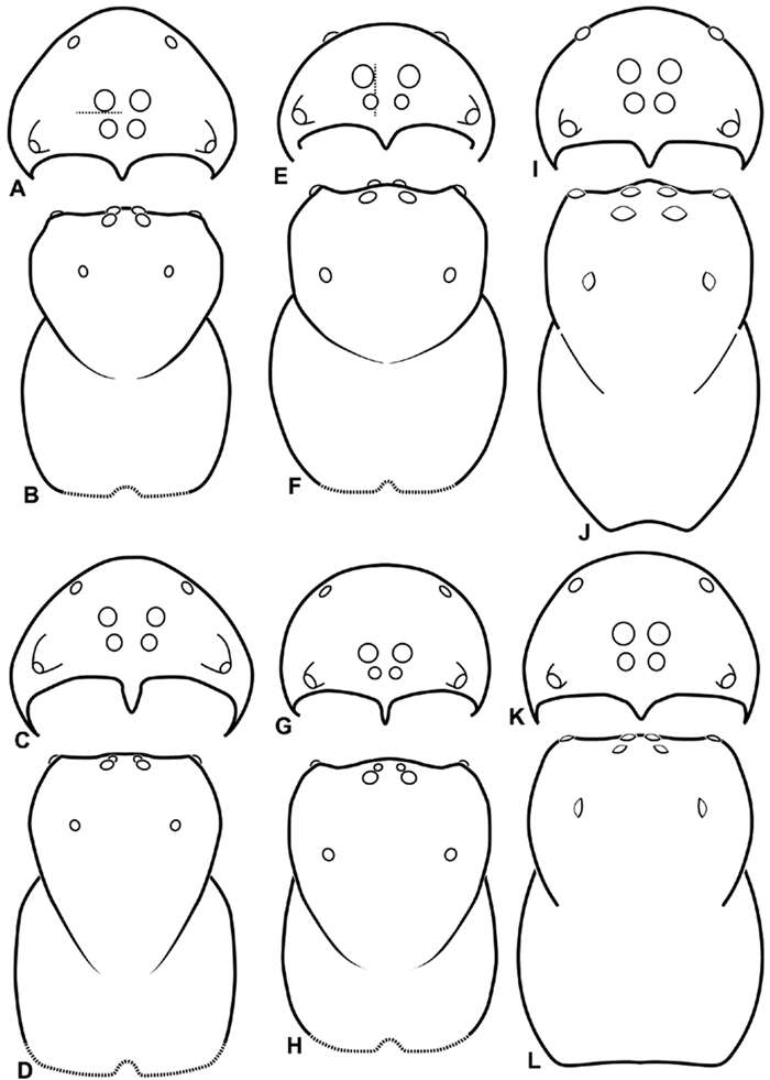
Figure 9.A–L Schematic illustrations of the carapace of assorted eresids. A–D Eresus kollari E–H Gandanameno sp. I–L Loureedia annulipes A–B, E–F, I–J male C–D, G–H, K–L female A, C, E, G, I, K anterior view B, D, F, H, J, L dorsal view. Dashed lines at posterior of carapace indicate uncertainty. Not to scale.
-
Jeremy A. Miller, Charles E. Griswold, Nikolaj Scharff, Milan Řezáč, Tamás Szűts, Mohammad Marhabaie
Zookeys
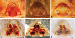
Figure 17.A–F Epigyna of Gandanameno sp., photomicrographs. A, D from Iringa, Tanzania (ZMUC 19970517, ZMUC) B, E from Kommetjie, Western Cape, South Africa (CASENT 9039241, CAS), note broken embolus left in female reproductive system C, F from Port Elizabeth, South Africa (port-3325, ZMHB) A–C ventral view D–F dorsal view, cleared. CD copulatory duct S spermatheca SH spermathecal head.
-
Jeremy A. Miller, Charles E. Griswold, Nikolaj Scharff, Milan Řezáč, Tamás Szűts, Mohammad Marhabaie
Zookeys
Figure 59.A–F Scanning electron micrographs of epigynum and vulva of Gandanameno sp. A, B from Iringa, Tanzania (ZMUC 19970530, ZMUC) C–F from Kommetjie, Cape Town, South Africa (CASENT 9039241, CAS) A epigynum, ventral view B detail of right copulatory opening, ventral view C cleared vulva, dorsal view D detail of right spermatheca and spermathecal head E detail, right spermatheca F detail, right spermathecal head. CD copulatory duct FD fertilization duct S spermatheca SH spermathecal head.
-
Jeremy A. Miller, Charles E. Griswold, Nikolaj Scharff, Milan Řezáč, Tamás Szűts, Mohammad Marhabaie
Zookeys
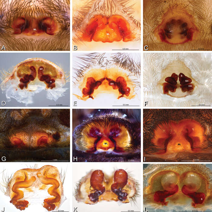
Figure 16.A–L Epigyna of eresid species, photomicrographs. A, D Adonea fimbriata; A from Mehav Am village, Israel (MR003, MR) D from Wadi Mashash, Israel (MR013, HUJ) B, E Dorceus fastuosus from Mashabim sand dunes, Israel (MR002, MR) C, F Dresserus sp. from Klein Kariba, South Africa (CASENT 9025745, CAS) G, J Eresus walckenaeri from 5 km south of Monemvasia, Lakonia, Greece (ZMUC 00012903, ZMUC) H, K Eresus kollari from res. Radotinske udoli, Czechia (MR016, MR) I, L Eresus sandaliatus from SE of Silkeborg, Denmark (CASENT 9039243, CAS) A–C, G–I ventral viewD–F, J–L dorsal view, cleared. CD copulatory duct ML median lobe S spermatheca SH spermathecal head.
-
Jeremy A. Miller, Charles E. Griswold, Nikolaj Scharff, Milan Řezáč, Tamás Szűts, Mohammad Marhabaie
Zookeys
Figure 29.A–F Dorceus fastuosus, female from Mashabim sand dunes, Israel (MR002, MR), scanning electron micrographs. A median eye group B prosoma, dorsal C epigynum, ventral view D cleared vulva, dorsal view E detail, left spermathecal head F detail, right spermatheca. ML median lobe S spermatheca SH spermathecal head.
-
Jeremy A. Miller, Charles E. Griswold, Nikolaj Scharff, Milan Řezáč, Tamás Szűts, Mohammad Marhabaie
Zookeys
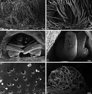
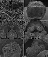
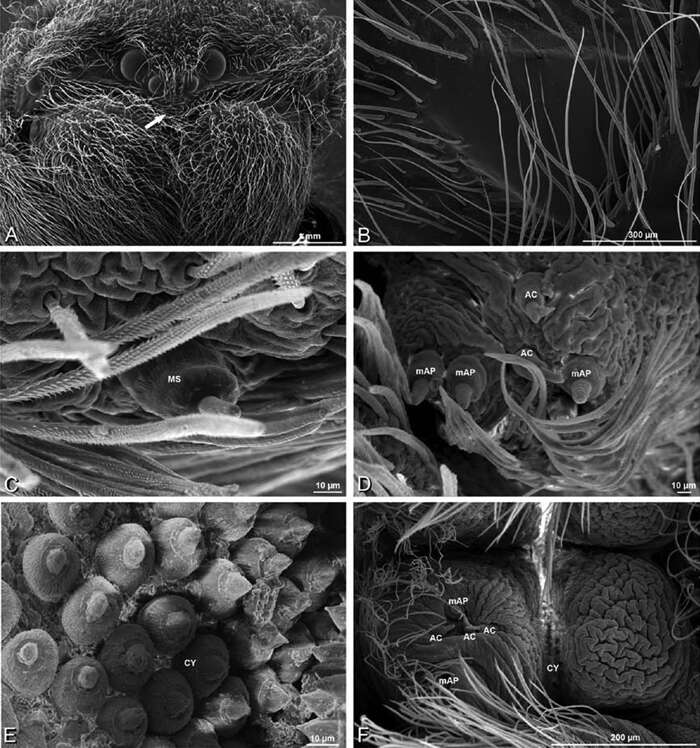
Figure 57.A–F Gandanameno sp. from Iringa, Tanzania (ZMUC 19970530, ZMUC), scanning electron micrographs of female spinnerets. A overview B right ALS C right PMS D left PLS E cribellum F cribellar spigots. AC aciniform gland spigot ALS anterior lateral spinneret CR cribellum CY cylindrical gland spigot MAP major ampullate gland spigot mAP minor ampullate gland spigot MS modified spigot n nubbin PI piriform gland spigot PLS posterior lateral spinneret PMS posterior median spinneret t tartipore.
-
Jeremy A. Miller, Charles E. Griswold, Nikolaj Scharff, Milan Řezáč, Tamás Szűts, Mohammad Marhabaie
Zookeys
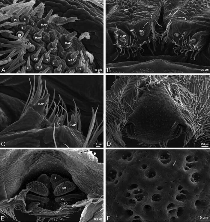
Figure 58.A–F Gandanameno sp., scanning electron micrographs. A–E female from Iringa, Tanzania (ZMUC 19970530, ZMUC) F female from Hanover, South Africa (SAM-ENW-B006896/9958) A, B prosoma C–F details of spinneret spigots A anterior view, arrow indicates clypeal hood B left cheliceral boss C detail of modified spigots on right female PLS D detail of spigots on anterior part of right female PMS E detail of cylindrical gland spigots on posterior part of left female PMS F right PMS. AC aciniform gland spigot CY cylindrical gland spigot MAP major ampullate gland spigot mAP minor ampullate gland spigot MS modified spigot n nubbin PI piriform gland spigot t tartipore.
-
Jeremy A. Miller, Charles E. Griswold, Nikolaj Scharff, Milan Řezáč, Tamás Szűts, Mohammad Marhabaie
Zookeys
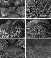
Figure 37.A–F Dresserus sp., scanning electron micrographs. A, C female from Mazumbai, Tanzania (CASENT 9025747, CAS) D, F female from Klein Kariba, South Africa (CASENT 9025745, CAS) A detail of spigots on right ALS B detail of spigots on anterior part of PMS C detail of spigots on anterior part of right PMS D epigynum, ventral view E vulva, dorsal view F detail of pores on right spermathecal head. AC aciniform gland spigot CD copulatory duct CY cylindrical gland spigot FD fertilization duct MAP major ampullate gland spigot mAP minor ampullate gland spigot PI piriform gland spigot S spermatheca SH spermathecal head t tartipore.
-
Jeremy A. Miller, Charles E. Griswold, Nikolaj Scharff, Milan Řezáč, Tamás Szűts, Mohammad Marhabaie
Zookeys
Figure 60.A–F Gandanameno sp. from Hanover, South Africa (SAM 9465, SAM), scanning electron micrographs of male spinnerets. A overview B left ALS C right PMS D left PLS E vestigial cribellum F detail of vestigial cribellum. AC aciniform gland spigot ALS anterior lateral spinneret MAP major ampullate gland spigot mAP minor ampullate gland spigot MS modified spigot PI piriform gland spigot PLS posterior lateral spinneret PMS posterior median spinneret n nubbin t tartipore.
-
Jeremy A. Miller, Charles E. Griswold, Nikolaj Scharff, Milan Řezáč, Tamás Szűts, Mohammad Marhabaie
Zookeys
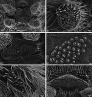
Figure 36.A–F Dresserus sp., female from Mazumbai, Tanzania (CASENT 9025747, CAS), scanning electron micrographs of spinnerets. A overview B left ALS C right PMS D detail, cylindrical gland spigots on right PMS E left PLS F cribellum. AC aciniform gland spigot ALS anterior lateral spinneret CR cribellum CY cylindrical gland spigot MAP major ampullate gland spigot mAP minor ampullate gland spigot MS modified spigot PI piriform gland spigot PLS posterior lateral spinneret PMS posterior median spinneret.
-
Jeremy A. Miller, Charles E. Griswold, Nikolaj Scharff, Milan Řezáč, Tamás Szűts, Mohammad Marhabaie
Zookeys
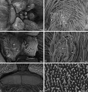
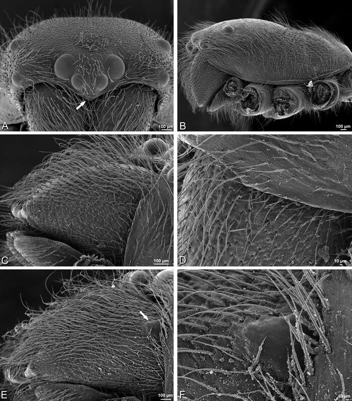
Figure 56.A–F Gandanameno sp., scanning electron micrographs of prosoma and chelicerae. A–D male from Harare, Zimbabwe (AcAT 2005/123, NCA) E, F male from Hanover, South Africa (SAM 9465, SAM) A prosoma, anterior view, arrow indicates clypeal hood B prosoma, lateral view C, E left chelicerae, lateral view, arrow in E indicates cheliceral boss D detail of left chelicerae showing absence of cheliceral boss F detail of left chelicerae showing cheliceral boss.
-
Jeremy A. Miller, Charles E. Griswold, Nikolaj Scharff, Milan Řezáč, Tamás Szűts, Mohammad Marhabaie
Zookeys
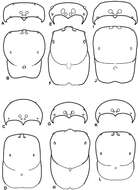
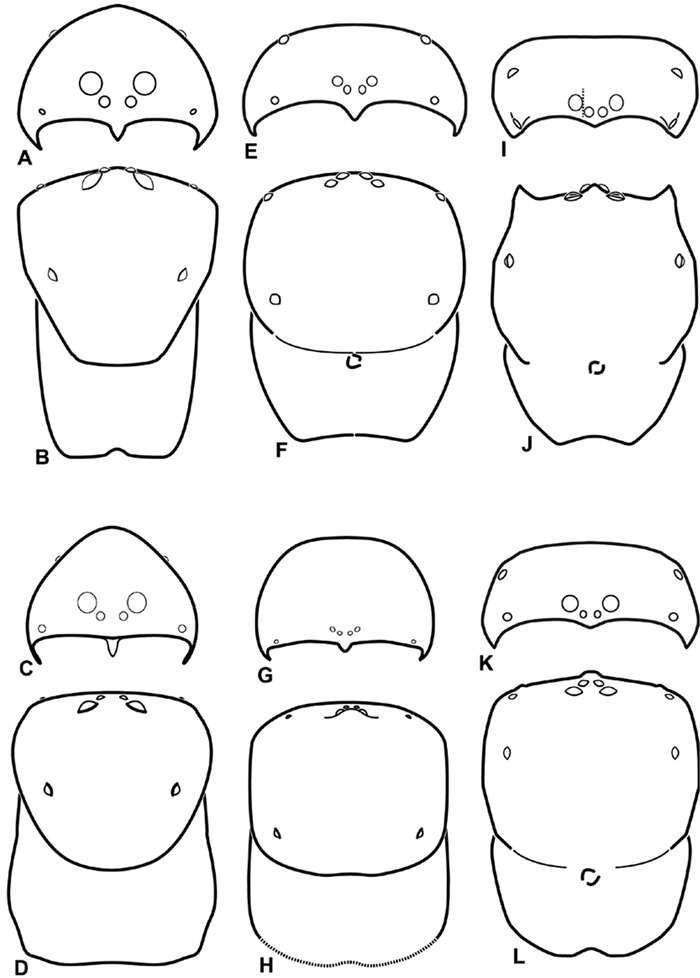
Figure 10.A–L Schematic illustrations of the carapace of assorted eresids. A–B Paradonea striatipes C–D Paradonea splendens E–H Paradonea variegata I–L Seothyra henscheli A–D, E–F, I–J male G–H, K–L female. A, C, E, G, I, K anterior view B, D, F, H, J, L dorsal view G illustrates example of median eyes overlapping on horizontal axis. Dashed lines at posterior of carapace indicate uncertainty. Not to scale.
-
Jeremy A. Miller, Charles E. Griswold, Nikolaj Scharff, Milan Řezáč, Tamás Szűts, Mohammad Marhabaie
Zookeys
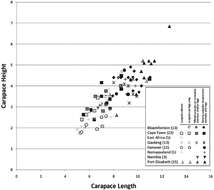
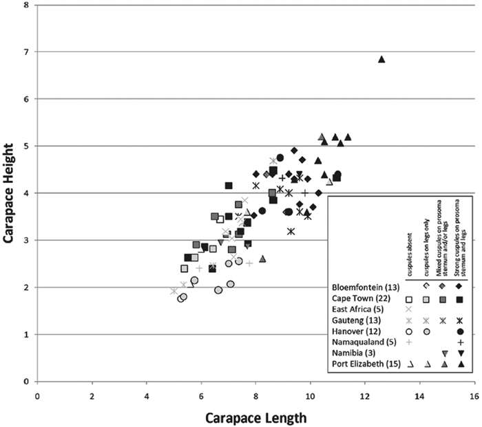
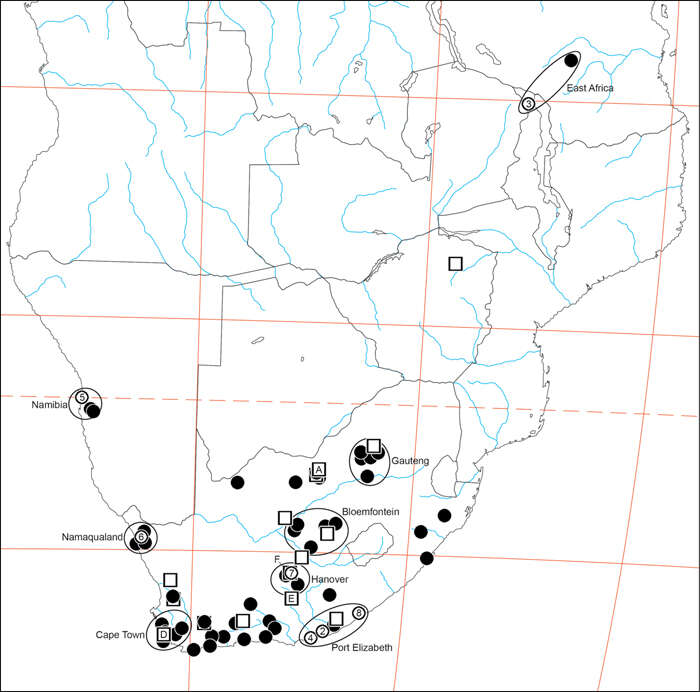
Figure 50.Carapace height plotted against carapace length for adult female Gandanameno specimens from eight regions: Bloemfontein, Cape Town, Gauteng, Hanover, Namaqualand, Namibia, and Port Elizabeth. Regions circumscribed in Fig. 49; sample size given in parentheses. Symbol shape indicates region while symbol darkness indicates presence and strength of cuspules. Specimens were scored as having cuspules absent, having medium to strong cuspules only on the legs, having a mixture of medium and strong cuspules on the prosoma, sternum, and/or legs, and having exclusively strong cuspules on the prosoma, sternum, and legs. As reflected in the legend, not all degrees of spinulation were observed in all regions.
-
Jeremy A. Miller, Charles E. Griswold, Nikolaj Scharff, Milan Řezáč, Tamás Szűts, Mohammad Marhabaie
Zookeys
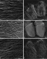
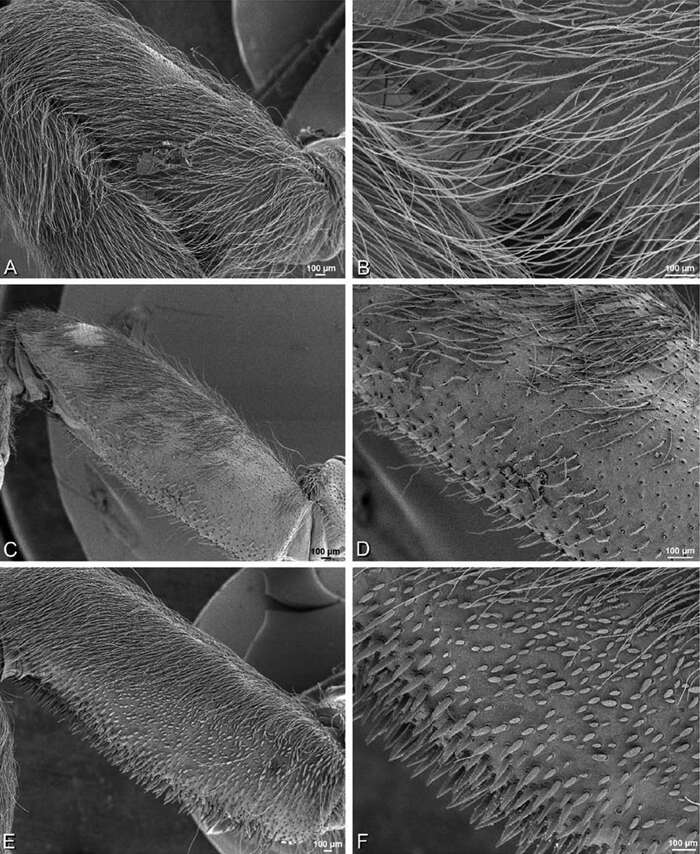
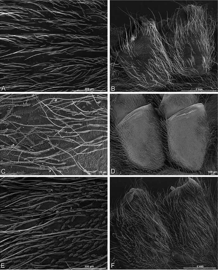
Figure 52.A–F Gandanameno sp., femur, left leg I of female, retrolateral view, scanning electron micrographs. A, C, E overview B, D, F detail of setae A, B from Iringa, Tanzania (ZMUC 19970530, ZMUC) C, D from Hanover, South Africa (SAM-ENW-B006896/9958, SAM) E, F from Eierfontein, Eastern Cape, South Africa (SAM-12823, SAM).
-
Jeremy A. Miller, Charles E. Griswold, Nikolaj Scharff, Milan Řezáč, Tamás Szűts, Mohammad Marhabaie
Zookeys
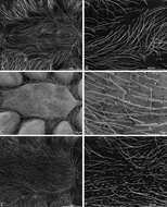
Figure 53.A–F Gandanameno sp., prosoma and coxae of female, scanning electron micrographs. A, C, E detail of setae on prosoma B, F right coxae I and II D left coxae I and II, image reversed to appear as right coxae A, B from Iringa, Tanzania (ZMUC 19970530, ZMUC) C, D from Hanover, South Africa (SAM-ENW-B006896/9958, SAM) E, F from Eierfontein, Eastern Cape, South Africa (SAM-12823, SAM).
-
Jeremy A. Miller, Charles E. Griswold, Nikolaj Scharff, Milan Řezáč, Tamás Szűts, Mohammad Marhabaie
Zookeys
Figure 54.A–F Gandanameno sp., sternum of female, scanning electron micrographs A, C, E overview of sternum B, D, F detail of setae on sternum A, B from Iringa, Tanzania (ZMUC 19970530, ZMUC) C, D from Hanover, South Africa (SAM-ENW-B006896/9958, SAM) E, F from Eierfontein, South Africa (SAM-12823, SAM).
-
Jeremy A. Miller, Charles E. Griswold, Nikolaj Scharff, Milan Řezáč, Tamás Szűts, Mohammad Marhabaie
Zookeys
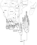
Figure 51.Bayesian phylogenetic tree of the spider family Eresidae based on mixed model analysis (eight data partitions, manually adjusted alignment; see Miller et al. 2010a); outgroups not shown, see Fig. S1. For the genus Gandanameno, DNA specimen codes are substituted for taxonomic name and specimens are linked to their collection locality in southern Africa. Male specimens indicated by male symbol, female specimens indicated either by a female symbol or a square, the darkness of which indicates the strength and presence of cuspules, scored as in Fig. 50. Branches drawn proportional to change. Numbers at nodes are percent posterior probabilities of 50 or greater.
-
Jeremy A. Miller, Charles E. Griswold, Nikolaj Scharff, Milan Řezáč, Tamás Szűts, Mohammad Marhabaie
Zookeys
Figure 49.Distribution of Gandanameno. Type localities are numbered circles, males are squares (if with letters, these refer to illustrations in Fig. 48), non-type females are filled circles. Type localities: circle 2 Eresus bubo L. Koch, 1865; circle 3 Eresus inornatus Pocock, 1898; circle 4 Eresus spenceri Pocock, 1900; circle 5 Eresus echinatus Purcell, 1908; circle 6 Eresus namaquensis Purcell, 1908; circle 7 Eresus depressus Tucker, 1920; circle 8 Eresus purcelli Tucker, 1920; type locality of Eresus fumosus C. L. Koch, 1837 is reported simply as “Afrika" and no type specimen is known (Lehtinen 1967: 235). Localities of males illustrated in Fig. 48: square A Fig. 48A–C square D Fig. 48D square E Fig. 48E square F Fig. 48F. Ellipsoids indicate regions for size chart Fig. 50, region names are for convenience only.
-
Jeremy A. Miller, Charles E. Griswold, Nikolaj Scharff, Milan Řezáč, Tamás Szűts, Mohammad Marhabaie
Zookeys
Figure 11.A–L Schematic illustrations of the carapace of assorted Stegodyphus species. A–D Stegodyphus lineatus E–H Stegodyphus mimosarum I–L Stegodyphus sarasinorum. A–B, E–F, I–J male C–D, G–H, K–L female A, C, E, G, I, K anterior view B, D, F, H, J, L dorsal view A illustrates example of median eyes separated on horizontal axis; E illustrates example of median eyes overlapping on vertical axis. Dashed lines at posterior of carapace indicate uncertainty. Not to scale.
-
Jeremy A. Miller, Charles E. Griswold, Nikolaj Scharff, Milan Řezáč, Tamás Szűts, Mohammad Marhabaie
Zookeys
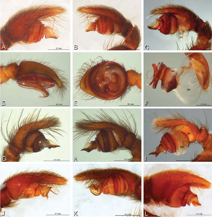
Figure 13.A–L Left male palpi of eresid species, photomicrographs. A–C Eresus kollari from res. Radotinske udoli, Czechia (MR007, MR) D–F Gandanameno sp. from Van Riebeeck Park, Western Cape, South Africa (CASENT 9023763, CAS) G–I Loureedia annulipes from Haluqim Ridge, Israel (PET03, MR) J, K Paradonea striatipes from Otjivasandu (NMN), Namibia L Paradonea splendens from Sunnyside, South Africa (C1076, SAM) A, D, G, J, L prolateral view B, H, K retrolateral view E ventral view C, F, I expanded palp. BH basal haematodocha MH median haematodocha.
-
Jeremy A. Miller, Charles E. Griswold, Nikolaj Scharff, Milan Řezáč, Tamás Szűts, Mohammad Marhabaie
Zookeys
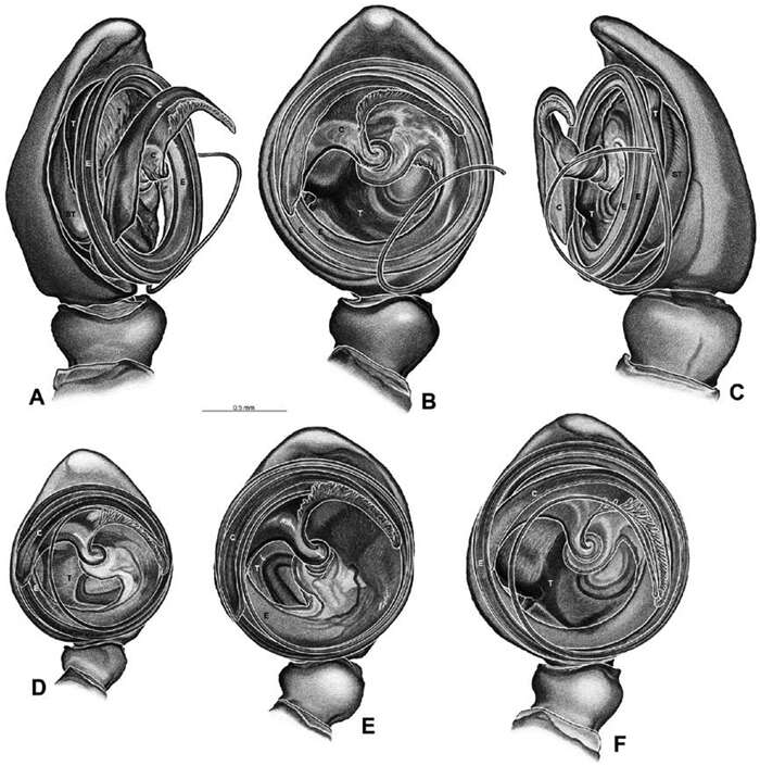
Figure 48.A–F Gandanameno sp., illustrations of left male palp. A–C from Naauwpoort, North West Province, South Africa (SAM 1600, SAM) D from Van Riebeeck Park, Western Cape, South Africa (CASENT 9023763, CAS) E from Graaff-Reinet, Eastern Cape, South Africa (SAM 12571, SAM) F from Hanover, South Africa (SAM 9465, SAM) A obliquely retrolateral view B, D–F ventral view C obliquely prolateral view. All images at the same scale. C conductor E embolus ST subtegulum T tegulum.
-
Jeremy A. Miller, Charles E. Griswold, Nikolaj Scharff, Milan Řezáč, Tamás Szűts, Mohammad Marhabaie
Zookeys
Figure 20.A–F Adonea fimbriata from Algeria-Morocco (MR012, MR), scanning electron micrographs of right male palp, images reversed to appear as left palp. A prolateral view B retrolateral view C detail of embolic division, prolateral view D detail of embolic division, retrolateral view E ventral view F apical view. C conductor E embolus ST subtegulum T tegulum.
-
Jeremy A. Miller, Charles E. Griswold, Nikolaj Scharff, Milan Řezáč, Tamás Szűts, Mohammad Marhabaie
Zookeys
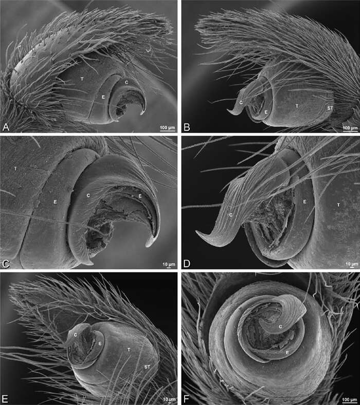
Figure 55.A–F Gandanameno sp. from Harare, Zimbabwe (AcAT 2005/123, NCA), scanning electron micrographs, right male palp, images reversed to appear as left palp. A prolateral view B retrolateral view C ventral view D apical view E detail of distal tip of conductor F palpal tibia, dorsal view. C conductor E embolus ST subtegulum T tegulum.
-
Jeremy A. Miller, Charles E. Griswold, Nikolaj Scharff, Milan Řezáč, Tamás Szűts, Mohammad Marhabaie
Zookeys
Figure 8.A–L Schematic illustrations of the carapace of assorted eresids A–D Adonea fimbriata E–H Dorceus fastuosus I–L Dresserus sp. A–B, E–F, I–J male C–D, G–H, K–L female A, C, E, G, I, K anterior view B, D, F, H, J, L dorsal view. Dashed line in I drawn tangential to the mesal margin of the PME does not intersect with the AME indicating median eyes separated on vertical axis. Dashed lines at posterior of carapace indicate uncertainty. Not to scale.