NMNH Plagiodinium belizeanum type specimen
Description:
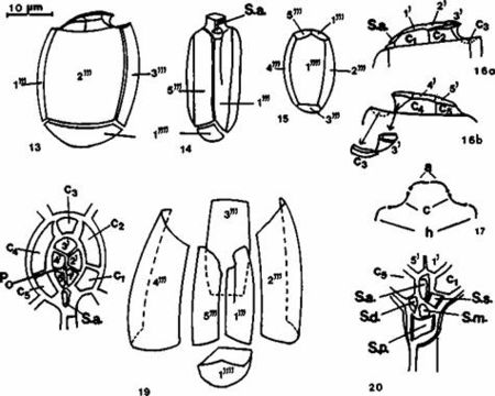
Figs 13-20. Line drawings of thecal plates of Plagiodinium belizeanum sp. nov. FIG. 13. Left lateral view; FIG. 14. Ventral view; FIG. 15. Antapical view; FIGS. 16. a, b) Plate composition of the epitheca and cingulum. Architecture of epithecal and cingular plates in a left view and right view; FIG. 17. Cross-section of the epitheca (a), cingulum (c), and hypotheca (h); A schematic representation of a section of a cell along the midplane parallel to the ventral surface. FIG. 18. Shape position and designation of plates. Relationship on epitheca plates is: (1’-5' and Po) cingulum (C1-C5), and sulcus (S.a.). A minute thecal element (PO), perhaps the 6th epithecal plate situated by the ventral internal border of the 4' plate, probably represents the rudiment of the PO plate. FIG. 19. Hypothecal plates (1'"-5'") and antapical plate 1"). FIG. 20. Designation, shape, and location of five sulcal plates: S.a., S.d., S.p., S.s., and S.m., surrounded by cingular plates Cl and C5 and epithecal plates 1' and 5'.
Included On The Following Pages:
- Life (creatures)
- Cellular (cellular organisms)
- Eukaryota (eukaryotes)
- SAR (Stramenopiles, Alveolates, Rhizaria)
- Alveolata (alveolates)
- Dinophyceae
- Prorocentrales
- Prorocentraceae
- Plagiodinium
- Plagiodinium belizeanum
- Dinoflagellata (dinoflagellates)
This image is not featured in any collections.
Source Information
- license
- cc-by-nc-sa-3.0
- copyright
- National Museum of Natural History, Smithsonian Institution
- original
- original media file
- partner site
- NMNH Marine Dinoflagellates
- ID