Image of American Levi tick
Description:
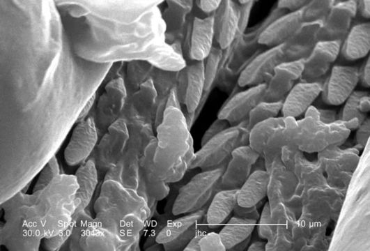
Under a magnification of 3043X, approximately 8 times greater than PHIL 9963, this scanning electron micrograph (SEM) depicted a dorsal view of an unidentified male Dermacentor sp. tick found upon a cat in the suburbs of Decatur, Georgia, which measured approximately 3.5mm from its gnathosoma (i.e., capitulum), which is where its mouthparts are located, to the distal abdominal margin (PHIL 9961). Note in PHIL 9959 and 9960, that the entire dorsum of this ticks abdomen is covered by its tough scutum, or shield, categorizing it as a male. In female Ixodid-species ticks, the scutum only partially covers the dorsal abdomen. Seen clearly in this image is the foliate covering of the ticks skin-piercing hypostome, which is located in what appears to be a trough between its pedipalps.
Created: 2006
Included On The Following Pages:
- Life (creatures)
- Cellular (cellular organisms)
- Eukaryota (eukaryotes)
- Opisthokonta (opisthokonts)
- Metazoa (Animal)
- Bilateria
- Protostomia (protostomes)
- Ecdysozoa (ecdysozoans)
- Arthropoda (arthropods)
- Chelicerata (chelicerates)
- Arachnida (arachnids)
- Acari (mites)
- Parasitiformes (parasitiform)
- Ixodida (ticks)
- Ixodoidea
- Ixodidae (hard ticks)
- Dermacentor (American Levi tick)
- Panarthropoda
This image is not featured in any collections.
Source Information
- license
- cc-publicdomain
- photographer
- Janice Carr
- provider
- Public Health Image Library
- original
- original media file
- visit source
- partner site
- Public Health Image Library
- ID