Image of American Levi tick
Description:
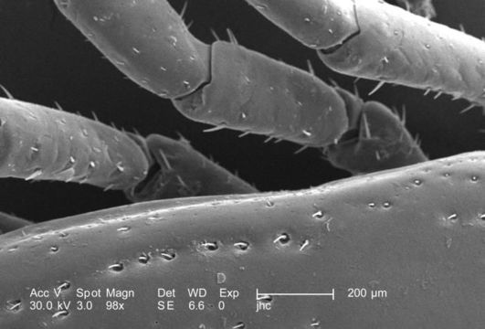
Under a relatively low magnification of 98X, this scanning electron micrograph (SEM) provided a closer view of this male Dermacentor sp. tick found upon a cat in the suburbs of Decatur, Georgia, which measured approximately 3.5mm from its gnathosoma (i.e., capitulum), which is where its mouthparts are located, to the distal abdominal margin (PHIL 9961). PHIL 9959 revealed all this ticks legs, placing it into the Phylum Arthropoda, i.e., from jointed ( Arthro), and legs (poda), as well as the Class Arachnida, for theyve eight of these legs, unlike insects, which use six appendages to move about. From proximal to distal location, each leg is comprised of a coxa, trochanter 1, trochanter 2, a femur, patella, tibia, a two-sectioned tarsus, and a two-part pretarsus, i.e., a pulvillus and claw. Here we see the trochantofemoral joints of the arachnids left 3rd and 4th legs, and the femoropatellar joints of its left 2nd and 3rd legs.
Created: 2006
Included On The Following Pages:
- Life (creatures)
- Cellular (cellular organisms)
- Eukaryota (eukaryotes)
- Opisthokonta (opisthokonts)
- Metazoa (Animal)
- Bilateria
- Protostomia (protostomes)
- Ecdysozoa (ecdysozoans)
- Arthropoda (arthropods)
- Chelicerata (chelicerates)
- Arachnida (arachnids)
- Acari (mites)
- Parasitiformes (parasitiform)
- Ixodida (ticks)
- Ixodoidea
- Ixodidae (hard ticks)
- Dermacentor (American Levi tick)
- Panarthropoda
This image is not featured in any collections.
Source Information
- license
- cc-publicdomain
- photographer
- Janice Carr
- provider
- Public Health Image Library
- original
- original media file
- visit source
- partner site
- Public Health Image Library
- ID