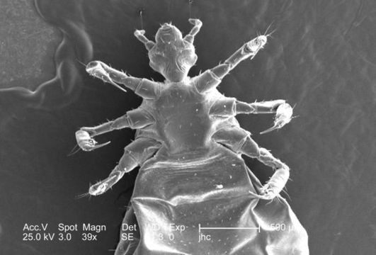
Image of Pediculus humanus Linnaeus 1758
Description:
This was one of five scanning electron micrographic (SEM) images (PHIL# 9243 9247), successively magnified at higher and higher values, which focused on the head region of a female body louse, Pediculus humanus var. corporis from a ventral perspective. At a relatively low magnification, this SEM revealed some of the insects exoskeletal morphology exhibited by its cephalic, or head region, thoracic, and proximal abdominal regions. Of interest is the jointed configuration of its six extremities, from which it derived its classification in the phylum of Athropoda, i.e., Arthro from "joint, and poda from "leg").
Created: 2006
Included On The Following Pages:
- Life
- Cellular
- Eukaryota (eukaryotes)
- Opisthokonta (opisthokonts)
- Metazoa (animals)
- Bilateria
- Protostomia (protostomes)
- Ecdysozoa (ecdysozoans)
- Arthropoda (arthropods)
- Pancrustacea
- Hexapoda (hexapods)
- Insecta (insects)
- Pterygota (winged insects)
- Neoptera (neopteran)
- Paraneoptera
- Psocodea (bark lice, book lice and true lice)
- Troctomorpha (book louse)
- Pediculidae (primate body lice)
- Pediculus
- Pediculus humanus
- Panarthropoda
- Nanopsocetae
This image is not featured in any collections.
Source Information
- license
- cc-publicdomain
- photographer
- Janice Carr
- provider
- Public Health Image Library
- original
- original media file
- visit source
- partner site
- Public Health Image Library
- ID