Image of book louse
Description:
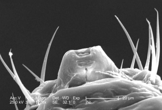
This was one of five scanning electron micrographic (SEM) images (PHIL# 9243 9247), successively magnified at higher and higher values, which focused on the head region of a female body louse, Pediculus humanus var. corporis from a ventral perspective. At a high magnification of 1228x, this SEM revealed some of the insects exoskeletal morphology exhibited by the cephalic region. Highlighted in this view is the insects cone-shaped mouth, which is surrounded by a number of setae, or sensorial hairs, which provide the organism with informational feedback about its environment such as chemistry and temperature.
Created: 2006
Included On The Following Pages:
- Life
- Cellular
- Eukaryota (eukaryotes)
- Opisthokonta (opisthokonts)
- Metazoa (animals)
- Bilateria
- Protostomia (protostomes)
- Ecdysozoa (ecdysozoans)
- Arthropoda (arthropods)
- Pancrustacea
- Hexapoda (hexapods)
- Insecta (insects)
- Pterygota (winged insects)
- Neoptera (neopteran)
- Paraneoptera
- Psocodea (bark lice, book lice and true lice)
- Troctomorpha (book louse)
- Pediculidae (primate body lice)
- Pediculus
- Pediculus humanus
- Panarthropoda
- Nanopsocetae
This image is not featured in any collections.
Source Information
- license
- cc-publicdomain
- photographer
- Janice Carr
- provider
- Public Health Image Library
- original
- original media file
- visit source
- partner site
- Public Health Image Library
- ID