Image of Streptococcus anginosus
Description:
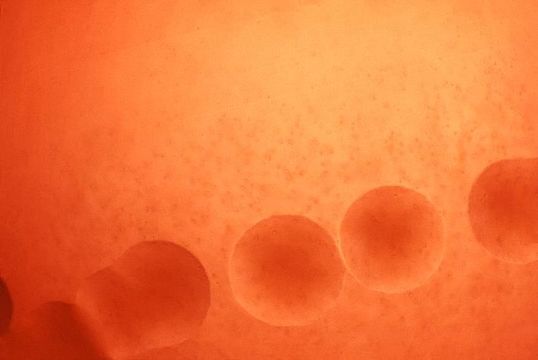
Magnified 100x, this 1977 photograph depicted a Petri dish filled with trypticase soy agar medium containing 5% defibrinated sheep's blood, i.e., blood agar plate (BAP). After having been inoculated with alpha-hemolytic Streptococcus anginosus bacteria, i.e., a member of the Gram-positive viridans group of streptococci (VGS), the BAP was incubated in a carbon dioxide enriched atmosphere at 35oC for 24 hours. The culture grew bacterial colonies, which were seen here. The characteristic color changes, i.e., a hazy, faded, indistinct region surrounding each colony in which some of the red blood cells (RBCs) were destroyed in the blood agar medium, or "hemolyzed", indicated that these bacteria were indeed alpha-hemolytic in nature.
It is the incomplete nature of the hemolytic reaction adjacent to the colonies, which spares numbers of RBCs in the blood agar medium, that is of qualitative importance when distinguishing alpha from beta-hemolysis
Created: 1977
Included On The Following Pages:
- Life (creatures)
- Cellular (cellular organisms)
- Bacteria
- Firmicutes (gram-positive bacteria)
- Bacilli
- Lactobacillales
- Streptococcaceae
- Streptococcus
- Streptococcus anginosus
This image is not featured in any collections.
Source Information
- license
- cc-publicdomain
- provider
- Public Health Image Library
- original
- original media file
- visit source
- partner site
- Public Health Image Library
- ID