Image of figeater beetle
Description:
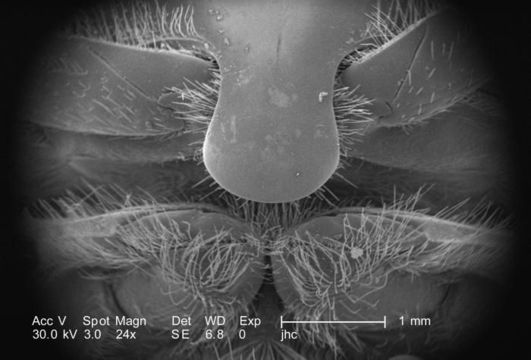
Under a low magnification of only 24x, this scanning electron micrograph (SEM) depicted some of the anatomical relationships found on the ventral surface of this adult figeater beetles, Cotinis mutabilis thorax, which is the body region situated between the insects head and abdomen. In this particular view, two of the three pairs of legs may be seen emanating adjacent to the prosternal process, which is a posterior extension of the sternum. The insect leg is comprised of a variable number of segments, however, there are usually six which predominate, including the most proximal, i.e., attaching the leg to the thorax, coxa, followed by the trochanter, femur, tibia, tarsus, and pretarsus, which in the case of this beetle is a claw, well visualized in PHIL 9943, 9944, and 9945.
Created: 2007
Included On The Following Pages:
- Life (creatures)
- Cellular (cellular organisms)
- Eukaryota (eukaryotes)
- Opisthokonta (opisthokonts)
- Metazoa (Animal)
- Bilateria
- Protostomia (protostomes)
- Ecdysozoa (ecdysozoans)
- Arthropoda (arthropods)
- Pancrustacea
- Hexapoda (hexapods)
- Insecta (insects)
- Pterygota (winged insects)
- Neoptera (neopteran)
- Endopterygota (endopterygotes)
- Coleoptera (beetles)
- Polyphaga
- Scarabaeiformia
- Scarabaeoidea (scarab beetle)
- Cotinis
- Cotinis mutabilis (figeater beetle)
- Scarabaeidae (scarab beetles)
- Panarthropoda
This image is not featured in any collections.
Source Information
- license
- cc-publicdomain
- photographer
- Janice Carr
- provider
- Public Health Image Library
- original
- original media file
- visit source
- partner site
- Public Health Image Library
- ID